Cem K. Parsak
1, Ulku Tuncer
2, Erol Kesiktas
3, Tolga Akcam
1, Gurhan Sakman
1,
Suleyman Ozdemir
2, Ozgur Tarkan
2, Ismail O. Kara
4Reconstruction of Cervical Esophagus Defects
by Free Jejunal Flap After Proximal Esophageal
Carcinoma Resections
Rekonstrukcja wad przełyku szyjnego za pomocą wolnego przeszczepu
jelitowego po proksymalnej resekcji z powodu raka
1 Department of General Surgery, Cukurova University Faculty of Medicine, Adana, Turkey 2 Department of Otolaryngology, Cukurova University Faculty of Medicine, Adana, Turkey
3 Department of Plastic and Reconstructive Surgery, Cukurova University Faculty of Medicine, Adana, Turkey 4 Department of Medical Oncology, Cukurova University Faculty of Medicine, Adana, Turkey
Abstract
Background. Surgical treatment of proximal esophageal defects remains one of the greatest challenges to sur-geons.
Objectives. The aim of this study was to evaluate the free jejunal flap surgical technique and the surgical outcomes in patients who underwent it for proximal esophageal carcinoma resections.
Material and Methods. The medical records of nine patients who underwent free jejunal flap reconstruction of the cervical esophagus for proximal esophageal cancer between January 2007 and December 2009 at the Cukurova University Faculty of Medicine, Adana, Turkey, were evaluated retrospectively. The patients’ age, gender, symp-toms, laboratory findings, diagnostic procedures, stage of disease, pathology, flap viability, length of hospitaliza-tion, post-operative ability to swallow, post-operative morbidity, mortality and recurrence were evaluated. The patients were followed up for 11 to 36 months.
Results. Although the free jejunal flap technique is a time-consuming and multi-step procedure, all of the free jejunal flaps survived. All of the patients exhibited good post-operative swallowing ability. One patient developed a salivary fistula. There was only one death, resulting from sepsis caused by late diagnosis of displacement of the jejunostomy tube.
Conclusions. All the patients regained their capacity to swallow, which improved their quality of life. The free jejunal flap procedure offers rapid rehabilitation and improved quality of life. The study involved only a limited number of patients, but confirmed that with the cooperation of the surgical teams, free jejunal flaps can be used successfully on suitable patients requiring laryngopharyngectomy with cervical esophagectomy (Adv Clin Exp Med 2011, 20, 6, 729–736).
Key words: esophageal cancer, free jejunal flap, reconstruction, microvascular.
Streszczenie
Wprowadzenie.Chirurgiczne leczeniewadprzełyku proksymalnego pozostaje jednymz największych wyzwań
dlachirurgów.
Cel pracy. Ocenatechniki chirurgicznej z użyciem wolnego płatajelita czczegoiwyniku leczenia chirurgicznego
u pacjentów, u których wykonano ten zabieg w celu proksymalnejresekcjirakaprzełyku.
Materiał i metody. Ocenionoretrospektywnie dokumentację medyczną 9 pacjentów, u których przeprowadzono rekonstrukcję szyjnej części przełyku wolnympłatem jelita czczego z powodu raka bliższego przełykuod stycznia
2007 do grudnia2009 r. naWydziale Lekarskim UniwersytetuCukurova, Adana, Turcja. Oceniono wiek pacjen -tów, płeć, objawy, wyniki badań laboratoryjnych, procedury diagnostyczne, stadium choroby, patologię, żywotność płata,czas pobytu w szpitalu, pooperacyjnązdolnośćdo przełykania,pooperacyjną zachorowalność, śmiertelność i występowanie nawrotów. Pacjencibyli obserwowani przez11–36 miesięcy.
Adv Clin Exp Med 2011, 20, 6, 729–736 ISSN 1230-025X
ORIGINAl PAPERS
Wyniki. Chociaż technika używająca wolnych płatów jelita czczegojestczasochłonna iwieloetapowa,wszyst
-kie wolnepłatyjelita czczegoprzeżyły.Wszyscy pacjenci wykazali się dobrą pooperacyjnązdolnościąpołykania. U jednego pacjentawystąpiłaprzetoka ślinowa.Tylko jedenpacjent zmarł z powodu sepsy, która wystąpiła przez późne rozpoznanieprzemieszczeniarurki dojejunostomii.
Wnioski. Wszyscy pacjenci odzyskali zdolność przełykania, co poprawiło jakość ich życia. Technika używająca wolnych płatów jelita czczego zapewnia szybką rehabilitację i poprawę jakości życia. W badaniu wzięła udział tylko ograniczona liczba pacjentów, ale autorzy potwierdzili, że przy współpracy zespołów chirurgicznych technika używająca wolnego płata jelita czczego może być z powodzeniem stosowana u odpowiednio dobranych pacjentów wymagających usunięcia krtani i gardła wraz z częścią szyjną przełyku (Adv Clin Exp Med 2011, 20, 6, 729–736).
Słowa kluczowe: rak przełyku, wolny płat jelita czczego, rekonstrukcja, mikrokrążenie.
In the literature there are reports of various reconstruction methods being used to achieve safe and functional reconstruction secondary to total pharyngolaryngoesophagectomy [1–5]. Deltopec-toral and musculocutaneous flaps are among these reconstruction methods, and are reported to be as-sociated with some disadvantages, including a pro-longed operation time, an increased rate of flap ne-crosis and other documented complications [3–6]. In pharyngoesophageal reconstruction operations with vascular anastomosis, gastrointestinal system continuity is maintained by transplanting a free intestinal flap or by using an intestinal segment with a pedicle [3]. The aim of the free jejunal flap is a single-stage reconstruction with low morbid-ity and mortalmorbid-ity, a short hospital stay and early restoration of swallowing. For many years this pro-cedure was infrequently used due to the low suc-cess rate of microvascular anastomosis. However, in 1992 Miller and lee reported a high success rate for reconstructions with jejunal free flaps [7]. With the advanced applications of microsurgery, almost all difficult defects of the esophagus can be reconstructed [7, 8], and increased experience and improved instrumentation have contributed the popularity of this technique.
Curative treatment of proximal esophageal cancer is a multistep process, involving pre-opera-tive chemoradiotherapy followed by extensive sur-gery, including laryngectomy and reconstruction of the upper part of the esophagus. The aim of this study was to evaluate the surgical technique, com-plications and results in a series of nine consecu-tive patients who underwent pharyngoesophageal resection for proximal esophageal cancer followed by reconstruction with a free jejunal flap.
Material and Methods
The authors retrospectively evaluated the medical records of nine patients (six males, three females) who underwent resection of cancer of the proximal esophagus followed by reconstruc-tion using free jejunal flap interposireconstruc-tion between January 2007 and December 2009 in the General
Surgery Department of the Cukurova University Faculty of Medicine, Adana, Turkey. The authors performed all the operations in collaboration with the Otolaryngology (ENT) Clinic and the Plastic and Reconstructive Surgery Clinic. All the patients were operated on by the same surgical team. The charts of all the patients were reviewed retrospec-tively. Data from these nine cases were evaluated for clinical characteristics, the stage of the disease, pathology, flap viability, length of hospitalization, post-operative ability to swallow and post-opera-tive morbidity and mortality.
The mean age of the patients was 64.9 years (ranging from 58 to 73 years). The male-to-female ratio was 6:3. All the patients were evaluated by means of endoscopy of the upper respiratory and gastrointestinal tract in order to determine the tumor spread and stage, and the diagnosis was confirmed by histological examination of the bi-opsy tissues. The histopathological diagnosis was
Table 1. Patient and tumor characteristics
Tabela 1. Charaterystyka pacjentów i guzów
Age – years
(Wiek – lata) mean: 64.9; range from 58 to 73 Gender (Płeć) 6 males/3 females Signs and symptoms (Objawy)
dysphagia weight loss
vomiting or regurgitation cough or hoarseness pain
9 (100%) 7 (77%) 3 (33%) 2 (22%) 2 (22%) Pathology (Patologia)
squamous cell carcinoma 9 (100%) Tumor stage – TNM (Stopień
zaawansowania nowotworu wg klasyfikacji TNM)
I II III
3 (33%) 2 (22%) 4 (44%) Pre-operative radiation therapy
squamous cell carcinoma (epidermoid carcinoma) in all cases; the clinical pre-operative staging ac-cording to the tumor-node metastasis (TNM) classification system is presented in Table 1. Ad-ditionally, the neck and thorax were evaluated with computed tomography, and the whole body with positron emission tomography. The patients’ su-pra-aortic branches were examined with Doppler ultrasonography to assess vascular anastomosis. After hospitalization, the patients’ nutritional sta-tus was established and a combination of enteral and parenteral nutritional support was started for all the patients.
The Surgical Technique
The operation was started by the ENT team: A to-tal pharyngolaryngectomy with bilateral modified neck dissection was performed on all the patients. Then the general surgery team started its involve-ment in the operation: A proximal esophagectomy with a negative surgical margin confirmed by in-traoperative frozen section was performed. For the free flap, a jejunum segment approximately 15 cm long was resected (from a location 20–30 cm from the Treitz ligament) with an appropriate ar-tery, vein and adequate intestinal arcade, using the transillumination technique and preserving the vascular pedicle of the remaining jejunum (Fig. 1). Just after the complete resection of the tumor, the vascular pedicle of the jejunal segment was excised and the recipient cervical vessels were prepared, following intravenous administration of 1500 IU of heparin sulphate. The proximal 3 cm of the re-sected segment was divided with bowel wall and mesentery to serve as a monitoring segment. A je-junojejunostomy was performed by end-to-end anastomosis between the proximal and distal jeju-num. Finally, the mesenteric window was closed. For early enteral nutrition, a Witzel enterostomy was performed distal to the bowel anastomosis in all patients and the abdomen was closed.
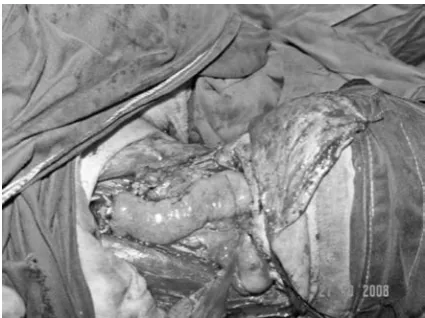
To prevent donor ischemia, the plastic and reconstructive surgical teams immediately began their participation in the operation. First, vascular anastomosis between the main venous branch of the jejunal segment and the vena jugularis interna was performed, using microvascular surgical tech-niques. Then the main jejunal artery was anasto-mosed with the arteria carotis externa. Peri-oper-ative Doppler ultrasonography was performed to confirm micro-vascular anastomosis. The viability and peristalsis of the jejunal segment were also carefully observed. After the vascular anastomosis, a 3 cm segment of the proximal jejunum was left attached to the revascularized mesenteric arcade and then externalized through the cervical wound
to serve as a monitor (Fig. 2). This “monitoring segment” has the same blood supply as the jeju-nal segment used for reconstruction but is not an integral part of the reconstruction. Enteral anas-tomosis between the proximal jejunum and the hypopharyngeal remnant of esophageal tissue and between the distal jejunum and distal esophagus was performed by interposing the free isoperistal-tic jejunal flap. Both proximal and distal bowel anastomosis was performed in one layer using ab-sorbable polyglactin sutures (Fig. 2). Anticoagula-tion treatment with subcutaneous low-molecular-weight heparin (enoxaparine sodium) was started in all patients on the first post-operative day and was continued for 15 days.
Fig. 1. Transillumination showing arches on the mes-enteric border of the jejunum
Ryc. 1. Transiluminacja ukazująca łuki na granicy krezki jelita czczego
Fig. 2. The completion of free jejunal flap anastomosis to the cervical esophagus. A jejunal monitoring seg-ment will be placed outside the wound
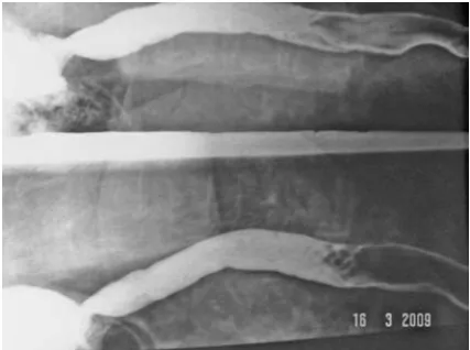
Urine output was monitored and hematologi-cal and biochemihematologi-cal examinations were performed every 24 hours. Post-operative complications were recorded, along with their management. Enteral feeding was started via jejunostomy on the first post-operative day and the amount was gradually increased to the full dose. The jejunal segment used as a monitor was checked every 6 hours, and obser-vation of flap viability lasted for five days (Fig. 3). On the fifth post-operative day, the monitoring segment was excised at the bedside under local an-esthesia by ligating and dividing the pedicle at skin level. On the seventh day, the jejunal flap passage was confirmed by fluoroscopy (Fig. 4) and patients were allowed oral intake if the examination results were normal. Oral alimentation was recommenced, first with liquids and then with semi-solids for the first 15 days. From the 15th day, a full range of food was allowed.
The length of hospitalization, mean oral li-quid starting time, early complications (in the first 15 days) and late complications (after 15 days) were recorded. The patients were followed up at 1, 6, 12, 18 and 24 months post-operatively in outpa-tient clinics. Their follow-up included endoscopy when required, evaluation of swallowing and of any changes in weight.
Results
The mean age of the female patients was 64.6 years (range: 61–68 years) and that of male patients was 65.1 years (range: 58–73 years). Of the nine patients, three had no previous treatment, while six had previously had radiotherapy due to local spread of the cancer. The most common complaint at the initial evaluation was dysphagia. All nine patients were treated by the same ENT, reconstructive and general surgery teams. All the patients were treated for epidermoid carcinoma of the hypopharynx with a laryngopharyngectomy and cervical esophagectomy. Tumor-free margins were achieved in all patients, as confirmed by fro-zen section evaluation, and all the operations were performed without any perioperative complica-tions.
During the early post-operative period, all the patients were managed in intensive-care-unit con-ditions. There were no flap failures or revisions in the series. There were no complications related to the donor site due to mobilization of the free je-junal transfer. The incidence of the post-operative complications in the series was low. One patient developed a salivary fistula due to an infection in the neck, and recovered with anti-inflammatory treatment and dressing changes. Other
post-op-erative medical complications included wound in-fection in two patients and pneumonia in one pa-tient. All of the complications were managed with conservative treatment. Unfortunately, one of the patients died in the early post-operative period due to peritoneal sepsis secondary to late diagnosis of jejunostomy tube displacement. Because of enteral nutrient leakage into the abdomen, the patient was re-operated to restore jejunal continuity and to insert a new jejunostomy tube. As a result of this complication the patient died seven days after his second operation (Table 2).
The average time period before resumption of oral feeding was 8.7 days after the operation (range: Fig. 3. The monitoring segment can be seen in the lat-eral aspect of the neck. On the fifth post-operative day, the monitoring segment is excised under local anesthe-sia at the bedside
Ryc. 3. Segment jelita do monitorowania jest widoczny w części bocznejszyi.W piątym dniupo operacji seg-ment do monitorowaniajest usuwanyw znieczuleniu
miejscowymprzy łóżku chorego
Fig. 4. On the seventh day, fluoroscopic examination revealed transit of a contrastmedium through the esophagus
Ryc. 4. Siódmego dnia badaniefluoroskopowe wyka
6–20 days). The longest period (20 days) was due to the patient with a salivary fistula. Swallowing was achieved in all patients after their recovery from the procedure. The mean length of hospitalization was 19.2 days (range: 15–27 days, Table 2). The patients were followed up in the outpatient clinic at 1, 6, 12, 18 and 24 months post-operatively. There were no late complications such as strictures (Table 3). For the eight patients for whom follow-up data
are available, the median survival time to the most recent control date is 17.2 months (range: 11–36 months). local recurrences of esophageal cancer have been detected in two patients
Discussion
The poor prognosis for hypopharyngeal and esophageal cancers have led surgeons to search for extensive surgical operations that maintain the physiological functions of the gastrointestinal sys-tem and also entail low morbidity and a short hos-pital stay. Increased experience and improvement of instrumentation for microvascular operations have contributed to the popularity of free jejunal flaps [4–9]. With technological improvements in microsurgery, long-segment supercharged ped-icled jejunal flaps are used for total esophageal reconstructions, and deep inferior epigastric per-forator flaps are used for partial reconstruction of the esophagus [10, 11].
Chen and Tang reported that in their experi-ence the best results in reconstructions of small seg-ments of the cervical esophagus might be achieved with advancement of the esophageal wall, when feasible [7]. Their second choice was substitution with another mucosal lining – e.g., a jejunal flap – because it heals the best. If the surgeon is not confident and not experienced with jejunal flaps, a musculocutaneous flap can be used. They ranked free skin flaps, such as free forearm flaps, as the last choice.
Table 2. Patients who underwent free jejunal flap reconstruction
Tabela 2. Pacjenci, u których wykonano rekonstrukcję za pomocą wolnego płata jelita czczego
Patient number, gender (Numer pacjenta, płeć)
Age – years (Wiek – lata)
Radio-therapy (Radiotera-pia)
Chemo-therapy (Chemio-terapia)
Complications
(Powikłania) Hospital stay – days (Czas pobytu w szpitalu – dni)
Time to oral liquid – days (Czas, jaki upłynął do przyjmowania pokarmów płynnych – dni)
1, M 58 + + – 16 7
2, M 67 – – – 15 6
3, F 60 + + – 17 8
4, M 73 – – pneumonia + wound infection 23 7
5, M 70 + – salivary fistula 27 20
6, F 63 + + – 16 7
7, M 66 + + wound infection 19 8
8, F 62 + – – 18 7
9, M 65 – – death 22 –
Table 3. Functional evaluation of patients at last follow-up (n = 8)
Tabela 3. Ocena czynnościowa pacjentów podczas ostatniej obserwacji (n = 8)
Regular diet (Typowa dieta) tube feeds
none 8 (100%)8 (100%) Dysphagia (Dysfagia)
none 8 (100%) Solids (Pokarmy stałe)
mild
severe 2 (25%)0 liquids (Pokarmy płynne)
mild
severe 2 (25%)0 Stricture-dilation
(Zwężenie, rozszerzenie) 0 Reflux (Refluks)
none mild severe
At the current authors’ institution, the free je-junal flap technique is used for the reconstruction of the proximal esophagus with the cooperation of the plastic and reconstructive surgery clinics. The revascularization success rate was 100%, which confirms the clinical usefulness and reliability of the procedure. This high rate of flap viability can be explained by the fact that the surgical team per-forms vascular anastomosis before enteral anasto-mosis and uses a monitoring segment for flap vi-ability. As far as the authors know, only one other free jejunal flap series (involving 14 patients) has been reported in the literature from Turkey [12]. Therefore the current series is important to dem-onstrate the feasibility of this surgical technique in the context of this country’s standards. The free jejunal flap technique ensures an excellent blood supply and one-stage reconstruction even in ir-radiated patients. In the series described, despite a high rate of pre-operative radiotherapy, there was no anastomotic leakage except one salivary fis-tula, and resumption of oral alimentation was pos-sible in all patients. After surgery, obvious weight gains were recorded during the patient follow-ups. Concerning the functional results, good swallow-ing has been preserved in all the patients, none of whom has had to change from a normal diet dur-ing the follow-up period, and no problems with gastric reflux have been noted.
Colon transfer is not a preferred technique for the reconstruction of defects, due to the high risk of post-anastomotic leakage in the donor area. The il-eum is narrow and its vascular structures are short. The jejunum is readily available, is generally free of intrinsic disease; it closely approximates the diam-eter of the esophagus, and might maintain intrinsic peristalsis after reconstruction. The jejunum, espe-cially the distal part of the duodeno-jejunal junc-tion, is a very good donor because it has a suitable vascular anatomy for the hypopharyngeal area [13].
It has been emphasized in the literature that jeju-nal free flaps, along with their vascular pedicles, have available anatomo-physiologic characteristics for the reconstruction of hypopharynx or cervical esophagus. The swallowing function may be re-es-tablished even earlier than with other flaps, owing to the secreting function of the mucosal layer [13]. The possibility of completing the surgery in one se-quence and reconstructing large defects, as well as early bowel movement are significant advantages of this procedure; visceral reconstruction without thoracic entry is a relative advantage of this proce-dure [14]. In addition to these advantages, as the current case series confirms, this technique can be used in patients who have undergone neoadjuvant and adjuvant radiotherapy without any complica-tions [4, 14, 15]. When the early- and late-onset
complications of the abdominal procedure are evaluated, the rates of anastomotic leakage, enteral obstruction, anastomotic stenosis and/or intra-abdominal abscess are very low [3].
The jejunal free flap technique involves specific complications related to the abdominal donor area, microvascular anastomosis or enteric anastomosis. Major complications of this procedure include flap insufficiency and bowel intussusceptions [5, 6]. In free flap reconstruction, the important point is the identification of vascular structures suitable for anastomosis. Depending on the patient’s vascular structures, the common carotid artery, transverse cervical artery, facial artery, lingual artery, superior thyroid artery, inferior thyroid artery, ascendant cervical artery, internal thoracic artery or brachio-cephalic artery might be used. The external jugular vein, internal jugular vein and facial vein are avail-able for venous anastomosis [7, 9]. The current authors preferred the external carotid artery and jugular vein for microvascular anastomosis in the operations described.
For the survival of free jejunal flaps, it is crucial to maintain the patency of the anatomized vessels. To ensure this, the jejunal flap should be frequently monitored post-operatively by fiber laryngoscope, Doppler ultrasonography or direct visualization in order to detect any disruption of the blood supply as early as possible. In the series described in this paper, the authors preferred to use a monitoring technique with a vascularized 3 cm jejunal segment externalized through the cervical wound to observe flap viability, similar to the technique described by Cho et al. [16] and Dionyssopoulos et al. [17].
Microvascular complications have decreased in the last 20 years [18]. A retrospective review by Disa et al. of 90 consecutive free jejunum transfers “demonstrated a 98% flap success rate. The mean hospital stay was 19 days. Nearly 65% of patients resumed an unrestricted diet and 88% maintained adequate peroral nutrition” [18]. Although the current study involves a limited number of cases, revascularization was successful in all of the recon-structions. In the current authors’ opinion, per-forming microvascular anastomosis before enteric anastomosis ensures good perfusion of the free flap and can provide early revision of vascular anasto-mosis in case of flap ischemia or congestion.
The most common complications of free je-junal reconstruction are wound healing disorders, hemorrhage, necrosis, pulmonary infections and fistula [19].Salivary fistulae and strictures are an-other risk in esophageal operations [14]. A salivary fistula occurred in one of the patients in the cur-rent study and was successfully managed conserva-tively. In the authors’ opinion, this salivary fistula occurrence might have been associated with the condition of the microvascular anastomosis rather than problems arising from the enteric anasto-mosis technique. The salivary fistula occurred in a 70-year-old patient with atherosclerotic cardio-vascular disease. This demonstrates that pre-oper-ative evaluation of co-morbid diseases is important to prevent complications.
The mean hospitalization time in the current study was acceptable: 19.2 days (range: 15–27 days), the longest stay being the patient with a sali-vary fistula. In a Danish report from 2010, the av-erage length of hospitalization was 24.1 days [20].
A potential limitation of this study is the small number of cases. However, as noted earlier, when considered along with those from the other clinical study with similar case numbers from Turkey, the combined results may provide a good picture of the procedure’s safety, utility and feasibility in this country [12].
The optimum reconstruction method should maintain alimentary comfort with low morbidity
and mortality [3]. Complications observed after surgery mainly affect the duration of hospitaliza-tion, the initiation of post-operative therapy (e.g. radiotherapy), nutritional status, quality of life and the cost of hospitalization. With advanced tech-niques and improved surgical equipment, the free jejunal flap has become almost a standard tech-nique in the reconstruction of hypopharyngeal and cervical esophageal defects. However, free je-junal flap reconstruction remains a procedure that requires a surgeon and/or surgical team skilled at microvascular techniques.
In this study, a high success rate was achieved with a low incidence of complications in the re-construction of the proximal esophagus. Col-laboration among different clinics has made it possible to use this technique, but it remains a technically demanding operation best suited for patients with defects in the cervical esophagus due to proximal cancers. Gastric tube interposi-tion or a supercharged pedicled jejunal flap may be more appropriate for reconstructing the hypo-pharynx and cervical esophagus, when the resec-tion extends below the thoracic inlet [21]. In the author’s opinion, based on a limited number of cases, the free jejunal flap technique can be used safely and successfully for the reconstruction of the hypopharynx or cervical esophagus in centers where cooperation among different surgical teams is possible.
References
[1] Suh JD, Sercarz JA, Abemayor E, Calcaterra TC, Rawnsley JD, Alam D, Blackwell KE: Analysis of outcome and complications in 400 cases of microvascular head and neck reconstruction. Arch Otolaryngol Head Neck Surg 2004, 130, 962–966.
[2] Nakatsuka T, Harii K, Asato H, Takushima A, Ebihara S, Kimata Y, Yamada A, Ueda K, Ichioka S: Analytic re-view of 2372 free flap transfers for head and neck reconstruction following cancer resection. J Reconstr Microsurg 2003, 19, 363–368.
[3] Hanson RP, Chow TK, Feehan E, Eadie PA, Timon CT, Keogh S: Analysis of functional results and quality of life following free jejunal flaps for reconstruction after upper aerodigestive neoplastic resection: the St James’s experi-ence. J Plast Reconstr Aesthet Surg 2007, 60, 577–582.
[4] Ikeguchi M, Miyake T, Matsunaga T, Yamamoto M, Fukumoto Y, Yamada Y, Fukuda K, Saito H, Tatebe S, Tsujitani S: Free Jejunal Graft Reconstruction After Resection of Neck Cancers: Our Surgical Technique. Surg To-day 2009, 39, 925–928.
[5] Wong CH, Wei FC: Microsurgical free flap in head and neck reconstruction. Head Neck 2010, 32, 1236–1245.
[6] Clark JR, Gilbert R, Irish J, Brown D, Neligan P, Gullane PJ: Morbidity after flap reconstruction of hypopharyn-geal defects. laryngoscope 2006, 116, 173–181.
[7] Chen HC, Tang YB: Microsurgical reconstruction of the esophagus. Semin Surg Oncol 2000, 19, 235–245.
[8] Archibald S, Young JE, Thoma A: Pharyngo-cervical esophageal reconstruction. Clin Plast Surg 2005, 32, 339– 346.
[9] Lam LK, Wei WI, Chan VS, Ng RW, Ho WK: Microvascular free tissue reconstruction following extirpation of head and neck tumour: experience towards an optimal outcome. J laryngol Otol 2002, 116, 929–936.
[10] Ascioti AJ, Hofstetter WL, Miller MJ, Rice DC, Swisher SG, Vaporciyan AA, Roth JA, Putnam JB, Smythe WR, Feig BW, Mansfield PF, Pisters PW, Torres MT, Walsh GL: long-segment, supercharged, pedicled jejunal flap for total esophageal reconstruction. J Thorac Cardiovasc Surg 2005, 130, 1391–1398.
[11] Louie O, Dickinson B, Granzow J, Boyd JB: Reconstruction of total laryngopharyngectomy defects with deep in-ferior epigastric perforator flaps. J Reconstr Microsurg 2009, 25, 555–558.
[13] Chen HC, Rampazzo A, Gharb BB, Wong MT, Mardini S, Chen HY, Salgado CJ: Motility differences in free colon and free jejunum flaps for reconstruction of the cervical esophagus. Plast Reconstr Surg 2008, 122, 1410– 1416.
[14] Disa JJ, Pusic AL, Mehrara BJ: Reconstruction of the hypopharynx with the free jejunum transfer. J Surg Oncol 2006, 94, 466–470.
[15] Ott K, Lordick F, Molls M, Bartels H, Biemer E, Siewert JR: limited resection and free jejunal graft interposition for squamous cell carcinoma of the cervical oesophagus.Br J Surg 2009, 96, 258–266.
[16] Cho BC, Shin DP, Byun JS, Park JW, Baik BS: Monitoring flap for buried free tissue transfer: its importance and reliability. Plast Reconstr Surg 2002, 110, 1249–1258.
[17] Dionyssopoulos A, Harris PG, Karagergou E, Ferrarro P, Guertin L, Danino AM: Monitoring of free jejunal transfer. J Plast Reconstr Aesthet Surg 2010, 63, e209–210.
[18] Disa JJ, Pusic AL, Hidalgo DA, Cordeiro PG: Microvascular reconstruction of the hypopharynx: defect classifica-tion, treatment algorithm, and functional outcome based on 165 consecutive cases. Plast Reconstr Surg2003, 111, 652–660.
[19] Zhao D, Gao X, Guan L, Su W, Gao J, Liu C, Luo X, Li X: Free jejunal graft for reconstruction of defects in the hypopharynx and cervical esophagus following the cancer resections. J Gastrointest Surg 2009, 13, 1368–1372.
[20] Wallentin RS, Sorensen HB, Bundgaard T, Pahle E, Nordsmark M, Pilegaard H: Reconstruction using free je-junal transfer after resection of cancer of the upper oesophagus. Dan Med Bul 2010, 57, 1–5.
[21] Triboulet JP, Mariette C, Chevalier D, Amrouni H: Surgical management of carcinoma of the hypopharynx and cervical esophagus: analysis of 209 cases. Arch Surg 2001, 136, 1164–1170.
Address for correspondence:
Cem K. Parsak
Çukurova Üniversitesi Týp Fakültesi, Genel Cerrahi Anabilim Dalý Balcalý, Adana
Turkey
Tel.: 0 322 338 60 60/3171 E-mail: [email protected]
Conflict of interest: None declared