gested influence on atherosclerosis development. Higher serum TSH could become easy to diag-nose and eliminate cardiovascular (CV) risk fac-tor, but the relationship between SCH and ad-verse CV outcomes are still uncertain. Although some interventional studies showed that L-thy-roxine replacement therapy in SCH is able to im-prove most CV structural and functional surro-gate markers [3–5], there is no clear evidence that restoration of euthyroidism in SCH reduces CV disease risk. Recent European Thyroid Associa-Subclinical hypothyroidism (SCH) is defined
as an elevated serum thyroid stimulating hormone (TSH) concentration in the presence of normal serum free thyroxine (fT4) and triiodothyronine (fT3) concentrations. This condition has been re-cently more commonly recognized due to thyroid function testing increase in primary care. The oc-currence of SCH is estimated from 4% to 10% of the population, being diagnosed more frequent-ly in women and elderfrequent-ly people [1, 2]. The im-portance of SCH results mainly from its
sug-Maria Adamarczuk-Janczyszyn
1, A–D, F,
Aleksandra Zdrojowy-Wełna
2, C, D, F, Natalia Rogala
2, D–F,
Katarzyna Zatońska
3, D–F, Grażyna Bednarek-Tupikowska
2, A, C, E, FEvaluation of Selected Atherosclerosis Risk Factors
in Women with Subclinical Hypothyroidism Treated
with L-Thyroxine
1 Endocrinological Outpatient Clinic, Bolesławiec, Poland
2 Department of Endocrinology, Diabetology and Isotope Therapy, Wroclaw Medical University, Poland 3 Department of Social Medicine, Wroclaw Medical University, Poland
A – research concept and design; B – collection and/or assembly of data; C – data analysis and interpretation;
D – writing the article; E – critical revision of the article; F – final approval of article
Abstract
Background. Subclinical hypothyroidism (SCH) is a common endocrine disorder, probably increasing cardiovas-cular (CV) risk. However, the relation between SCH and atherosclerosis risk factors remains unclear.
Objectives. The aim of the study was to evaluate selected atherosclerosis risk factors in women with SCH in com-parison to a group of healthy women and women with overt hypothyroidism, as well as to investigate the influence of L-thyroxine replacement on those risk factors.
Material and Methods. The study group consisted of 187 obese women aged between 50 and 70 years: 100 women with SCH, 45 women with overt hypothyroidism and 42 women with TSH level in reference ranges. Anthropometric parameters were evaluated. Laboratory tests included thyroid hormones concentrations, lipid profile with apoli-poproteins, CRP, homocysteine. Atherosclerotic indexes were calculated: LDL C/HDL C ratio, apoA1/apoB ratio and Castelli risk index. Women with hypothyroidism were given L-thyroxine treatment and after 6 months in euthyroidism the evaluation was repeated.
Results. Total cholesterol, LDL-cholesterol and triglycerides concentrations as well as LDL-C/HDL-C ratio and Castelli index were higher in SCH than in controls and decreased after L-thyroxin substitution. All of the calculated atherosclerosis indexes showed significant positive correlations with TSH concentration in SCH group. Also in this group the systolic and diastolic blood pressure decreased significantly after treatment.
Conclusions. Dyslipidemia in obese SCH women is not severe, but if untreated for many years, it may lead to atherosclerosis. Substitution therapy improves the lipid profile, changing the relations between protective and proatherogenic fractions of serum lipids, and optimises blood pressure (Adv Clin Exp Med 2016, 25, 3, 457–463).
Key words: risk factors, atherosclerosis, subclinical hypothyroidism, cardiovascular disease, L-thyroxine treatment.
ORIGINAL PAPERS
Adv Clin Exp Med 2016, 25, 3, 457–463
tion Guidelines recommend individual approach to each patient [6].
The aim of the study was to evaluate selected atherosclerosis risk factors in women with SCH in comparison to a group of healthy women and women with overt hypothyroidism, as well as to investigate the influence of L-thyroxine replace-ment on those risk factors in SCH and overt hypo-thyroidism groups.
Material and Methods
The study group consisted of 187 postmeno-pausal women aged between 50 and 70 years (mean 61.2 ± 9.7 years), recruited in Endocrino-logical Outpatient Clinic in Bolesławiec in years 2005–2008. Written informed consent was ob-tained from each participant. The study protocol was approved by the ethical committee.
Specific anamnesis and general physical exam-inations were done. Exclusion criteria were drugs interfering with TSH, fT4, serum lipids and homo-cysteine (HCY) serum levels and other causes of elevated serum TSH concentration.
Body weight and height were measured. The BMI was calculated as body weight in kilo-grams divided by height in square meters (kg/m2). Waist circumference was measured at the umbilical level, hip circumference was measured at the great-er trochantgreat-er, waist-to-hip ratio (WHR) was also calculated. Blood pressure was measured on the left arm after 10 min of rest. Ultrasonography of the thyroid gland was performed with ALOKA appa-ratus, transducer frequency 7.5 mHz. Blood sam-ples were drawn after an overnight fast (12 h) for plasma TSH, fT4, anti-thyroid peroxidase antibod-ies (TPO-Ab), triglycerides (TG), HDL cholesterol (HDL-C), LDL cholesterol (LDL-C), apoliporoteins – apoA1 and apoB, HCY, C-reactive protein (CRP).
The serum TSH, FT4 and TPO-Ab levels were measured by immunofluorescence (Immu-lite 1000; DPC). The levels of TC, TG and HDL-C were measured with a colorimetric method (Flex-or E, Vitalab). LDL-C was calculated by means of the Friedewald equation. ApoA1, apoB and CRP were measured with the use of nephelometric methods (Siemens) and HCY with enzyme con-version immunoassay (AxSYM test, Abbott).
For each patient atherosclerotic indexes were calculated: LDL-C/HDL-C ratio, Castelli risk index (TC-HDL-C/HDL-C) and apoA1/apoB ratio.
The patients were divided into subgroups based on the thyroid function tests:
– One hundred women with SCH, with fT4 in reference ranges and TSH level above 4.0 µIU/mL (ranged from 4.1 to 12 µIU/mL with mean value
8.6 ± 4.0 µIU/mL). All patients after confirming SCH received L-thyroxine therapy and were ob-served for 6 months. During that time all became euthyroid and the examinations were repeated.
S1 – SCH women before treatment, S2 – the same group after treatment.
– Fourty-five women with overt hypothy-roidism, TSH level over 10 µIU/mL (mean val-ue 44.6 ± 21.6) and fT4 below below 0.8 ng/mL. All patients after confirming hypothyroidism re-ceived L-thyroxine therapy and were observed for 6 months. During that time all became euthyroid and the examinations were repeated.
H1 – women with hypothyroidism before treatment, H2 – the same group after treatment.
– Fourty-two healthy women with TSH level in reference ranges – 0.4 to 4.0 µIU/mL (mean val-ue 1.5 ± 0.8) and normal fT4 in reference ranges – 0.8 to 1.9 ng/dL (mean value 1.3 ± 0.2 ng/dL).
C – control group.
Variables were described with the use of ele-ments of descriptive statistics and included: mini-mum and maximini-mum value, mean and standard de-viation.
Planned comparisons between two of three pairs of independent continuous variables were made with the Mann-Whitney U test. The 5% sig-nificance level was set to each individual compar-ison, than the whole family of comparisons as in multiple comparisons, i.e. the Bonferroni correc-tion to adjust the significance level was not ap-plied. This approach has more power to detect true differences [7].
Differences between dependent continuous variables were evaluated by the Wilcoxon signed-rank test.
In order to examine the correlation between the analyzed indices, Spearman’s rank correlation coefficient was calculated.
Results
Thyroid Function
As a result of subgroups definition, before treatment TSH concentrations were significantly higher in SCH and overt hypothyroidism groups than in controls. After treatment TSH levels de-creased significantly and showed no difference with TSH in controls (Table 1).
RR, BMI, WHR
vs. 138.3/84.2 mm Hg in S2). RRs and RRd were al-so higher in S1 group in comparial-son to controls, but the difference was not statistically significant. RRs, RRd were significantly higher in H1 group than in control group. After treatment with L-thyroxine
RRs and RRd decreased and there was no differ-ence between groups H2, S2 and controls (Table 2). Study participants were mostly obese or over-weight (mean BMI over 30). There was no signifi-cant difference in BMI and WHR between groups before and after treatment and in comparison to control group (Table 2).
Serum Lipids and
Atherosclerosis Indexes
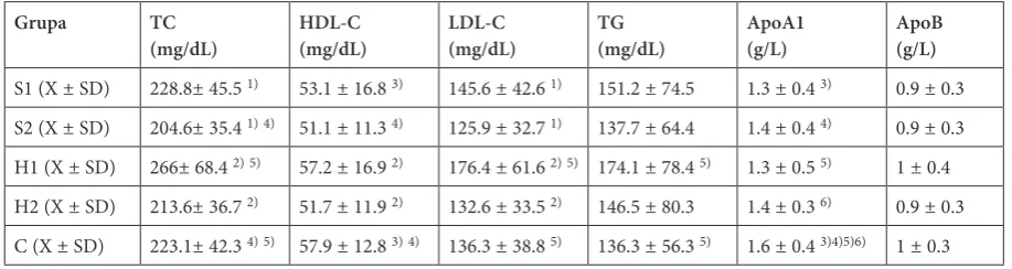
TC and LDL-C concentrations were higher in S1 and H1 groups than in controls and decreased significantly after L-thyroxin substitution. After 6 months of treatment both parameters were low-er in S2, H2 than in controls (statistically signif-icant only TC in S2). TG concentration was also higher in S1 and H1 than in controls (but signif-icantly only in group with overt hypothyroidism) and decreased after treatment, but the differenc-es between S1 and S2, as well as H1 and H2 did not reach statistical significance. HDL-C levels were lower in S1 and H1 than in the control group and decreased after treatment in both groups
(Ta-Table 1. TSH and fT4 concentrations before and after treatment
Group TSH (µIU/mL) fT4 (ng/dL) S1 (X ± SD) 8.6 ± 4.0 1)3) 1.1 ± 0.2 1)3) S2 (X ± SD) 1.9 ± 1.0 1) 1.4 ± 0.3 1)3) H1 (X ± SD) 44.6 ± 21.6 2)5) 0.6 ± 0.2 2)5) H2 (X ± SD) 1.4 ± 0.9 2) 1.6 ± 0.3 2)6) C (X ± SD) 1.6 ± 0.8 3)4) 1.3 ± 0.2 3)4)5)6) X – mean value; SD – standard deviation; S1 – SCH group before treatment; S2 – SCH group after treatment; H1 – group with overt hypothyroidism before treatment; H2 – group with overt hypothyroidism after treatment; C – control group; statistically significant differences between groups (p < 0.05): 1) S1 and S2; 2) H1 and H2; 3) S1 and C; 4) S2 and C; 5) H1 and C; 6) H2 and C.
Table 2. Characteristic of the groups before and after the treatment with L-thyroxine
Group BMI
(kg/m2) WHR RRs (mm Hg) RRd (mm Hg) V thyroid (mL) TPO-Ab (IU/mL)
S1 (X ± SD) 30.1 ± 5.7 0.9 ± 0.1 144.4 ± 2.01) 87.1 ± 9.81) 12.9 ± 12.3 225 ± 356.7 S2 (X ± SD) 30.5 ± 5.9 0.9 ± 0.1 138.3 ± 18.81) 84.2 ± 10.41)
H1 (X ± SD) 31 ± 5.7 0.9 ± 0.1 152.3 ± 22.22)3) 90.9 ± 9.42)3) 12.8 ± 16.7 471.7 ± 446.6 H2 (X ± SD) 30.9 ± 5.7 0.9 ± 0.1 138 ± 20.82) 84.9 ± 9.32)
C (X ± SD) 29.9 ± 5.1 0.9 ± 0.1 141.7 ± 15.83) 84.5 ± 9.63) 14 ± 9.2 78.5 ± 232.1
X – mean value; SD – standard deviation; RRs – systolic blood pressure; BMI – body mass index; RRd – diastolic blood pressure; WHR – waist-hip ratio; V thyroid – thyroid volume; S1 – SCH group before treatment; S2 – SCH group after treatment; H1 – group with overt hypothyroidism before treatment; H2 – group with overt hypothyroidism after treatment; C – control group; statistically significant differences between groups (p < 0.05): 1) S1 and S2, 2) H1 and H2, 3) H1 and C.
Table 3. Serum lipids and apolipoproteins concentrations before and after treatment Grupa TC
ble 3). There was also a significant positive corre-lation between fT4 and HDL-C concentrations in S1 and H2 groups (p-values 0.02 and 0.004, respec-tively) – data not shown.
ApoA1 concentration was significantly lower in each group with subclinical and overt hypothy-roidism before and after treatment in comparison to the control group. L-thyroxine treatment did not influence significantly the apoA1 level in any group. ApoB concentrations in examined groups did not differ significantly (Table 3).
LDL-C/HDL-C ratio was in SCH group sig-nificantly higher than in the controls. It also de-creased after treatment, the difference before and after L-thyroxin substitution was not statistically significant (S1 vs. S2); however, the value between S2 and controls did not differ significantly. In the group with overt hypothyroidism, the LDL-C/ /HDL-C ratio was significantly higher before treat-ment than in the controls and after treattreat-ment. Cas-telli risk index (TC-HDL-C/HDL-C) was signifi-cantly higher in S1 and H1 than in the controls, but the treatment decreased it only insignificant-ly. L-thyroxin substitution influenced apoB/apoA1 ratio only in the group with overt hypothyroid-ism (Table 3). All of the calculated
atherosclero-sis indexes showed statistically significant positive correlations with TSH concentration in S1 group (p-values for LDL-C/HDL-C: 0.002, Castelli risk index: 0.02, apoB/apoA1: 0.03). There were also statistically significant negative correlations be-tween fT4 and two indexes - LDL-C/HDL-C and Castelli risk index in S1 group (p-values 0.03 and 0.02, respectively) – data not shown.
Homocysteine
HCY concentration was the highest in the group with overt hypothyroidism before treat-ment, but the differences between groups and af-ter treatment were not significant (Table 5). There was also no correlation between HCY and TSH or fT4 (data not shown).
CRP
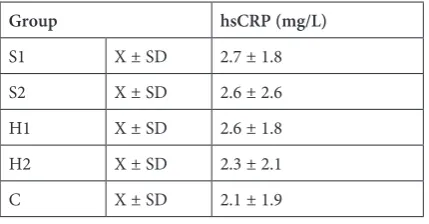
CRP concentrations were higher before treat-ment both in SCH and overt hypothyroidism groups than after 6 months of L-thyroxine replace-ment and in comparison to controls, but the dif-ferences were not statistically significant (Table 6). There were no significant correlations between CRP and TSH or fT4 (data not shown).
Table 4. Atherosclerosis indexes before and after treatment
Group LDL-C/HDL-C Castelli risk index apoB/apoA1
S1 (X ± SD) 2.9 ± 1.2 2) 3.6 ± 1.3 2) 0.7 ± 0.2
S2 (X ± SD) 2.6 ± 0.8 3.1 ± 0.9 0.7 ± 0.2
H1 (X ± SD) 3.2 ± 1.3 1)3) 4 ± 1.7 3) 0.9 ± 0.3 1)3)
H2 (X ± SD) 2.6 ± 0.8 1) 3.3 ± 1.0 0.7 ± 0.2 1)
C (X ± SD) 2.5 ± 0.8 2)3) 3 ± 0.9 2)3) 0.6 ± 0.2 3)
X – mean value; SD – standard deviation; S1 – SCH group before treatment; S2 – SCH group after treatment; H1 – group with overt hypothyroidism before treatment; H2 – group with overt hypothyroidism after treatment; C – control group; sta-tistically significant differences between groups (p < 0.05): 1) H1 and H2, 2) S1 and C, 3) H1 and C.
Table 5. Homocysteine concentrations before and after treatment
Group Homocysteine (HCY)(µmol/L) S1 X ± SD 13.0 ± 3.2
S2 X ± SD 13.6 ± 4.1 H1 X ± SD 15.2 ± 3.4
H2 X ± SD 14.4 ± 7
C X ± SD 13.5 ± 4.4
X – mean value; SD – standard deviation; S1 – SCH group before treatment; S2 – SCH group after treatment; H1 – group with overt hypothyroidism before treatment; H2 – group with overt hypothyroidism after treatment; C – control group.
Table 6. CRP concentrations before and after treatment Group hsCRP (mg/L)
S1 X ± SD 2.7 ± 1.8
S2 X ± SD 2.6 ± 2.6
H1 X ± SD 2.6 ± 1.8
H2 X ± SD 2.3 ± 2.1
C X ± SD 2.1 ± 1.9
Discussion
SCH is a common finding, often causing a treatment dilemma in endocrine practice. Euro-pean guidelines recommend a L-thyroxine substi-tution in younger patients (< 65 years), especially with some symptoms suggestive of hypothyroid-ism, with goiter, after hemithyroidectomy or with serum TSH > 10 mU/L. However, patients with TSH < 10 mU/L and lack of complaints might be also monitored without treatment [6]. Such ambi-guity arises from unanswered questions, including whether SCH increases CV risk or mortality and whether it negatively influences metabolic param-eters.
Our patient group was classified to a catego-ry, where no clear indications are given – aged 50–70 years, with median TSH 8.6 ± 4.0 µIU/mL, without obvious symptoms of hypothyroidism. The most important reason to treat such individu-als would be to improve their metabolic profile, es-pecially taking into consideration obesity affecting most of them. We assessed the influence of SCH on clinical and laboratory markers of CV risk as well as the changes of those parameters after treat-ment with L-thyroxine. The limitation of our study is the number of patient groups, because the ex-amined parameters (such as body weight or serum lipids) are influenced by many other factors, espe-cially lifestyle. That might be the reason why some correlations did not reach statistical significance, but some tendencies were observed.
The first examined CV risk factor was blood pressure. It was higher in SCH group than in con-trols, and interestingly enough both RRs and RRd decreased significantly after the treatment (Ta-ble 2). Before L-thyroxine supplementation mean RRs value was over 140 mm Hg – allowing us to diagnose hypertension, and after 6 months it fell below this limit. Our results correspond with ma-ny other studies in this field. Adrees et al. ob-served a reduction of blood pressure and intima media thickness together with a normalization of glomerular filtration rate in SCH women treated 18 months with L-thyroxine, suggesting its hemo-dynamic effect [8]. Dernellis et al. showed that hy-pothyroidism is connected to aortic stiffness, and hormone replacement therapy eliminated hyper-tension in 50% of patients [9]. The role of hypo-thyroidism, even subclinical, in hypertension de-velopment probably stems from increasing the systemic arterial resistance and influence mostly RRd. Even the elevation of TSH in normal rang-es might be related to higher blood prrang-essure [10].
The second measured parameter was BMI and WHR. Most of the patients in our study were obese or overweight, and there was no significant
differ-ence between SCH vs. control or overt hypothy-roidism group (Table 2). Hormone substitution did not influence body weight or WHR. In con-trast to our results, there are some studies show-ing that elevated TSH, even in upper reference ranges, may lead to weight gain [11]. Also Knud-sen et al. observed a positive correlation between TSH and BMI; however, this relationship disap-peared when BMI exceeded 30 kg/m2 [12]. Au-thors explain that when BMI reaches the limit of obesity, lifestyle factors like diet or physical activity have a greater influence on body weight than thy-roid hormones. The European Thythy-roid Associa-tion declares: “There is no evidence for a favorable effect of L-thyroxine therapy on body weight in obese subjects with serum TSH levels < 10 mU/L and normal fT 4 concentrations” [6]. Our results remain in accordance with this claim.
de-crease was only statistically significant in overt hy-pothyroidism group in LDL-C/HDL-C ratio. Some authors reported a similarly beneficial influence of hormone therapy on LDL-C/HDL-C ratio [17], but others failed to show such relations [18]. As for atheroprotective HDL particles, their concentra-tion was lower in both hypothyroidism groups vs. controls in our study. Surprisingly, HDL-C level fell even lower after the treatment, which remains in contrast to the general advantageous effect of L-thyroxine therapy on lipid profile. The possi-ble explanation might be the decrease of TC lev-el. Moreover, the relations between LDL and HDL cholesterol improved after treatment, so we can conclude that decrease of HDL-C in this situation does not cause negative effect on CV profile.
A case-control INTERHEART study empha-sized the influence of apoB/apoA ratio on CV risk in every ethnic and age group [19]. In our study apoA1 concentration was significantly lower in groups with subclinical and overt hypothyroid-ism than in controls and tended to increase af-ter treatment (Table 3). ApoB concentration did not differ significantly among the studied groups. The proatherogenic apoB/apoA ratio was the high-est in patients with overt hypothyroidism and the lowest in euthyroid group (Table 4). Once again, SCH group presented intermediate profile among studied groups. There was a significant correlation between apoB/apoA ratio and TSH level in SCH group before treatment. However, L-thyroxine treatment caused a significant decrease of this in-dex only in the group with overt hypothyroidism, not in SCH group. The data concerning this prob-lem in literature is scarce. According to Turhan et al., there was no difference in apoB concentra-tion between SCH and control group [18], while
some authors reported not only higher apoB level, but also its positive correlation with TSH in SCH patients [17]. The observations concerning apoA1 are also unclear. In some studies L-thyroxine sub-stitution caused increase of apoA1 level [20], while in some it remained unchanged [21].
Generally, most of the authors note a positive influence of thyroid hormone supplementation on lipid profile in patients with SCH. Our work remains consistent with this statement. The re-ported changes in absolute values may not be very large, but the relations between protective and proatherogenic factors changed in a beneficial way and they seem to be more important in predict-ing CV risk.
Hiperhomocysteinemia, which is an indepen-dent CV risk factor, was also reported to be associ-ated with SCH [22]. However, some studies denied this correlation [23]. We did not observe differenc-es in HCY concentration between SCH and con-trol group or significant changes after treatment (Table 5). Probably, a greater effect of factors such as age, body weight and vitamin B status limited the influence of SCH on HCY.
We also observed no clear association between SCH or thyroid hormone substitution in SCH with CRP concentration. Therefore, our results are sim-ilar to some large studies [24, 25]. Those findings imply that SCH probably does not play any impor-tant role in inflammation associated with athero-sclerosis.
Summarizing, our results indicate that SCH in postmenopausal women causes a negative effect on CV risk, mainly due to increase of blood pres-sure and disturbances in serum lipid profile. Treat-ment with L-thyroxine may reverse some of those phenomena.
References
[1] Canaris GJ, Manowitz NR, Mayor G, Ridgway EC: The Colorado thyroid disease prevalence study. Arch Intern Med 2000, 160, 526–534.
[2] Vanderpump MP, Tunbridge WM, French JM: The incidence of thyroid disorders in the community: A twenty-year follow-up of the Whickham Survey. Clin Endocrinol 1995, 43, 55–68.
[3] Razvi S, Ingoe L, Keeka G, Oates C, McMillan C, Weaver JU: The beneficial effect of L-thyroxine on cardiovas-cular risk factors, endothelial function, and quality of life in subclinical hypothyroidism: Randomized, crossover trial. J Clin Endocrinol Metab 2007, 92, 1715–1723.
[4] Monzani F, Caraccio N, Kozakowa M, Dardano A, Vittone F, Virdis A, Taddei S, Palombo C, Ferrannini E:
Effect of levothyroxine replacement on lipid profile and intima-media thickness in subclinical hypothyroidism: A double-blind, placebo-controlled study. J Clin Endocrinol Metab 2004, 89, 2099–2106.
[5] Taddei S, Caraccio N, Virdis A, Dardano A, Versari D, Ghiadoni L, Salvetti A, Ferrannini E, Monzani F:
Impaired endothelium-dependent vasodilatation in subclinical hypothyroidism: Beneficial effect of L-thyroxine therapy. J Clin Endocrinol Metab 2003, 88, 3731–3737.
[6] Pearce S, Brabant G, Duntas L, Monzani F, Peeters RP, Razvi S, Wemeau JL: 2013 ETA Guideline: Management of subclinical hypothyroidism. Eur Thyroid J 2013, 2, 215–228.
[7] Keppel G, Wickens TD: Design and Analysis: A Researcher’s Handbook, 4th Edition. Upper Saddle River, NJ:
Pearson Prentice Hall, 2004.
[9] Dernellis J, Panaretou M: Effects of Thyroid replacement therapy on arterial blood pressure in patients with hyperten-sion and hypothyroidism. Am Heart J 2002, 143, 718–724.
[10] Asvold BO, Bjøro T, Nilsen TI, Vatten LJ: Association between blood pressure and serum TSH concentration within the reference range: A population based study. J Clin Endocrinol Metab 2007, 92, 841–845.
[11] Fox CS, Pencina MJ, D’Agostino RB, Murabito JM, Seely EW, Pearce EN, Vasan RS: Relations of thyroid func-tion to body weight. Arch Intern Med 2008, 268, 587–592.
[12] Knudsen N, Laurberg P, Rasmussen LB, Bülow I, Perrild H, Ovesen L, Jørgensen T: Small differences in thyroid function may be important for body mass index and the occurrence of obesity in the population. J Clin Endocrinol Metab 2005, 90, 4019–4024.
[13] Boekholdt SM, Titan SM, Wiersinga WM, Chatterjee K, Basart DC, Luben R, Wareham NJ, Khaw KT: Initial thyroid status and cardiovascular risk factors: The EPIC-Norfolk prospective population study. Clin Endocrinol 2010, 72, 404–410.
[14] Bindels AJ, Westendorp RG, Frolich M, Seidell JC, Blokstra A, Smelt AH: The prevalence of subclinical hypo-thyroidism at different total plasma cholesterol levels in middle-aged men and women: A need for case-finding? Clin Endocrinol 1999, 50, 217–220.
[15] Meier C, Staub JJ, Roth CB, Guglielmetti M, Kunz M, Miserez AR, Drewe J, Huber P, Herzog R, Müller B:
TSH-controlled L-thyroxine therapy reduces cholesterol levels and clinical symptoms in subclinical hypothyroid-ism: A double blind, placebocontrolled trial (Basel Thyroid Study). J Clin Endocrinol Metab 2001, 86, 4860–4866.
[16] Manninen V, Elo MO, Frick MH: Lipid alterations and decline in the incidence of coronary heart disease in the Helsinki Heart Study. JAMA 1988, 260, 641–651.
[17] Caraccio N, Ele F, Monzani F: Lipoprotein profile in subclinical hypothyroidism: Response to levothyroxine replacement, a randomized placebo-controlled study. J Clin Endocrinol Metab 2002, 87, 1533–1538.
[18] Turhan S, Sezer S, Erden G, Guctekin A, Ucar F, Ginis Z, Ozturk O, Bingol S: Plasma homocysteine concentra-tions and serum lipid profile as atherosclerotic risk factors in subclinical hypothyroidism. Ann Saudi Med 2008, 28, 96–101.
[19] McQueen MJ, Hawken S, Wang X, Ounpuu S, Sniderman A, Probstfield J, Steyn K, Sanderson JE, Hasani M, Volkova E: Lipids, lipoproteins, and apoliporoteins as risk markers of myocardial infarction in 52 countries. (The INTERHEART study): A case-control study. Lancet 2008, 372, 224–233.
[20] Caron P, Calazel C, Parra HJ, Hoff M, Louvet JP: Decreased HDL cholesterol in subclinical hypothyroidism: The effect of levothyroxine therapy. Clin Endocrinol 1990, 33, 519–523.
[21] Razvi S, Shakoor A, Vanderpump M, Weaver JU, Pearce S: The influence of age on the relationship between sub-clinical hypotyroidism and ischemic heart disease: A metaanalysis. J Clin Endocrinol Metab 2008, 93, 2998–3007.
[22] Evrengul H, Tanriverdi H, Enli Y, Kuru O, Seleci D, Bastemir M, Kilic A, Kaftan A, Kilic M: Interaction of plasma homocysteine and thyroid hormone concentrations in the patogenesis of the slow coronary flow phenom-enon. Cardiology 2007, 108, 186–192.
[23] Christ-Crain M, Meier C, Guglielmetti M, Huber P.R, Riesen W, Staub JJ, Müller B: Elevated C-reactive protein and homocysteine values: Cardiovascular risk factor in hypothyroidism? A cross-sectional and a double-blind, placebo-controlled trial. Atherosclerosis 2003, 166, 379–386.
[24] Hueston WJ, King DE, Geesey ME: Serum biomarkers for cardiovascular inflammation in subclinical hypothy-roidism. Clin Endocrinol 2005, 63, 582–587.
[25] Bell RJ, Rivera-Woll L, Davison SL, Topliss DJ, Donath S, Davis SR: Well-being, health-related quality of life and cardiovascular disease risk profile in women with subclinical thyroid disease – a community-based study. Clin Endocrinol 2007, 66, 548–556.
Address for correspondence:
Aleksandra Zdrojowy-Wełna
Department of Endocrinology, Diabetology and Isotope Therapy Wroclaw Medical University
ul. Pasteura 4 50-367 Wrocław Tel.: +48 71 784 25 59
E-mail: [email protected]
Conflict of interest: None declared