type includes 3 immunohistochemical subgroups, namely germinal center B cell-like (GCB), non-germinal center B cell-like (non-GCB) and, least frequently, CD5-positive DLBCL [1].
CD5-positive DLBCL remains a poorly-de-scribed, relatively new subgroup of lymphomas, constituting about 10% of DLBCL cases [1]. Sparse reports suggest worse prognosis in this popula-Diffuse Large B Cell Lymphoma (DLBCL) is
the most common non-Hodgkin lymphoma. It oc-curs most frequently in the elderly and is typical-ly characterized by rapid progression and thus re-quires immediate treatment. The current 2008 WHO classification distinguishes multiple types and variants of DLBCL, the most common of which being DLBCL, not otherwise specified (NOS). This
Nina Woźnialis
1, A–D, Beata Gierej
1, 2, B, C, E, Lidia Popławska
3, B,
Mateusz Ziarkiewicz
4, B, D, Ewa Wolińska
2, B, C, Elżbieta Kulczycka
1, B,
Bogna Ziarkiewicz-Wróblewska
1, 2, A, C, E, FAngiogenesis in CD5-positive Diffuse
Large B Cell Lymphoma: A Morphometric Analysis
1 Department of Pathology and Laboratory Diagnostics, Maria Sklodowska-Curie Institute and Oncology Center,
Warszawa, Poland
2 Department of Pathology, Center for Biostructure Research, Medical University of Warsaw, Poland 3 Department of Lymphoid Malignancies, Maria Sklodowska-Curie Institute and Oncology Center, Poland 4 Department of Hematology, Oncology and Internal Medicine, Medical University of Warsaw, Poland
A – research concept and design; B – collection and/or assembly of data; C – data analysis and interpretation;
D – writing the article; E – critical revision of the article; F – final approval of article
Abstract
Background. CD5-positive diffuse large B cell lymphoma (DLBCL) is the least frequent immunohistochemical subgroup of DLBCL. The relatively little available data suggests a worse outcome in this population, resulting from a resistance to chemotherapy.
Objectives. The aim was the comparative assessment of angiogenesis in both CD5-positive and CD5-negative DLBCL, as well as in lymphatic tissues without lymphoproliferative diseases.
Material and Methods. The analysis included 36 cases of CD5-positive DLBCL (19 females and 17 males) aged 29–87 years (mean age 69), diagnosed and treated in the Maria Sklodowska-Curie Institute and Oncology Center and Medical University of Warsaw in 2002−2013. The control group comprised 28 cases of CD5-negative DLBCL (14 females and 14 males) aged 24–82 years (mean age 58.5). The secondary control group (13 cases) consisted of normal lymphatic tissue obtained from patients without lymphoproliferative diseases. The level of angiogenesis was assessed on the basis of immunohistochemical CD34, vWF and HIF1α expression measured using morpho-metric methods.
Results. CD5-positive DLBCL, in comparison to CD5-negative DLBCL, was characterized by: (1) higher mean of total blood vessel area, (2) higher mean total ratio of blood vessel area and staining intensity, (3) higher mean of total blood vessel area in regions defined as hot spots, (4) higher mean of total ratio of blood vessel area and staining intensity in hot spots. The measurements in lymph nodes without lymphoproliferative diseases lay between the values obtained in both DLBCL subgroups.
Conclusions. We observed a significant exacerbation of angiogenesis in CD5-positive DLBCL in comparison to the CD5-negative subgroup, possibly explaining its more aggressive clinical course. Our data does not substantiate the hypothesis that angiogenesis is more pronounced in frequent CD5-negative DLBCL subgroup in comparison to benign lymphatic tissue (Adv Clin Exp Med 2016, 25, 6, 1149–1155).
Key words: CD5-positive DLBCL, angiogenesis, CD34, vWF, HIF1α.
ORIGINAL PAPERS
Adv Clin Exp Med 2016, 25, 6, 1149–1155
tion, resulting mainly from resistance to chemo-therapy [2]. It is still unclear whether CD5-posi-tive DLBCL constitutes an independent clinical entity or merely an immunophenotypic variant of DLBCL.
Angiogenesis is a complex process of the for-mation of new blood vessels on the basis of the al-ready existing network. An initial step is the pro-teolytic degradation of the extracellular matrix (ECM) by matrix metalloproteinases (MMPs). Subsequent steps are the migration and prolifer-ation of endothelial cells, as well as the formprolifer-ation and maturation of blood vessels [3]. Judah Folk-man in 1971 was the first to point out the poten-tial role of angiogenesis in cancer development [4]. There are multiple factors either promoting or inhibiting angiogenesis. The supremacy of one group of factors results in either induction or in-hibition of the angiogenesis, respectively. The for-mation of new vessels is stimulated by various cy-tokines like vascular endothelial growth factor (VEGF), platelet-derived growth factor (PDGF), fibroblast growth factor (bFGF), transforming growth factor β (TGFβ), etc. On the contrary, an-tithrombin, thrombospondin, angiostatin, plate-let factor 4 (PF4) and metalloproteinase inhibitors possess antiangiogenic properties [5].
Rapid tumor growth is frequently accompa-nied by hypoxia, which stimulates the expression of genes encoding proangiogenic factors and thus promotes formation of new vessels, enabling both the survival and proliferation of malignant cells. A hypoxic environment is associated with activa-tion of a transcripactiva-tion factor – hypoxia inducible factor 1α (HIF1α), which strongly enhances signal-ization via VEGF protein [6].
The aim of the present study was the com-parative assessment of angiogenesis in both CD5-positive and CD5-negative DLBCL, as well as in lymphatic tissues without lymphoproliferative dis-eases.
Material and Methods
The analysis included 36 specimens of CD5-positive DLBCL (27 lymph nodes, 5 stomachs, 2 brains, 1 intestine, 1 testis) obtained from 19 women (52.8%) and 17 men (47.2%), aged 29–87 years (mean age 69), diagnosed and treated in the Maria Sklodowska-Curie Institute and On-cology Center and the Medical University of War-saw in 2002−2013.
The primary control group comprised 28 specimens of CD5-negative DLBCL (23 lymph nodes, 3 stomachs, 1 intestine, 1 liver) obtained
from 14 women (50%) and 14 men (50%), aged 24–82 years (mean age 58.5). The secondary con-trol group included normal lymphatic organs (11 lymph nodes, 2 tonsils) obtained from persons without lymphoproliferative diseases.
Histological Procedures
Tissue samples for histological analysis were fixed in a 4% formalin buffer, embedded in par-affin blocks according to standard procedure and then cut into slices 3−4 m thick. The slices were subsequently stained with hematoxylin and eosin (H&E).
Immunohistochemical
Procedures
Immunohistochemical staining (IHC) was performed in an Autostainer Link 48 automatic staining system manufactured by Dako or manu-ally, at room temperature (20−25°C). The staining protocols were based on standard methodology and the manufacturers’ recommendations in the case of the following antibodies used to confirm DLBCL diagnosis: CD20, CD3, CD5, CD10, ki67, MUM1, Bcl2, Bcl6, CyclinD1, CD138 and CD30. The assessment of angiogenesis was performed by means of 3 immunostains: CD34 (QBEnd10, IR632, Dako), vWF (IR527, Dako) and HIF1α (H1alpha67, MS-215, ThermoScientific). In the case of HIF1α, an individual staining procedure was developed, including the selection of a buffer for antigen retrieval, concentration and duration of incubation (pH 6.0, dilution 1/50, incubation time 20 min).
All specimens were evaluated by two experi-enced hematopathologists (Bogna Ziarkiewicz-Wróblewska and Beata Gierej).
Morphometric Analysis
Analysis of CD34, vWF and HIF1α expres-sion was performed using morphometric meth-ods, namely the automatic image analyzer and Im-age ProPlus software (Media Cybernetics, USA). The evaluation included both the total area of vi-sualized blood vessels and the staining intensi-ty. Ten representative visual fields were assessed in each specimen; the final result was the mean of all values. The study parameters included the to-tal blood vessel area in μm2, as well as total ratio of
Statistical Methods
Statistical analysis was performed using IBM SPSS Statistics 19 software. The Fisher exact test with 2x2 contingency tables was used to assess the significance of the observed distribution of study parameters between CD5-positive and CD5-neg-ative DLBCL. The comparison of morphomet-ric indexes between study groups (CD5-positive DLBCL, CD5-negative DLBCL and the additional control group without hematologic disorders) was performed in two steps: firstly, analysis of variance with multiple-comparison Tukey’s test was imple-mented; secondly, linear regression analysis was performed. The results were regarded as statisti-cally significant if the p-values were below 0.05 for each of the aforementioned tests.
Results
The intensity of staining differed greatly for each investigated antibody. Reaction with CD34
was more pronounced than with vWF, whereas the reaction with HIF1α was distinctly weaker in com-parison to both remaining stains (Fig. 1).
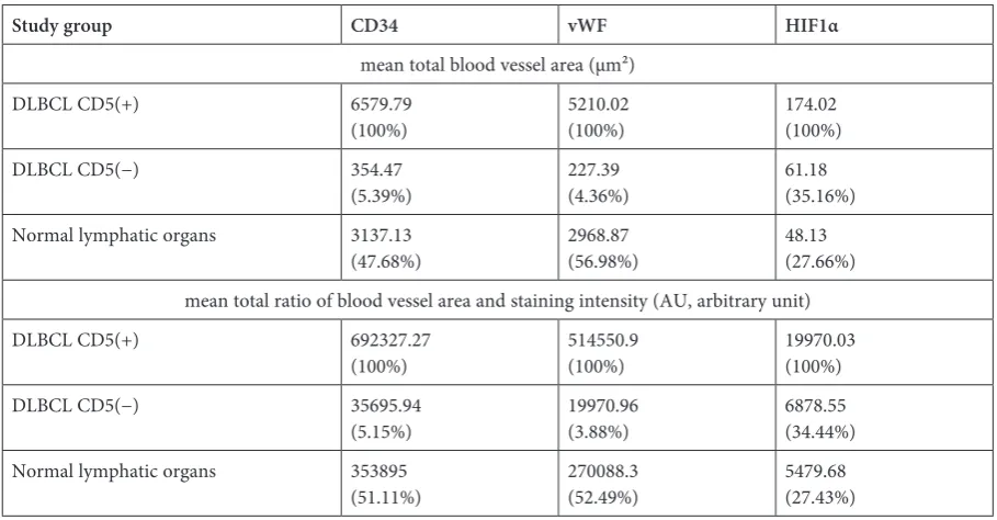
Mean total blood vessel area (µm²) and mean total ratio of blood vessel area and staining intensi-ty (AU), measured by means of CD34 and vWF ex-pression, was highest in the CD5-positive DLBCL group, whereas the lowest values were noted in the CD5-negative DLBCL group. The respective val-ues observed in lymphatic organs acquired from patients without lymphoproliferative diseases lay between both these extremes. In the case of HIF1α, both tested parameters were distinctly lower in comparison to the values obtained using CD34 and vWF. Similarly, the highest HIF1α expression was noted in the CD5-positive DLBCL subgroup. In patients with CD5-negative DLBCL, the study parameters were borderline higher than in patients without lymphoproliferative diseases (Table 1).
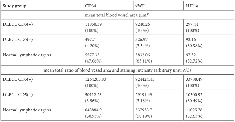
Likewise, the analysis of areas with the highest blood vessel density (hot spots) showed the high-est values in CD5-positive DLBCL, intermediate values in patients without lymphoproliferative
dis-Table 1. Angiogenesis parameters in three study groups
Study group CD34 vWF HIF1α
mean total blood vessel area (µm²)
DLBCL CD5(+) 6579.79
(100%) 5210.02(100%) 174.02(100%)
DLBCL CD5(−) 354.47
(5.39%) 227.39(4.36%) 61.18(35.16%) Normal lymphatic organs 3137.13
(47.68%) 2968.87(56.98%) 48.13(27.66%) mean total ratio of blood vessel area and staining intensity (AU, arbitrary unit) DLBCL CD5(+) 692327.27
(100%) 514550.9 (100%) 19970.03(100%)
DLBCL CD5(−) 35695.94
(5.15%) 19970.96(3.88%) 6878.55(34.44%) Normal lymphatic organs 353895
(51.11%) 270088.3(52.49%) 5479.68(27.43%)
eases, and lowest in patients with CD5-negative DLBCL. This general tendency was also observed in the HIF1α analysis, although the values noted in CD5-negative DLBCL and in the group with-out lymphoproliferative diseases were very similar (Table 2).
The observed differences in the expression of markers of angiogenesis were statistically signifi-cant in all three study groups (Table 3).
Discussion
The results of the present study show that CD5-positive DLBCL is characterized by a more abundant blood vessel network in comparison to the DLBCL subtype without CD5 expression, as well as normal lymphatic organs (Table 1, 2). To our knowledge, in the available literature there are no reports on angiogenesis intensity in CD5-positive DLBCL.
CD5-positive DLBCL is considered a DLBCL subtype with aggressive clinical course. It occurs
in the elderly with female sex predominance, more frequently in extranodal localization. In compari-son to the CD5-negative DLBCL subtype, patients with CD5-positive DLBCL have more disseminat-ed disease at diagnosis, worse performance status, higher serum lactate dehydrogenase (LDH) ac-tivity, and a propensity towards central nervous system involvement, as well as worse response to treatment [7].
The high blood vessel density promotes pro-gression of the neoplastic process and may be an indirect indicator of high malignant poten-tial. Cardesa-Salzmann et al. assessed the vascula-ture of DLBCL using antibodies specific to CD31 and computerized picture analysis and conclud-ed that high angiogenesis intensity was correlatconclud-ed with malignant clinical behavior and shorter over-all survival (OS). The authors showed significantly lower values of microblood vessel density (MVD): (11.8 × 10³; 78.67%) in the GCB subtype vs. ABC
subtype (15 × 103; 100%), regarded as a more
ag-gressive variant of DLBCL [8]. Similarly, Vac-ca et al. documented richer tumor vasculature in
Table 2. Hot spot analysis in three study groups
Study group CD34 vWF HIF1α
mean total blood vessel area (µm²)
DLBCL CD5(+) 11850.39
(100%) 9240.26(100%) 297.44(100%)
DLBCL CD5(−) 497.71
(4.20%) 326.97(3.54%) 92.16(30.98%) Normal lymphatic organs 5577.35
(47.06%) 5832.06(63.11%) 97.32(32.72%) mean total ratio of blood vessel area and staining intensity (arbitrary unit, AU)
DLBCL CD5(+) 1264203.83
(100%) 924424.41(100%) 33788.49(100%)
DLBCL CD5(−) 50112.23
(3.96%) 29194.49(3.16%) 10300.92(30.49%) Normal lymphatic organs 643884.9
(50.93%) 537933.7(58.19%) 11025.78(32.63%)
Table 3. The analysis of variance for differences of means in CD5-positive DLBCL, CD5-negative DLBCL and normal lymph nodes subgroups
Parameter tested by the analysis of variance for differences of means in CD5-
-positive DLBCL, CD5-negative DLBCL and normal lymph nodes subgroups CD34 vWFP-values HIF1α
Mean total blood vessel area 0.0001 0.0001 0.026
aggressive lymphomas in comparison to subtypes with an indolent clinical course [9]. The number of blood vessels per 0.78 mm2 ± SD in lymphomas
with low, intermediate and high malignant po-tential was 7 ± 2, 12 ± 3 and 14 ± 5, respectively. This observation was also confirmed by other in-vestigators: Arias et al., Crivelatto et al. and Ribatti et al. [10−12]. The increased angiogenesis in CD5-positive DLBCL observed in the present study may thus be associated with worse clinical course of this subtype of lymphoma.
Surprisingly, the intensity of angiogenesis was higher in normal lymphatic organs obtained from patients without lymphoproliferative diseases than in specimens of CD5-negative DLBCL (Table 1, 2). The results of other studies undertaking this ques-tion are inconsistent. Some authors found lower blood vessel density in lymphomas than in reactive lymph nodes, as in the present study [13, 14]. Kad-owaki et al. performed an MVD analysis in normal lymph nodes and in various types of lymphomas and obtained the following results: 7818 ± 3533 and 6243 ± 4027, respectively. On the basis of CD34 immunostain, the researchers found that angiogenesis in DLBCL in general (without divi-sion into subtypes) is less prominent than in nor-mal lymph nodes: 3243 ± 1453 and 7818 ± 3533, respectively [14]. Korkolopoulou et al. [15] and Mazur et al. [16] did not observe any relationship between blood vessel density and the morphologi-cal variant of lymphoma.
According to other reports, new vessel forma-tion in nodes involved with lymphoma is more pronounced in comparison with non-neoplastic lymph nodes [17−19]. Vacca et al. analyzed speci-mens from 30 patients with benign lymphadenop-athy and 71 patients with various lymphomas and assessed the number of blood vessels per area of 0.78 mm2 ± SD, obtaining the following values:
3 ± 1 and 11 ± 5, respectively [17].
It should be noted that proliferation of small blood vessels remains a classic feature of T zone reaction in lymphatic organs, frequently observed as an unspecific response to various stimuli. It is probably important to precisely select the control group with exclusion of all lymph nodes with even minor histological abnormalities.
In line with the results presented by others, CD34 was the most sensitive marker for the de-tection of angiogenesis in the present study [20], whereas the staining reaction was weakest in the case of HIF1α (Fig. 1). HIF1α induces the
bio-synthesis of over forty protein regulators prepar-ing cells for hypoxic conditions [21], which also increase the propensity of the tumor to develop distant metastases [22−24]. Owing to the uneven distribution of hypoxia, the neoplastic tissue is characterized by heterogeneous HIF1α expression, which is in turn correlated with regional blood vessel density [25, 26]. The morphometric analy-sis of the study parameters using antibodies specif-ic to both CD34 and vWF gave comparable results. The analysis using HIF1α did not show any differ-ences in angiogenesis intensity between lymphat-ic organs obtained from patients without lympho-proliferative diseases and CD5-negative DLBCL. This observation may be explained by the low in-tensity of the staining with this antibody, below the threshold of sensitivity of the implemented meth-od.
Identification of a subgroup of patients with high risk lymphomas on the basis of increased an-giogenesis forms a mainstay for the introduction of antiangiogenic therapies. Currently, multiple clinical trials assessing antiangiogenic molecules are ongoing. These are inhibitors of integrins and metalloproteinases, as well as soluble receptors of proangiogenic cytokines and others. These agents have already become standard treatment of certain malignancies, like sorafenib in unresectable hepa-tocellular carcinoma [27]. Another effective anti-angiogenic treatment indicated in metastatic colon cancer is bevacizumab, a recombinant monoclo-nal antibody directed against vascular endothelial growth factor (VEGF) [28].
Therefore, inhibitors of angiogenesis may be a very promising therapeutic option in the treat-ment of certain lymphoma subtypes, which may substantially complement the standard chemo-therapy and improve its effects [8, 29]. However, further clinical trials are mandatory to confirm the preliminary results presented in this report.
The study showed a higher intensity of angio-genesis in CD5-positive DLBCL in comparison to the CD5-negative variant, which may contribute to its more aggressive clinical behavior. The re-sults do not substantiate the hypothesis that the more frequent CD5-negative DLBCL subtype was better vascularized than non-neoplastic lymphat-ic organs.
References
[1] Swerdlow SH, Campo E, Harris NL, Jaffe ES, Pileri SA, Stein H, Thiele J, Vardiman JW: WHO Classification of Tumours of Haematopoietic and Lymphoid Tissues, 4th ed., IARC Press, Lyon 2008, 9−15.
[2] Niitsu N, Okamoto M, Tamaru J, Yoshino T, Nakamura N, Nakamura S, Ohshima K, Nakamine H, Hirano M:
Clinicopathologic characteristics and treatment outcome of the addition of rituximab to chemotherapy for CD5- -positive in comparison with CD5-negative diffuse large B-cell lymphoma. Ann Oncol 2010, 21, 2069−2074.
[3] Dvorak HF: Angiogenesis: Update 2005. J Thromb Haemost 2005, 3, 24−28.
[4] Cao Y, Langer R: A review of Judah Folkman’s remarkable achievements in biomedicine. Proc Natl Acad Sci USA 2008, 105, 13203−13205.
[5] Carmeliet P: Angiogenesis in health and disease. Nat Med 2003, 9, 653−660.
[6] Ferrara N: Vascular endothelial growth factor: Basic science and clinical progress. Endocr Rev 2004, 25, 581−611.
[7] Jain P, Fayad LE, Rosenwald A, Young KH, O’Brien S: Recent advances in de novo CD5+ diffuse large B cell lymphoma. Am J Hematol 2013, 88, 798−802.
[8] Cardesa-Salzmann TM, Colomo L, Gutierrez G, Chan WC, Weisenburger D, Climent F, Gonzalez-Barca E, Mercadal S, Arenillas L, Serrano S, Tubbs R, Delabie J, Gascoyne RD, Connors JM, Mate JL, Rimsza L, Braziel R, Rosenwald A, Lenz G, Wright G, Jaffe ES, Staudt L, Jares P, Lopez-Guillermo A, Campo E: High microvessel density determines a poor outcome in patients with diffuse large B-cell lymphoma treated with ritux-imab plus chemotherapy. Haematologica 2011, 96, 996−1001.
[9] Vacca A, Ribatti D, Ruco L, Giacchetta F, Nico B, Quondamatteo F, Ria R, Iurlaro M, Dammacco F: Angiogenesis extent and macrophage density increase simultaneously with pathological progression in B-cell non-Hodgkin’s lymphomas. Br J Cancer 1999, 79, 965−970.
[10] Arias V, Soares FA: Vascular density (tumor angiogenesis) in non-Hodgkin’s lymphomas and florid follicular hyperplasia: A morphometric study. Leuk Lymph 2000, 40, 157−166.
[11] Crivellato E, Nico B, Vacca A, Ribatti D: B-cell non-Hodgkin’s lymphomas express heterogeneous patterns of neovascularization. Hematologica 2003, 88, 671−678.
[12] Ribatti D, Vacca A, Marzullo A, Nico B, Ria R, Roncali L, Dammacco F: Angiogenesis and mast cell density with tryptase activity increase simultaneously with pathological progression in B-cell non-Hodgkin’s lymphomas. Int J Cancer 2000, 85, 171−175.
[13] Ridell B, Norrby K: Intratumoral microvascular density in malignant lymphomas of B-cell origin. APMIS 2001, 109, 66−72.
[14] Kadowaki I, Ichinohasama R, Harigae H, Ishizawa K, Okitsu Y, Kameoka J, Sasaki T: Accelerated lymphangio-genesis in malignant lymphoma: Possible role of VEGF-A and VEGF-C. Br Journal Haematol 2005, 130, 869−877.
[15] Korkolopoulou P, Thymara I, Kavantzas N, Vassilakopoulos TP, Angelopoulou MK, Kokoris SI, Dimitriadou EM, Siakantaris MP, Anargyrou K, Panayiotidis P, Tsenga A, Androulaki A, Doussis-Anagnostopoulou IA, Patsouris E, Pangalis GA: Angiogenesis in Hodgkin’s lymphoma: A morphometric approach in 286 patients with prognostic implications. Leukemia 2005, 19, 894−900.
[16] Mazur G, Wróbel T, Dzięgiel P, Jeleń M, Kuliczkowski K, Zabel M: Angiogenesis measured by expression of CD34 antigen in lymph nodes of patients with non-Hodgkin’s lymphomas. Folia Histochem Cytobiol 2004, 42, 241−243.
[17] Vacca A, Ribatti D, Ruco L, Giacchetta F, Nico B, Quondamatteo F, Ria R, Iurlaro M, Dammacco F: Angiogenesis extent and macrophage density increase simultaneously with pathological progression in B-cell non-Hodgkin’s lymphomas. Br J Cancer 1999, 79, 965−970.
[18] Ribatti D, Vacca A, Nico B, Fanelli M, Roncali L, Dammacco F: Angiogenesis spectrum in the stroma of B-cell non-Hodgkin’s lymphomas. An immunohistochemical and ultrastructural study. Eur J Haematol 1996, 56, 45−53.
[19] Wołowiec D: Angiogeneza i limfangiogeneza w chłoniakach złośliwych nieziarniczych. Acta Haematol Pol 2011, 42, 357−365 [in Polish].
[20] Gryczyński M, Pietruszewska W: Angiogeneza jako nowy czynnik rokowniczy u chorych na raka krtani. Otorynolaryngologia 2002, 1, 13−20 [in Polish].
[21] Hewiston KS, Schofield CJ: The HIF pathway as a therapeutic target. Drug Discovery Today 2004, 9, 704−710.
[22] Hockel M, Vaupel P: Tumor hypoxia: Definitions and current clinical, biologic, and molecular aspects. JNCI 2001, 93, 266−276.
[23] Semenza GL, Nejfelt MK, Chi SM, Antonarakis SE: Hypoxia-inducible nuclear factors bind to an enhancer ele-ment located 3’ to the human erythropoietin gene. Proc Natl Acad Sci USA 1991, 88, 5680−5684.
[24] Gasińska A, Biesaga B: Dwa oblicza hipoksji nowotworów. Nowotwory 2010, 60, 332−340 [in Polish].
[25] Maxwell PH: The HIF pathway in cancer. Sem Cell Dev Biol 2005, 16, 523−530.
[26] Ravi R, Mookerjee B, Bhujwalla ZM, Sutter CH, Artemov D, Zeng Q, Dillehay LE, Madan A, Semenza GL, Bedi A: Regulation of tumor angiogenesis by p53-induced degradation of hypoxia-inducible factor 1 alfa. Genes Dev 2000, 1, 34−44.
[27] Rimassa L, Santoro A: Sorafenib therapy in advanced hepatocellular carcinoma: The SHARP trial. Exp Rev Anticancer Ther 2009, 9, 739−745.
[28] Olbryt M, Szala S: Białkowe inhibitory angiogenezy w terapii nowotworów. Współcz Onkol 2005, 9, 48−53 [in Polish].
Address for correspondence:
Nina Woźnialis
Department of Pathology and Laboratory Diagnostics Maria Sklodowska-Curie Institute and Oncology Center ul. Roentgena 5
02-781 Warszawa Poland
Tel: +48 504 159 041
E-mail: [email protected]
Conflict of interest: None declared