R E S E A R C H
Open Access
Digital breast tomosynthesis image
reconstruction using 2D and 3D total variation
minimization
Metin Ertas
1, Isa Yildirim
2,3*, Mustafa Kamasak
4and Aydin Akan
1* Correspondence: [email protected]
2Electrical and Electronics
Engineering Department, Istanbul Technical University, Maslak, 34469 Istanbul, Turkey
3College of Engineering
Department, University of Illinois at Chicago, Chicago, IL 60607, USA Full list of author information is available at the end of the article
Abstract
Background:Digital breast tomosynthesis (DBT) is an emerging imaging modality which produces three-dimensional radiographic images of breast. DBT reconstructs tomographic images from a limited view angle, thus data acquired from DBT is not sufficient enough to reconstruct an exact image. It was proven that a sparse image from a highly undersampled data can be reconstructed via compressed sensing (CS) techniques. This can be done by minimizing the l1norm of the gradient of the image which can also be defined as total variation (TV) minimization. In tomosynthesis imaging problem, this idea was utilized by minimizing total variation of image reconstructed by algebraic reconstruction technique (ART). Previous studies have largely addressed 2-dimensional (2D) TV minimization and only few of them have mentioned 3-dimensional (3D) TV minimization. However, quantitative analysis of 2D and 3D TV minimization with ART in DBT imaging has not been studied. Methods:In this paper two different DBT image reconstruction algorithms with total variation minimization have been developed and a comprehensive
quantitative analysis of these two methods and ART has been carried out: The first method is ART + TV2Dwhere TV is applied to each slice independently. The other method is ART + TV3Din which TV is applied by formulating the minimization problem 3D considering all slices.
Results:A 3D phantom which roughly simulates a breast tomosynthesis image was designed to evaluate the performance of the methods both quantitatively and qualitatively in the sense of visual assessment, structural similarity (SSIM), root means square error (RMSE) of a specific layer of interest (LOI) and total error values. Both methods show superior results in reducing out-of-focus slice blur compared to ART. Conclusions:Computer simulations show that ART + TV3Dmethod substantially enhances the reconstructed image with fewer artifacts and smaller error rates than the other two algorithms under the same configuration and parameters and it provides faster convergence rate.
Keywords:Breast tomosynthesis, Compressed sensing, Total variation
Background
Several imaging modalities such as mammography, ultrasound and magnetic resonance have been extensively used in breast imaging for decades. Digital breast tomosynthesis (DBT) is an imaging technique using limited range of view angles, hence it has
limitations in implementation and challenges in image reconstruction [1]. Recently, digital tomosynthesis has become an emerging imaging modality for breast imaging where the breast is projected onto a flat panel detector from a limited view angle and a few number of projections is acquired to produce a three dimensional image of the breast. It is now currently being used for both diagnosis and screening purposes [1]. This technique overcomes the overlapping problem which is the most present artifact in 2D mammographic imaging. However, due to the limited range of view angles, the number of projections may not be sufficient to fully reconstruct the image in the Fou-rier space [2]. More specifically, the FouFou-rier space is not fully sampled which causes streaking artifacts. In order to acquire tomosynthesis images, algebraic reconstruction technique (ART) is adapted to acquire 3D objects from two dimensional projection im-ages [3]. Although several studies has shown that iterative algorithms can show satisfac-tory results over filtered back projection (FBP), modified FBP and matrix-inversion algorithms in missing data image reconstruction, they still do not give acceptable re-sults in tomosynthesis imaging [2,4,5].
DBT image reconstruction algorithms have shown significant improvements over the years. Shift and add (SAA) which is based on shifting and adding projections to sharpen the plane focus was the first idea of DBT image reconstructions [6]. However, significant amount of out of focus slice blur occurs in SAA. FBP, modified FBP and iterative methods have also been widely used in DBT image reconstruction perspective [7-9] to overcome the blurring problem. Among these techniques, iterative methods are generally suitable for complete data sets or nearly complete data sets. However, in tomosynthesis, acquired data is highly incomplete thus acquiring the exact image can be severely affected by this incompleteness. This problem can be addressed in a compressed sensing (CS) framework which allows the reconstruction of a signal or image from a highly undersampled observa-tion [10]. Hence after introducobserva-tion of the CS approach, the number of studies addressing sparse image reconstruction has drastically increased.
Iterative methods and FBP methods are not sufficient enough to preserve the edges which play an important role in identifying objects and fundamental features in the image. However, total variation (TV) minimization was proven to be applicable in edge preserving image denoising processes [11,12]. This algorithm is based on minimizing the l1 norm of the sparsified image. TV minimization has recently been adapted to DBT imaging reconstruction problems and better results were obtained than FBP and iterative methods [13,14]. By adding different constraints, such as prior image con-strained CS, improved results were obtained over CS and FBP algorithms [13]. More-over, compressed imaging based on wavelet sparsity has been investigated in limited tomographic x-ray imaging [15] and MRI [16] to reconstruct the image from fewer pro-jections and promising results have been obtained. Another alternative approach was introduced in [17] where, curvelets were used for reconstruction at a limited angular range and the results showed that the method was stable and edge-preserving.
different DBT image reconstruction algorithms with total variation minimization have been developed and a comprehensive quantitative analysis of these two methods and ART has been carried out:
i) ART + TV2Dmethod: TV2Dwas implemented layer by layer along the axial dimension to fully cover the entire image.
ii) ART + TV3Dmethod: TV3Dwas implemented to the entire space to get a reconstructed image in a single step.
A specific 3D phantom which roughly simulates a breast tomosynthesis image was designed to compare the reconstruction performances of ART, ART + TV2Dand ART + TV3Dmethods in the sense of root mean square errors (RMSE) of reconstructed 3D image and a specific layer of interest (LOI). The visual perception assessment was done by means of structural similarity (SSIM) curves.
Reconstruction methods
In linear imaging problems, the following model is used:
Y ¼AXþn; ð1Þ
where Y shows the measured or observed data, X is the original image, A shows the system matrix which gives the data measurement process in the image and n is the additive noise. Consistency condition in (1) should be fulfilled in image reconstruction algorithms. Image reconstruction is an inverse problem which is based on estimation of X from the given Y and A. In (1), the system matrix may vary according to the imaging problem such as Fourier transform in MRI or Radon transform in tomographic reconstruction.
Algebraic reconstruction technique (ART)
In tomosynthesis imaging, limited view angle creates a large portion of missing data which makes the exact image reconstruction more difficult. Thus, iterative image re-construction techniques are introduced to estimate the exact object. ART is one of the most commonly used iterative reconstruction technique in image processing [3,21,22]. In ART, an image is estimated in an iterative manner while satisfying the consistency condition in (1). The idea that lays behind the reconstruction is that the voxel inten-sities are updated ray by ray for each projection. A 3D image is updated with ART re-construction by using the following formulation:
Xðkþ1Þ
j ¼Xð Þjk þ
Yi−
XN
k¼1Aik:X
k ð Þ j
XN
k¼1A 2
ik
Aij;ij¼¼11;;22;;……;;NM; ð2Þ
of the related projection based on the Siddon’s coefficients. Error backprojection pro-cedure is continued for all projections to finish one iteration. This iteration process is repeated until a convergence criterion is satisfied. However, the measured projection data in DBT is not sufficient enough to reconstruct an exact image due to the limited view angle.
Total variation (TV) minimization
Out-of-focus slice blur and streaking artifacts arise as a result of missing data in DBT. There-fore, in order to improve the quality of image acquired from highly undersampled data, im-proved methods are needed. It was proven that a sparse image can be recovered from a highly undersampled data set which is called compressed sensing (CS) [10]. The compressed sensing can be represented as minimization of l1norm of the sparsified image X:
minjΨXj1 such that Y ¼AX; ð3Þ
whereΨrepresents a linear operator called the sparsifying transform. Discrete gradient and wavelet transforms are the most commonly used sparsifying transforms in CS theory. In this study, discrete gradient transform is used and applied in two different forms as 2D and 3D. Considering the discrete gradients for each pixel in the image, the problem turns into min-imizing the TV(X). Thus, TV of a 2D image can be shown as:
TV2DXð Þi;j¼
XK
i¼1
XL
j¼1
ffiffiffiffiffiffiffiffiffiffiffiffiffiffiffiffiffiffiffiffiffiffiffiffiffiffiffiffiffiffiffiffiffiffiffiffiffiffiffiffiffiffiffiffiffiffiffiffiffiffiffiffiffiffiffiffiffiffiffiffiffiffiffiffiffiffiffi jXð Þi;j−Xðiþ1;jÞj j2þXð Þi;j−Xði;jþ1Þj2 q
ð4Þ
where, X(i,j)is the intensity value at pixel (i,j), {i = 1,2,…..,K; j = 1,2,……,L}. Applying TV2D layer by layer to a 3D image shows significant improvements in image quality [24]. But ap-plying the TV term in such manner ignores the neighborhood of a 3D image along axial direction. Considering this neighborhood relation, the same discrete gradient transform may also be adapted in 3D form. TV of a 3D image can be shown as:
TV3DXði;j;kÞ¼
XK
i¼1
XL
j¼1
XM
k¼1
ffiffiffiffiffiffiffiffiffiffiffiffiffiffiffiffiffiffiffiffiffiffiffiffiffiffiffiffiffiffiffiffiffiffiffiffiffiffiffiffiffiffiffiffiffiffiffiffiffiffiffiffiffiffiffiffi
DxX ð Þ2þ D
yX
2
þðDzXÞ2 q
: ð5Þ
where X(i,j,k)is the intensity value at voxel (i,j,k), {i = 1,2,…,K, j = 1,2,…..,L and k = 1,2,…..,M}. (DxX) =X(i,j,k) −X(i+ 1,j,k), (DyX) =X(i,j,k) −X(i,j+ 1,k), (DzX)X(i,j,k) =X(i,j,k+ 1). Implementing TV
minimization as a penalty term has been shown to give improved results [2,6,12-14]. Thus, a widely used CS-based constrained minimization problem has been formulated as:
minX h
∇X 1
i
;such that AX¼Y:
ð6Þ This constrained minimization problem can be reformulated as an unconstrained minimization problem as:
minX h
Y−AX 2þα ∇X 1
i ;
ð7Þ
1. Applying ART to create the 3D image: Reconstructed image is acquired applying the following steps:
a. An initial image is selected or assigned.
b. System matrix in (1) is calculated using the Siddon’s ray-driven algorithm. c. The measured detector data and the forward projection of the image are compared. d. New image is obtained using ART (2).
2. Total variation minimization: (8) is minimized in order to acquire total variation minimized of image reconstructed by ART:
minXh X−X^ 2þα ∇X 1
i
;
ð8Þ
whereX^ is reconstructed image by ART.
Initialization:
GivenNi, Nr, Np,α,Ԑo, S(x,y,z), D(x,y,z),Ds, Y
Xpk¼Zeroes initial
System Matrix Calculation:
Reconstruction Algorithm Step:
for ite = 1,2,3...,Ni
TV Minimization using (8) for TV3D:
The pseudo code for the ART + TV3Dalgorithm is shown in Algorithm of ART + TV3D reconstruction where S(x,y,z)and D(x,y,z)represent the coordinates of X-ray source and de-tector, Dsis the size of the detector, Ni, Np, Nr, Nvshow number of iterations, projections, rays and voxels respectively and k is the voxel coordinate. Measured data is shown as Y.α is the regularization parameter in TV minimization step andεoshows the value for stop-ping criterion of TV. By using this algorithm, in the present work, two different TV methods were investigated called as TV2Dand TV3D. In the first method, TV2Dinclusion is applied layer by layer to cover the entire image. In the second method, in order to apply TV3Dinto ART, TV minimization step is applied to the entire phantom at once after the reconstructed image obtained at each iteration. There are several factors in the algorithm which may increase the speed of convergence and the efficiency of the optimization prob-lem. The first one is the initial guess of the image. Prior image constrained algorithm was conducted better results than classical ART, CS and FBP [13]. However the image registra-tion needs to be taken into account in order to achieve a faster convergence speed. In this study, the initial image is chosen to be zeros. The second point is the stopping criterion of the iterative steps. The iteration is continued until the difference between two consecutive updated images is smaller than a certain value. The last item is to choose the appropriate regularization parameter in the objective function (8) which plays a critical role in the per-formance of TV minimization [24].
Results
TV2D and TV3D have been extensively used in various medical imaging problems [11-14,18-20]. Most of the studies have addressed TV2Din minimizing the total vari-ation and only few studies have addressed TV in 3D manner. However their perform-ance comparison has not been fully investigated. In our study, in order to observe the performance of three reconstruction algorithms used in DBT, a 3D phantom data was created and a tomosynthesis system was designed.
Phantom with a size of 71*71*10 was specifically designed to demonstrate the most common overlapping tissue problem in tomosynthesis imaging. In this study, since our aim is to study if ART + TV3D has a superiority over ART + TV2D on limited view angle 3D image reconstruction in terms of reconstruction quality, the size of the phan-tom and the detector matrix we use are only chosen for the simplicity and to speed up the simulations.
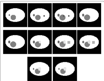
The phantom consists of 10 layers in axial dimension which gives a closer match with real tomosynthesis imaging. There are some smaller objects with lower x-ray absorp-tion rates located in the lower slices of the phantom. These small objects are obscured by objects with high x-ray absorbance in the upper slices of the phantom. 3rd layer of the image was chosen to be the LOI of the image due to its structure for the possible screening problem in 2D mammography imaging. The small square in the right side of the LOI represents an object with a very low x-ray absorption rate. In order to imitate the screening effect in 2D imaging a larger object with a higher x-ray absorption rate was located in the layers 7 and 8. Objects in the phantom or the phantom itself can be modified and extended for different purposes. The layers of the phantom used in the study are demonstrated in Figure 1.
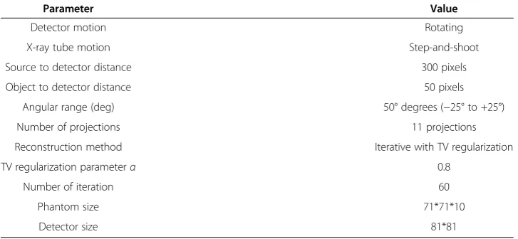
In DBT imaging the out-of-focus-slice blur in the layer of interest is the most domin-ant artifact, thus the simulations are performed with noiseless projection data. The same reconstruction parameters have been selected for ART, ART + TV2Dand ART + TV3Dmethods. An experience-based fixed regularization parameter α is set to 0.8 for ART + TV2Dand ART + TV3Dmethods in our experiments. All simulations were per-formed in MATLAB® software. The system configurations which are used for the simu-lations are Intel(RM) Core(TM) i7-2630 QM CPU @ 2.00 GHz CPU, 6 GB Memory, Windows 7 64 Bits operating system.
Performances of three reconstruction algorithms were compared both qualitatively and quantitatively. For qualitative assessment, the visual observation was conducted and the structure similarity value which shows the visual quality was used. For quanti-tative assessment, RMSE values of the LOI and 3D image were compared.
In this study the projections were acquired from 11 view angles and the reconstruc-tion algorithms were performed for 60 iterareconstruc-tions. Figure 2 shows the results of the re-construction algorithms for the LOI and 7th layer. Second column shows images
Table 1 Simulation parameters
Parameter Value
Detector motion Rotating
X-ray tube motion Step-and-shoot
Source to detector distance 300 pixels
Object to detector distance 50 pixels
Angular range (deg) 50° degrees (−25° to +25°)
Number of projections 11 projections
Reconstruction method Iterative with TV regularization
TV regularization parameterα 0.8
Number of iteration 60
Phantom size 71*71*10
reconstructed by ART. Compared to the original image the blur in images recon-structed with ART is apparent and in the 7th layer visible distortion makes the reading difficult. Compared with the reconstructed image by ART, reconstructed images using ART + TV2Dand ART + TV3D(the third and fourth columns respectively) are signifi-cantly improved. Both ART + TV2D and ART + TV3D methods preserve the edges, however it is clearly seen in images reconstructed with ART + TV2D that the blur is still existent. In images reconstructed with ART + TV3D the noise is lower than the
Figure 2Results of the reconstruction algorithms for the LOI and 7th layer.
other two methods. ART + TV3Dreconstructs images closer to the original images than ART and ART + TV2D.
Since the human visual perception is highly adapted for extracting structural infor-mation from images it was introduced an alternative complementary framework for quality assessment based on the structure similarity (SSIM) between two images [26]. For image quality assessment, SSIM index is used locally rather than globally. Thus, pa-rameters used to calculate the SSIM are computed within a local window which moves pixel by pixel through the image. For a single overall image quality index between two images the following equation is used:
MSSIM Xð ;YÞ ¼M1 XM i¼1
SSIM xð i;yiÞ; ð10Þ
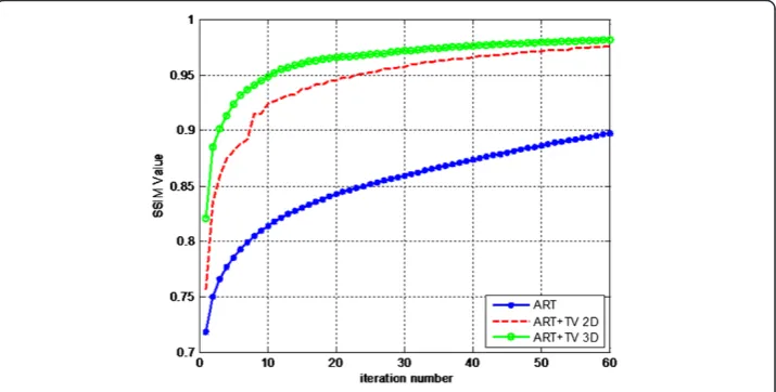
where, X and Y are the reference image and reconstructed image respectively; xiand yi are the image index at the i-th local window in X and Y respectively. M shows the number of local windows. This equation calculates mean of SSIM values in all local windows. MSSIM values show much better consistency with the qualitative results. MSSIM values of the three reconstruction methods for the LOI are shown in Figure 3.
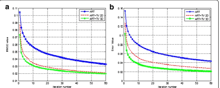
The best similarity is provided between reconstructed image and reference image at the value of 1. The overall SSIM values at 60th iteration are 0.8973, 0.9765, 0.9816 for ART, ART + TV2Dand ART + TV3Drespectively. In this sense, SSIM values for ART + TV2D and ART + TV3D show superior results compared to the ART reconstruction. Although the values for ART + TV2Dand ART + TV3Dare very similar to each other, the convergence speed of ART + TV3D is faster than ART + TV2D. ART + TV3D reaches to the value of 0.955 in 12th iteration while ART + TV2D can reach to that value in the 27th iteration. Thus, ART + TV3Dshows better results than the other two reconstruction methods. Also notice that the SSIM curve for ART + TV2Dshows fluc-tuations unlike the other two reconstruction methods because of the absence of neigh-borhood relationship in axial dimension. These fluctuations can also be seen for ART + TV2Din Figure 4a. Comparison of RMSE values of the methods for the LOI is shown in Figure 4a. The RMSE values at 60th iteration are 0.0327, 0.0212, 0.0198 for ART,
ART + TV2Dand ART + TV3Drespectively. ART + TV3Dgives better RMSE perform-ance than ART + TV2Dand much better RMSE performance than ART.
For further performance analysis of the methods, the total RMSE values of ART, ART + TV2D and ART + TV3D are compared in Figure 4b. ART shows the highest error value when it is compared to the other two methods. After 60th iteration, the error value for ART is 0.0433. ART + TV2Dgives lower error value than ART with the error value at 60th iteration 0.0273. ART + TV3D shows the best performance as giving 0.0206 total RMSE value at 60th iteration. The error difference between ART + TV2Dand ART + TV3D is slightly closer in Figure 4a than in Figure 4b. Increasing the number of layers in axial direc-tion may also increase the gap in performances of these two reconstrucdirec-tion methods.
Though both ART + TV2Dand ART + TV3Dmethods improved the results of ART considerably, ART + TV3Dis preferred over ART + TV2Ddue to its faster convergence rate. Reaching an error value of 0.043 for ART can only be achieved at 60th iteration. The same value is achieved for ART + TV2Dat 16th iteration and for ART + TV3Dat 6th iteration.
The computational cost of ART is considerably much more than analytic image re-construction methods. Thus, improving the speed and effectiveness of ART is crucial while keeping the computational time in reasonable levels. In this study, the recon-struction time spent for a single iteration of ART + TV3Dis %1.8 more than the time spent for a single iteration of ART. Thus, implementing TV2Dor TV3Dwith ART does not dramatically increase the time spent for the reconstructions while reducing the out of focus slice blur substantially.
Conclusion
Breast tomosynthesis imaging problem has been studied by implementing CS into ART in two different manners: 1) ART + TV2D: TV minimization step was done by applying TV layer by layer in 2D form along the axial dimension, 2) ART + TV3D: implementa-tion of TV in 3D form to fully cover the phantom in a single step. The numerical re-sults were conducted to compare the performances of ART, ART + TV2D, ART + TV3D by designing a breast phantom to simulate the overlapping tissue problem in breast tomosynthesis imaging. Results of this study demonstrated that including TV
after ART in the reconstruction algorithm significantly reduced out-of-focus slice blur in the reconstructed images compared to ART. ART + TV3D provided better results than two other reconstruction methods both quantitatively by giving smaller RMSE values of the LOI and 3D images and qualitatively by generating higher MSSIM values. The computational costs per iteration for the tested methods were almost the same due to the simplicity of total variation minimization step. However, ART + TV3D pro-vided the fastest convergence rate among all three methods. In conclusion, in tomo-synthesis imaging due to high amount of missing data, improved reconstruction techniques need to be developed to have better reconstructed images. In this paper, it was shown that a 3D breast tomosynthesis image can be reconstructed much faster and less artifact-free with ART + TV3Dmethod than ART and ART + TV2Dmethods.
Abbreviations
DBT:Digital breast tomosynthesis; CS: Compressed sensing; TV: Total variation; ART: Algebraic reconstruction technique; 2D: 2 Dimensional; 3D: 3 Dimensional; ART + TV2D: Algebraic reconstruction technique with 2 dimensional
total variation; ART + TV3D: Algebraic reconstruction technique with 3 dimensional total variation; SSIM: Structure
similarity; RMSE: Root mean squared error; LOI: Layer of interest; FBP: Filtered back projection; SAA: Shift and add; MRI: Magnetic resonance imaging; MSSIM: Modified structure similarity; TV2D: 2 Dimensional total variation;
TV3D: 3 Dimens0069onal total variation.
Competing interest
The authors declare that they have no competing interests.
Authors’contribution
ME carried out the reconstruction simulations, performed analysis of the simulation results and drafted the manuscript. IY conceived of the study, participated in the design of the study, and helped in drafting the manuscript. MK participated in the design of phantom and system, and helped in drafting the manuscript. AA participated in the coordination and helped in drafting the manuscript. All authors read and approved the final manuscript.
Acknowledgement
This work has been supported by TUBITAK, The Scientific and Research Council of Turkey, under the grant 111E086.
Author details
1Electrical and Electronics Engineering Department, Istanbul University, Avcilar, 34320 Istanbul, Turkey.2Electrical and
Electronics Engineering Department, Istanbul Technical University, Maslak, 34469 Istanbul, Turkey.3College of
Engineering Department, University of Illinois at Chicago, Chicago, IL 60607, USA.4Computer Engineering Department,
Istanbul Technical University, Maslak, 34469 Istanbul, Turkey.
Received: 24 July 2013 Accepted: 9 October 2013 Published: 31 October 2013
References
1. Karellas A, Giger MA:Advances in breast imaging: physics, technology, and clinical applications, Categorical course in diagnostic radiology physics. Oak Brook IL: RSNA; 2004:149–165.
2. Smith A:Full-field breast tomosynthesis.Radiol Manage2005,27:25–31.
3. Wu T, Moore RH, Rafferty EA, Kopans DB:A comparison of reconstruction algorithms for breast tomosynthesis.
Med Phys2004,31:2636–2647.
4. Zhang Y, Chan HP, Sahiner B, Wei J, Goodsitt MM, Hadjiiski LM, Ge J, Zhou C:Digital tomosynthesis mammography: improvement of artifact reduction methods for high attenuation.Proc SPIE2008,6913:181–186.
5. Wu T, Moore RH, Rafferty EA, Kopans DB:A comparative study of limited-angle cone beam reconstruction methods for breast tomosynthesis.Med Phys2006,33(10):3781–3795.
6. Grant G:Tomosynthesis: a three dimensional radiographic imaging technique.IEEE Trans Biomed Eng1972,19:20–28. 7. Nielsen T, Hitziger S, Grass M, Iske A:Filter calculation for X-ray tomosyhnthesis reconstruction.Phys Med Biol
2012,57(12):3915–3930.
8. Zhao B, Zhao W:Three dimensional linear system analysis for breast imaging.Med Phys2002,29(11):2655–2671. 9. Ludwig J, Mertelmeier T, Kunze H, Harer W:A novel approach for filtered backprojection in tomosynthesis
based on filter kernels determined by iterative reconstruction technique.Proc IWDM2008,5116:612–620. 10. Donoho DL:Compressed sensing.IEEE Trans Inform Theory2006,52(2):1289–1306.
11. Rudin LI, Osher S, Fatemi E:Nonlinear total variation based noise removal algorithms.Physica D1992,60:259–268. 12. Velikina J, Leng S, Chen GH:Limited view angle tomograhpic image reconstruction via total variation
minimization.Proc SPIE Med Imag2007,6510:651020.
13. Chen GH, Tang J, Leng S:Prior image constrained compressed sensing (PICCS): a method to accurately reconstruct dynamic CT images from highly undersampled projection data sets.Med Phys2008,35(2):660–663. 14. Sidky EY, Reisera I, Nishikawa RW, Pana X, Chartrandb C, Kopans DB, Moore RH:Practical iterative image
15. Rantala M, Vanska S, Jarvenpaa S, Kalke M, Lassas M, Moberg J, Siltanen S:Wavelet-based reconstruction for limited angle X-ray tomography.IEEE Trans Med Imag2006,25(2):210–217.
16. Huang J, Yang F:Compressed magnetic resonance imaging based on wavelet sparsity and nonlocal total variation.Proc IEEE ISBI2009:968–971.
17. Frikel J:A new framework for sparse regularization in limited angle x-ray tomography.IEEE ISBI Nano Macro 2010:824–827.
18. Sidky EY, Reiser IS, Nishikawa RM, Pan XC:Image reconstruction in digital breast tomosynthesis by total variation minimization.Proc SPIE2007,6510:651027.
19. Sidky EY, Pan X, Reiser IS, Nishikawa RM, Moore RH, Kopans DB:Enhanced imaging of microcalcifications in digital breast tomosynthesis through improved image-reconstruction algorithms.Med Phys2009,36(11):4920–4932. 20. Joshi SH, Marquina A, Osher SJ, Dinov I, Van Horn JD, Toga AW:MRI resolution enhancement using total
variation regularization.Proc IEEE ISBI2009:161–164.
21. Colsher JG:Iterative three-dimensional image reconstruction from tomographic projections.Comput Graph Imag Proc1977,6:513–537.
22. Dobbins JT, Godfrey DJ:Digital x-ray tomosynthesis: current state of the art and clinical potential.Physics Med Biol2003,48(19):R65–R107.
23. Siddon RL:Fast calculation of the exact radiological path for a three-dimensional CT array.Med Phys1985,
12:252–255.
24. Ertas M, Akan A, Cengiz K, Kamasak M, Sayyedi S, Yildirim I:3-D tomosynthesis image reconstruction using total variation.Proc ASE Biomed Com2012:1–5.
25. Sechopoulos I:A review of breast tomosynthesis. Part I. The image acquisition process.Med Phys2013,
40(1):1–12. 014301.
26. Wang Z, Bovik AC, Sheikh HR, Simoncelli HP:Image quality assessment: from error visibility to structural similarity.IEEE Trans Med Imag2004,13(4):600–612.
doi:10.1186/1475-925X-12-112
Cite this article as:Ertaset al.:Digital breast tomosynthesis image reconstruction using 2D and 3D total variation minimization.BioMedical Engineering OnLine201312:112.
Submit your next manuscript to BioMed Central and take full advantage of:
• Convenient online submission
• Thorough peer review
• No space constraints or color figure charges
• Immediate publication on acceptance
• Inclusion in PubMed, CAS, Scopus and Google Scholar
• Research which is freely available for redistribution