Iranian Journal of Virology, Volume 4, Number 2, 2010 17
Original Article
Construction of an Expression Vector Containing a Novel Fusion
Sequence from Middle Region of NS3 and Truncated Core Genes
of Hepatitis C Virus
Hosseini SY 1, Sabahi F1*, Moazzeni SM2, Modarressi MH3, Saberi-Firoozi M4, Ravanshad M1
1. Department of Virology, Tarbiat Modares University, Tehran, Iran. 2. Department of Immunology, Tarbiat Modares University, Tehran, Iran.
3. Department of Medical Genetic, Tehran University of Medical Sciences, Tehran, Iran.
4. GastroenteroHepatology Research Center, Shiraz University of Medical Sciences, Shiraz, Iran.
Abstract
Background and Aims: DNA constructs containing HCV antigens have become one of the vaccine candidates for induction of anti-HCV cellular and humoral immunity. In this study, we constructed a novel expressing vector harboring a fusion sequence derived from an overlapping fragment in the middle of NS3 and a truncated core fragment to avoid troubles reported to be associated with full gene expression.
Methods: The partial NS3 (pNS3) and core genes were amplified by RT-PCR method using serum of HCV infected patient harboring genotype 1a of virus. After purification and cloning the genes into TA-cloning vector, they were evaluated by sequencing and restriction digestion analysis. The resultant pNS3 and core gene subcloned into expression vector separately followed by expression evaluation using RT-PCR and western blotting. The core expressing vector exploited for amplification of a new truncated core (50-160aa) sequence using PCR. Truncated core fragment was first cloned into TA vector at a natural restriction site downstream of pNS3 fragment. The resulting fused sequence was cut and subcloned into expression vector. The integrity and ability of expression of this fused sequence was evaluated by sequencing followed by RT-PCR analysis after DNA transfection into 293 cells.
Results: The repeated sequencing data showed sequence integrity among the gene fragments as well as homology among them and reference 1a sequences. The colony-PCR, RT-PCR and western blotting confirmed insertion of genes into expressing vector, expression of genes in 293 cell line and production of protein in 293 respectively.
Conclusion: This new expressing vector harboring a novel fused fragments of NS3 and core genes may overcome shortcomings in vaccine design in the setting of HCV disease.
Keywords: Viral Hepatitis Vaccines; NS3; core gene
Introduction
esign and preparation of effective therapeutic and/or prophylactic vaccine for resolving Hepatitis C virus
infection remains a big challenge but improving area of vaccine development. Due to the importance of cellular immune responses, development of effective immunogens with the ability to induce both CD4+ and CD8+ T cell responses against HCV is an essential task in combating this virus (1, 2).
DNA based vaccines have been shown to evoke a suitable and long lasting T-cell
D
*Corresponding author: Farzaneh Sabahi, PhD, Department of Virology, Faculty of Medical Science, Tarbiat Modares University, P.O Box 14115-111, Tehran, Iran.
Tel: (+98) 21 82 88 38 80, Fax: (+98) 21 82 88 45 55 Email: [email protected]
18 Iranian Journal of Virology, Volume 4, Number 2, 2010 responses against a number of viral immunogens especially HCV antigens and are currently being used for vaccine trials in humans (3-5). DNA immunization methods are growing for prophylactic or therapeutic purposes in HCV infection. Two DNA vaccine candidates have already reached clinical evaluation, and are well tolerated and immunogenic in HCV-chronically infected individuals (5-9). Rather than the big advantages in comparison with other vaccine formulation, the ability to introduce virus antigen to the host immune system, enabling it to elicit strong Th1 type CD4+ T cells and CD8+ cytotoxic T cells, is an important feature of DNA immunization (5, 9). In DNA immunization procedure, by intracellular expression of recombinant HCV proteins, antigens would be endogenously synthesized and processed via MHC class I pathway and presented on the cell surface similar to that of natural infection (7, 10).
Non-structural protein 3 (NS3) is a relatively conserved protein with both protease and RNA helicase enzymatic activities (11). Previous publications showed that HCV specific cellular immune responses against NS3 is associated with viral clearance in acute infection, whereas absence of T cell response leads to viral persistence (12). Thus, NS3 is an important option for the purpose of anti-HCV vaccine design (4, 6, 7, 10, 13, 14). Theoretically, endogenously expressed NS3 protein may disrupt the functional integrity of antigen presenting cells (APCs) due to its protease and/or helicase enzymatic activity (15-17). Therefore, removing or reducing the protease function from NS3 may also be a way to boost immune induction by avoiding enzymatic function in APCs (18).
HCV core protein is the most conserved antigen among different HCV genotypes and immunization with this protein has resulted in induction of specific antibody and cytotoxic T lymphocyte (CTL) responses (12). Some previous studies have evaluated induction of immune response by core gene as an immunogen (12, 19-23). However recently published studies demonstrated that both C-
and N-terminal domain of HCV core contain autoimmune inducing sequences that maybe harmful for vaccine design (24). Also other studies had shown the immune suppression effect of core protein in animal models and defect of full core gene on immune induction in addition to usefulness of truncated form of core for vaccine purposes (18, 25-29). In this project, we prepared new expressing vector containing a truncated core gene without N- and C-terminal domain to avoid autoimmunity and/or immune modulatory effects of full length gene.
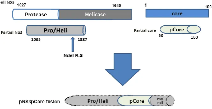
Therefore, constructing the DNA vaccine expressing specific parts of HCV antigens like overlapping middle region of NS3 or truncated core genes may overcome the present shortcomings of full length genes expression in order to avoid the autoimmunity, immune modulatory and other probably shortcoming effects of the full length protein in APCs. In this project, our aim was to design, construct and confirm a DNA construct containing fusion of novel overlapping region of NS3 with truncated core genes for future vaccine purposes as demonstrated in a schematic form in figure 1.
Methods
Reagents, vectors and cell Lines
The restriction enzymes (NdeI, BglII), InsTAclone™ PCR Cloning kit, Pfu DNA polymerase in addition to other routine molecular reagents prepared from Fermentas company(Slovenia). Plasmid and DNA isolation/purification kit from Bioneer (S.Korea), and LipofectaminTM 2000 from Invitrogen (USA), culture media and other culture related materials prepared from Gibco (USA). All primary antibodies were purchased from Abcam Company and stored at -70oC for long time storage.
pAdenovator-CMV5-IRES-EGFP expression vector was purchased from Qbiogene Corporation.The Low-passage HEK 293 cell line was provided from National Cell Bank of Iran, were grown in Dulbecco’s modified
Iranian Journal of Virology, Volume 4, Number 2, 2010 19 Eagle’s medium (DMEM) supplemented with
100 IU penicillin ml−1, 100mg streptomycin ml−1, 10% (v/v) fetal bovine serum, 20mM HEPES and non-essential amino acids.
Gene amplification by Reverse Transcription-Polymerase chain reaction (RT-PCR)
New overlapping region of NS3 gene covering incomplete protease/helicase domains (aa 1095-1387) of HCV genotype 1a was amplified and well characterized elsewhere previously (30). In the case of core gene similar method was employed for amplification. For Primer design, HCV reference sequences obtained from gene bank then by utilization of Generunner and Mega4 softwares, after multiple alignment 2 pairs of primers were selected for amplification of full core (aa 1-197) and 2 additional ones for new truncated core fragments, pCore (aa 50-160). The primers so designed to avoid false amplification of similar sequences from other HCV genotypes. Also, BglII or NdeI restriction sites, a stop codon (TGA) and a protein expression initiation codon (ATG) were introduced at both ends of primers for further manipulation. Also as a Glycin codon, GGT trinucleotides were inserted into primer backbone in the case of pcore-NdeF primer to make NS3-core fusion sequence (Table 1). Four Iranian patients infected with genotype 1 of HCV were enrolled for screening in the study. The sera had been confirmed as HCV positive sample and genotype 1 in Shariati
Hospital, Gastroenterology and Hepatology Research Center, Tehran. Viral RNA extracted by viral RNA extraction kit based on recommended protocol and RNA kept in -70oC until use.
First RT-PCR was done on four different extracted RNA samples using specific primer. After selection of one serum based on band density as the best sample, nested-PCR was developed to amplify a nearly 610 base pair sequence of full core.
PCR was performed in a total volume of 25 μl consisting of 0.2 μM each primer, Pfu DNA polymerase enzyme, and standard PCR constituents. The samples amplifed for 20 cycles in first round and 30 cycles for the second round at 60oC annealing temperature. The PCR products were then purified from the agarose gel using DNA Gel purification kit with respect to company instruction.
TA-genes Construction
The resulting extracted amplicons including full core gene was mixed with proper amount of pTZ57R/T cloning vector and ligation reaction was done as recommended by company with the help of T4 ligase. The ligation product was transformed into competent DH5-α bacteria and then the properly cloned core gene in pTZ57R/T vector (as a TA-core), were selected based on Blue/White screening and Ampicillin sensitivity on LB agar.
Insertion of gene into vector then was evaluated by colony-PCR using specific primers. The integrity of these constructs and
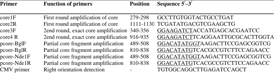
Primer Function of primers Position Sequence 5/–3/
core1F First round amplification of core 279-298 GCCTTGTGGTACTGCCTGAT
core2R First round amplification of core 1111-1130 TCGATATGACGTCGAAGCTG
core3F 2end round, exact core amplification 340-356 GGAAGATCTACCATGAGCACGAATCC
core4 R 2end round, exact core amplification 916-935 GGAAGATCTTCAGGAATTGCGCACTTGGTA
pcore-BglF Partial core fragment amplification 489-508 GGACATATGGTAAGACTTCCGAGCGGTCG
pcore-BglR Partial core fragment amplification 810-838 GGACATATGTCACGCCGTCTTCCAGAACC
pcore-Nde1F Partial core fragment amplification 489-508 GGACATATGGTAAGACTTCCGAGCGGTCG
pcore-Nde1R Partial core fragment amplification 810-838 GGACATATGTCACGCCGTCTTCCAGAACC
CMV primer Right orientation detection - TGTGGCAGGCTTGAGATCCAGCT
Table 1. The sequences of primers used in this study. The primers sequences and positions are based on HCV reference sequence strain H77 (GenBank accession number AF009606), underlined sequences demonstrate the modified sequence.
20 Iranian Journal of Virology, Volume 4, Number 2, 2010 insertion of gene were assayed using restriction analysis with BglII and sequencing as well. To make sure that the construction and manipulation processes did not affect the base sequences of the gene, selected plasmids were submitted for bidirectional sequencing using specific primers by the application of an automated DNA Sequencer ABI PRISM® 3700DNA.
Construction of TA-NS3 vector described previously (30). These confirmed TA-core and TA-pNS3 vectors were digested by BglII followed by extraction of the single band of each gene from agarose gel using a gel extraction kit.
Expressing vector construction
The sequences of core and pNS3 gene fragments were digested and purified then were mixed with high pure linearized pAdenovator-CMV5-IRES-EGFP, an IRES expressing vector and the ligation reaction drove them to join covalently together by exploiting T4ligase and also pretreatment with alkaline phosphatase. This vector contains strong CMV enhancer-promoter to drive substantial expression of genes of interest as well as an IRES sequence that induced GFP expression as a marker for transfection efficiency as well as evaluation of gene expression. The best constructs in bacteria were selected based on kanamycin resistance, Colony PCR and restriction analysis by BglII and designated as IR-core and IR-pNS3.
A new truncated core sequence harboring amino acid sequence 50-160 that is both N- and C-terminal domains deleted fragment prepared by PCR amplification on a TA-core and was named pCore. Briefly, 2 pairs of primers entitled pCore-Bgl and pCore-Nde (Table 1) exploited in 30 cycles standard PCR on TA-core containing full core and then the resultant amplicons prepared by core160-Bgl primers inserted directly into the IRES expressing vector. Also in addition to introduced NdeI restriction site at the ends of pCore-Nde primer, an additional GT nucleotide designed next to the coding sequence that added a new glycin code between pNS3 and pCore sequence as a spacer. These amplicons as well as TA-pNS3 were
digested by NdeI enzyme following gel extraction. In ligation mixture as shown in figure 1, TA-pNS3 linear plasmid will accept purified pCore sequence at NdeI restriction site so that pCore sequence followed first ¾ pNS3 region with a glycin amino acid between them as the spacer and named as TA-pNS3pCore. The proper vector designated as TA-pNS3pCore were selected based on ampicillin resistance and colony PCR.
The resultant TA-pNS3pCore was digested by BglII restriction enzyme then purified fusion sequences were inserted into linear IRES expressing vector by using T4 ligase. The proper clones were selected based on kanamycin resistance and colony PCR.
In all screening experiments, a new primer specific for CMV promoter was also employed to assess the correct orientation of genes inside the IR-gene expressing vectors (Table 1). The orientation of genes in plasmids was verified by special touchdown PCR in 65-55oC annealing temperature range using CMV-promoter primer as forward and core4-R, pcoreBglR and pcoreNdeR primers as reverse ones for detection of IR-core, IR-pCore and IR-pNS3pCore, similar to IR-pNS3 described previously.
To ensure the fidelity of the final fusion sequences, the gene was digested with BglII enzyme and also introduced into an ABI PRISM® 3700DNA analyzer automated sequencer using a set of forward and reverse primers.
Blast analysis of sequences
For sequence analysis, edited sequences of 2 genes were extracted from received data then submitted to online Blast and alignment tools for better understanding the sequence. After alignment of prepared sequences with reference sequences from NCBI gene bank, the degree of homology and difference of genes and amino acids was estimated.
Analyses of gene expression
Transfection of plasmids into 293 cells. The 293 cells were propagated in DMEM, supplemented with 10% FCS and antibiotics (penicillin 50 U/ml and streptomycin 50 μg/ml) and all cultures were maintained at 37°C in a moist atmosphere containing 5% CO2. They
Iranian Journal of Virology, Volume 4, Number 2, 2010 21 seeded in 6 well plates the day before
transfection so that at the transfection time they would reach to 70% confluency that is a marker for starting the logarithmic growth of the cells. Based on company instructions, a mixture of Lipofectamine 2000 and plasmid was prepared and then added to the cells gently. Following incubation at 37°C for 12 h, the cells were re-fed with fresh complete DMEM medium.
All expression vectors including IR-Core, IR- pNS3pCore and IR-pNS3 were transfected and compared with an empty plasmid as negative control for analysis of specific gene expression. These cells harvested up to 48 hours after transfection.
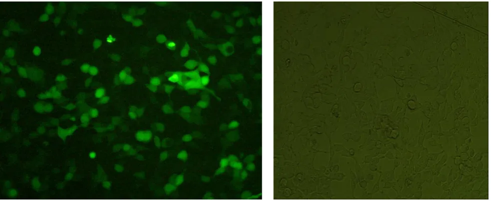
After brief screening by GFP expression under fluorescent microscopy, genomic RNA was extracted using total RNA extraction kit and Trizol method following DNase-I treatment to remove perturbing residual DNA and avoid false interpretation. Fluorescent images were captured at 490 nm using a Nikon Eclipse E1000 microscope after changing the medium with new DMEM and before harvesting the cells.
Reverse transcription. The 20 μl reaction
mixture for each gene contained 5 μl of extracted RNA, 4 μl of 5X RT buffer, 2 μl dNTP (2.5 mM of each dNTPs), 1 μl (10 pmol/μl) of reverse or random hexamer primer, 0.5 μl (40 unit/μl) of RNase inhibitor, 1 μl (40 unit/μl) of M-muLV reverse transcriptase and 6.5 μl of RNase free distilled water. Reverse transcription was carried out at 42ºC for 50 min followed by incubation at 70ºC for 10 min. PCR was done with above corresponding specific primers for each gene. The reaction was performed in a total volume of 25 μl consisting of 0.5 μM each primer, Taq DNA polymerase, and standard PCR constituents. The samples were amplified for 35 cycles at 56o c annealing temperature. The PCR products were then run on 1% agarose gel.
Western Blotting. For SDS–PAGE analysis, cell lysate samples were mixed in SDS–gel
sample buffer, containing 2% SDS and 2-mercaptoethanol and heated at 100 °C for 10 min. The proteins were separated in 12% SDS– polyacrylamide gels. Western blotting was performed as described earlier (31) using mouse monoclonal antibodies against core (aa 70-90) and commercially rabbit polyclonal antibodies for NS3. Proteins were blotted onto nitrocellulose membrane and it was blocked by incubation in 3% BSA in TBS buffer at 40 C overnight and subsequently exposed to diluted primary antibodies in 10ml of TBS for 2 h. The membrane was washed, and then incubated in 10 ml of TBS containing 2 U HRP-labeled goat anti-rabbit IgG or rat anti-mouse IgG (Sigma–Aldrich Corp.). The membrane was washed as in the previous step and then the chromophore 3, 3-diaminobenzidine (DAB) was used in order to develop the color-based signal.
Results
Amplification of genes
The electrophoretic results on agarose gel from RT-Nested-PCR product revealed 610 and 930 base pair fragments for full core and pNS3 amplification respectively. During preparation of truncated core from IR-core after standard PCR there was a 330 bp amplified fragment after colony PCR on pNS3pCore and IR-pCore as screening tool (fig. 2). The primers for amplification of two core truncated forms were designed such that they would amplify probable immunogenic regions in addition to avoiding harmful or unnecessary sequences.
Blast analysis of sequences
For evaluation the fidelity of the sequences, all cloning and expression vectors were sequenced by an automated sequencer. The sequence analysis of the full core gene showed more than 99% similarity with those of other 1a sequences using multiple alignment survey by online blast software. It is presumed that core is the most conserved sequence among HCV
22 Iranian Journal of Virology, Volume 4, Number 2, 2010
Fig. 1. Schematic representation of construction the novel fusion sequence including overlapping fragment of NS3 gene and N-, C-terminal deleted fragment of core gene. The truncated core inserted into naturally occurred NdeI restriction site near the ¼ end of pNS3 but did not disturb its reading frame.
Fig. 2. Colony-PCR for detection of insertion and right orientation of Core and pCore genes inside the TA or IRES vectors.
A: detection of core in IRES-core, from the left: 1- Marker, 1kb 2- near 750 bp fragment indicated right oriented gene, 3- 610 bp fragment showing insertion of gene
B: detection of pCore inside the TA-pNS3pCore and IR-pCore. from the left, 1 and 3 many nonspecific bands showed negative sample result. 4, 5, 6 a 330 bp fragment showing insertion of gene 7- marker: 1kb ladder.
Fig. 3. Restriction analysis of of IR-pNS3pCore (A) and TA-core (B) vectors by BglII. (A) Digestion of IR-pNS3pCore by BglII showed a 1250 bp fragment as pNS3pCore fragment and vector backbone with near 9000 bp size. (B) Digestion of TA-core by BglII showed a 610 bp fragment in addition to TA vector backbone with near 3000 bp size. Marker: 1 kb ladder.
genotypes and this data also confirmed it. High homology with H77 strain in analysis was significant. Phylogenetic analysis showed that NS3 sequence belonged to genotype 1a of HCV virus as demonstrated elsewhere (30).
Expressing vector construction
The percentage of positive colonies in colony-PCR method during the screening was significantly high as we expected. The colony PCR showed amplification of 330 bp as presence of partial core in pNS3pCore fusion construct and 610 bp for full core as shown in figure 2 rather than 930 bp fragments for pNS3. Restriction analysis of corresponding IR-pNS3pCore and TA-core constructs by
A B
1 2 3 1 2 3 4 5 6 7
A B
A B
Iranian Journal of Virology, Volume 4, Number 2, 2010 23
Fig. 4. GFP expression in 293 cells after transfection by IR-pNS3pCore in contrast to negative cells. After 2 days past transfection,fluorescent microscopy showed bright green in more than half of the cells as the sign of GFP expression, plasmid integrity and expression of gene of interest, fusion pNSpCore fragment.
Fig. 5. RT-PCR results of gene expression by IR-pNS3pCore fusion construct (A) and IR-core (B). 293 cells transfected with a mixture of respected plasmid and lipofectamine 2000 then 2 days past transfection total RNA extracted and analyzed by RT-PCR.
(A) 1- a 1250 bp fragment showing expression of pNS3pCore sequence 2- 293 cells as negative control 3- Positive control including IR- pNS3pCore plasmid 4- PCR negative control. Ladder 1 kb.
(B) 1- 610 bp fragment showing expression of core. 2, positive control. 3, 293 cells as negative control, ladder 1kb.
Fig. 6. The western blot analysis of IR-pNS3 (A) and IR-core (B) expression vector transfection (A): IR-pNS3 transfection of 293 cells. 1- IR-pNS3 expressing protein with estimated weight of 30 kd, M: Prestained Protein marker Cat. Number: SM1811 (Fermentas cop.) (B) Left, Control purified Core protein revealed 3 distinct bands indicating 3 different expressed size of protein. Right, IR-core plasmid expressing a sharp band for full core in 293 cells that showed by arrow head.
BglII showed the right excision of 610 and 1250 base pair amplicons as well as presence of near 9000 and 3000 base pair vector
backbone at the top that means its integrity (fig. 3).
New pair of primers specific for CMV
1 2 3 4
A
1 2 3 4
A
1 2 3
B
1 2 3
B
1 2 3
B
~30 KD
B
A
Construction of an Expression Vector Containing a Novel Fusion Sequence …
24 Iranian Journal of Virology, Volume 4, Number 2, 2010
promoter also employed to determine the correct orientation of genes inside the IRES expression vector. True amplification of right inserted gene inside the IRES plasmid made corresponding amplicons a little bigger than specific amplicons, with the sizes around 1150 bp for pNS3, 750 bp for core and 1350 bp for pNS3pCore fusion sequences after PCR (fig. 2).
Reverse transcription and fluorescent microscopy
At 2 days post transfection fluorescent images were captured by fluorescent microscopy. As shown in figure 4, about half of the cells showed the sign of GFP expression in compare to negative control that is nearly the indicator of gene expression as well.
Total RNA extracted from the 293 cell line showed suitable quality by detecting special common pattern for eukaryotic ribosomal RNA on the agarose gel. The RT-PCR results showed detectable expression of these genes that had been achieved after transfection of IRES expression vector containing gene fragments into 293 cell line. For pNS3, IR-core and IR-pNS3pCore, there were the band sizes of 930, 610 and 1250 bp respectively as shown in figure 5. To avoid false positive, especial care was paid for removing the residual DNA to prevent misinterpreting the data.
Western Blotting
A low density but distinct protein bands were seen after Western blot analysis in cells transfected by IR-pNS3 (figure 6). The putative molecular weight of the protein was estimated to be approximately 30-31 kDa by SDS–PAGE, consistent with 70 kDa size for the full length sequence and also other similar studies (32).
In the case of core protein, a sharp band on 293 cells transfected with IR-Core expressing plasmids detected on membrane. As positive staining control, a purified core protein in concentration of 10ug was used that revealed a three band patterns demonstrating the 3 different expressed size of core in vitro.
Discussion
DNA Immunization has been shown to elicit suitable immune response against a number of viral antigens especially HCV and is currently used even for vaccine trials in humans (3, 7). The vectors expressing partial or full length of core and NS3 genes from HCV have been exploited for vaccine purposes routinely due to their suitable immunogenicity and high homology among HCV genotypes (3, 7, 33, 34).
Recent publications reported significant controversy regarding to usefulness of full length core protein as vaccine antigen (20, 21, 24, 26, 29, 35). Some groups of scientists started to use partial fragments of core gene instead of full length genome to overcome shortcomings reported to be associated with the full size core gene such as immunomodulatory and autoimmunity effects (18, 24, 26). In this project, we prepared a new expression vector containing core sequence without N- and C-terminal domain to avoid autoimmunity and/or immune modulatory effect of the core full gene sequence that was evaluated by others (24).
Because of both protease and helicase activities of HCV NS3 gene, theoretically, expression of its full protein in APCs may diminish the immune induction due to disruption of antigen processing or presentation pathway (17). So, we designed the NS3 primers in such a way that could amplify a new overlapping fragment covering both but incomplete domains based on a previously reported study (32). This sequence harbored CTL epitopes suitable for establishment of the cellular immune responses.
All of the primers used in this study were specifically designed for genotype 1 in order to prohibit false amplification of other sequences. In order to better interpret the sequencing results, Blast analysis was carried out and it was shown high homology with references genotype 1a in both genes. The sequencing results showed a very high similarity between the amplified core genes with the other 1a reference sequences as expected for the most conserved genes.
1 2 3
B
1 2 3
B
Iranian Journal of Virology, Volume 4, Number 2, 2010 25 RT-PCR and Western blot showed suitable
expression of full core and pNS3 mother plasmids. RT-PCR also showed expression of fusion pNS3pCore fragments by expression plasmid. GFP screening assay confirmed the plasmids integrity and concomitant expression of proteins of interest in 293 cells.
In this work, new truncated core and an overlapping region covering incomplete helicase/protease of NS3 genes were amplified and fused together in an expression vector as a novel fusion construct. Theoretically, this new fusion vector containing the above sequences would be useful in future studies by avoiding hortcomings like the immunomodulatory/ autoimmunity effects usually associated with full length gene expression.
Acknowledgements
The authors would also like to acknowledge Tarbiat Modares University and Shiraz University of Medical Sciences for their financial supports.
References
1. Arribillaga L, Arina A, Gorraiz M, Ruiz J, Prieto J, et al. Enhancement of CD4 and CD8 immunity by anti-CD137 (4-1BB) monoclonal antibodies during hepatitis C vaccination with recombinant adenovirus. Vaccine. 2005;23:3493-9.
2. Neumann-Haefelin C, Blum HE, Chisari FV, Thimme R. T cell response in hepatitis C virus infection. J Clin Virol. 2005;32:75-85
3. Alvarez-Lajonchere L, Dueñas-Carrera S. Advances in DNA immunization against hepatitis C virus infection Opportunities and challenges. Human Vaccines. 2009;5(8):568-71.
4. Encke J, Geissler M, Stremmel W, Wands JR. DNA-Based Immunization Breaks Tolerance in a Hepatitis C Virus Transgenic Mouse Model. Human Vaccines. 2006;2(2):78-83.
5. Ulmer JB, Wahren B, Liu MA. Gene-based vaccines: recent technical and clinical advances. Trends Mol Med. 2006;12(5):216-22.
6. Frelin L, Alheim M, Chen A, Soderholm J, Rozell B, Barnfield C, et al. Low dose and gene gun immunization with a hepatitis C virus
nonstructural3 NS3 DNA-based vaccine containing NS4A inhibit NS3/4A-expressing tumors in vivo. Gene Ther. 2003;10(8):686-99.
7. Sallberg M, Weiland M, Frelin L. Therapeutic vaccines: challenges of chronic viral infections. Drug Discov Today, Therapeutic Strategies. 2007;4(4):254-66.
8. Castellanos M, Cinza Z, Dorta Z, Veliz G, Vega He, Lorenzo I, et al. Immunization with a DNA vaccine candidate in chronic hepatitis C patients is safe, well tolerated and does not impair immune response induction after anti-hepatitis B vaccination. J Gene Med. 2010;(12):107–16. 9. Weiner D. DNA vaccines: crossing a line in the sand. Introduction to special issue. Vaccine. 2008;26:5073-4.
10. Simon BE, Cornell KA, Clark TR, Chou S, Rosen HR, Barry RA. DNA vaccination protects mice against challenge with Listeria monocytogenes expressing the hepatitis C virus NS3 protein. Infect Immunol. 2003;71(11):6372– 80.
11. Lemon SM, Walker C, Alter MJ, M Y. Hepatitis C virus. Fields Virology. USA: Lippincott Williams &Wilkins. 2007. p. 1253-304. 12. El-Gogo S, Staib C, Lasarte JJ, Sutter G, Adler H. Protective vaccination with hepatitis C virus NS3 but not core antigen in a novel mouse challenge model. J Gen Med. 2008;10:177–86. 13. Lang K, Yan J, Draghia-Akli R, Khan A, Weiner D. Strong HCV NS3- and NS4Aspecific cellular immune responses induced in mice and Rhesus macaques by a novel HCV genotype 1a/1b consensus DNA vaccine. Vaccine. 2008;26:6225-31.
14. Folgori A, Capone S, Ruggeri L, Meola A, Sporeno E, Ercole BB, et al. A T-cell HCV vaccine eliciting effective immunity against heterologous virus challenge in chimpanzees. Nat Med. 2006;12(2):190–7.
15. Johnson CL, Owen DM, Gale M. Functional and therapeutic analysis of hepatitis C virus NS3.4A protease control of antiviral immune defense. J Biol Chem. 2007;282(14):10792-803. 16. Li XD, Sun L, Seth RB, Pineda G, Chen ZJ. Hepatitis C virus protease NS3/4A cleaves mitochondrial antiviral signaling protein off the
26 Iranian Journal of Virology, Volume 4, Number 2, 2010 mitochondria to evade innate immunity. Proc Natl Acad Sci USA. 2005;102(49):17717-22.
17. Krishnadas DK, Ahn JS, Han J, Kumar R, Agrawal B. Immunomodulation by hepatitis C virus-derived proteins: targeting human dendritic cells by multiple mechanisms. Int Immunol. 2010;22(6):491-502.
18. Haller AA, Lauer GM, King TH, Kemmler C, Fiolkoski V, Lua Y, et al. Whole recombinant yeast-based immunotherapy induces potent T cell responses targeting HCV NS3 and Core proteins. Vaccine. 2007;25:1452–63
19. Alekseeva E, Sominskaya I, Skrastina D, Egorova I, Starodubova E, Kushners E, et al. Enhancement of the expression of HCV core gene does not enhance core-specific immune response in DNA immunization: advantages of the heterologous DNA prime, protein boost immunization regimen. Genet Vaccines Ther. 2009;7:7.
20. Hartoonian C, Ebtekar M, Soleimanjahi H, Karami A, Mahdavi M, Rastgoo N, et al. Effect of immunological adjuvants: GM-CSF (granulocyte-monocyte colony stimulating factor) and IL-23 (interleukin-23) on immune responses generated against hepatitis C virus core DNA vaccine. Cytokine. 2009;46(1):43-50.
21. Matsui M, Moriya O, Abdel-Aziz N, Matsuura N, Miyamura T, Akatsuka T. Induction of hepatitis C virus-specific cytotoxic T lymphocytes in mice by immunization with dendritic cells transduced with replication-defective recombinant adenovirus. Vaccine. 2002(21):211– 20.
22. Roohvand F, Aghasadeghi M-R, Sadat SM, Budkowska A, Khabiri A-R. HCV core protein immunization with Montanide/CpG elicits strong Th1/Th2 and long-lived CTL responses. Biochemical and Biophysical Research Communications. 2007;5:641–9.
23. Thammanichanonda D, Moneera S, Yotnda P, Aitken C, Earnest-Silveirad L, a DJ, et al. Fiber-modified recombinant adenoviral constructs encoding hepatitis C virus proteins induce potent HCV-specific T cell response. Clin Immunol. 2008; 128: 329-339.
24. Liu Y, Zhou W, You C, Zheng H, al; e. An autoimmune domain-reduced HCV core gene
remains effective in stimulating anti-core cytotoxic T lymphocyte activity. Vaccine. 2006;24:1615–24. 25. Large MK, Kittlesen DJ, Hahn YS. Suppression of Host Immune Response by the Core Protein of Hepatitis C Virus: Possible Implications for Hepatitis C Virus Persistence. J Immunol. 1999;162:931-8.
26. Alvarez-Obrego´n JCs, Duen˜as-Carrera S, Valenzuela C, Grillo JM. A truncated HCV core protein elicits a potent immune response with a strong participation of cellular immunity components in mice. Vaccine 2001;19:3940–6. 27. Szabo G, Dolganiuc A. Hepatitis C core protein- the "core" of immune deception? J Hepatol. 2008;48(1):8-11.
28. O'Beirne J, Mitchell J, Farzaneh F, Harrison PM. Inhibition of major histocompatibility complex Class I antigen presentation by hepatitis C virus core protein in myeloid dendritic cells. Virology. 2009;389:1–7.
29. Zhu W, Chang Y, Wu C, Han Q, Pei R, Mengji Lu, et al. The Wild-Type Hepatitis C Virus Core Inhibits Initiation of Antigen-Specific T- and B-Cell Immune Responses in BALB/c Mice. Clin Vaccine Immunol 2010;17(7):1139-47.
30. Hosseini S-Y, Sabahi F, Moazzeni S-M, Modarressi MH, Saberi-Firoozi M, Ravanshad M, et al. Cloning and evaluation of expression of a novel overlapping region of NS3 gene of Hepatitis C virus by expressing vector. Modares Journal of Medical Sciences: Pathobiology. 2011;13(4):21-32. 31. Sambrook J, Fritsch EF, Maniatis T. Molecular cloning: a laboratory manual edition n, editor: NY: Cold Spring Harbor Laboratory Press. 1989. 32. Haller AA, Lauer GM, King TH, Kemmler C, Fiolkoski V, Lua Y ,et al. Whole recombinant yeast-based immunotherapy induces potent T cell responses targeting HCV NS3 and Core proteins. Vaccine. 2007;25:1452–63.
33. Houghton M, Abrignani S. Prospect for vaccine against the hepatitis C virus. Nat Rev Microbiol. 2005 Aug 18;436(7053):961-6.
34. Martin P, Inchauspe G. Hepatitis C vaccines. Drug discovery today. 2006;203:2090-6.
35. Dolganiuc A, Kodys K, Kopasz A, Marshall C, Do T, Romics L, Jr., et al. Hepatitis C virus core and NS3 proteins induce pro- and
Iranian Journal of Virology, Volume 4, Number 2, 2010 27 inflammatory cytokines and inhibit dendritic cell differentiation. J Immunol. 2003;170:5615–24.