A Biologically Inspired ELM-based Framework for
Classification of Brain MRIs
https://doi.org/10.3991/ijoe.v16i10.15653
Sarada Prasanna Pati, Debahuti Mishra
Sikhsa ‘O’ Anusandhan Deemed to be University, Odisha, India
Samarjeet Borah ()
Sikkim Manipal University, Sikkim, India [email protected]
Abstract—Use of medical images for clinical analysis of various critical dis-eases have become increasingly predominant in modern health care systems. Ap-plication of machine learning technique in this context evolves as a potential so-lution in terms providing faster output with high diagnostic accuracy. In this work, we propose an Extreme Learning Machine (ELM) based classifier SFLA-ELM for detection of normal and pathological brain condition from brain Mag-netic Resonance Images (MRIs). ELM is known for its speed and accuracy whereas the proposed method uses a swarm based evolutionary technique Shuf-fled Frog Leaping Algorithm (SFLA) and 10-fold cross validation method to op-timally determine the network parameter of the ELM for better classification per-formance. The proposed model is experimented on three different brain MRI da-tasets of three different brain diseases. To get better approximation accuracy and generalization ability for the base ELM classifier, the suitable activation function and the appropriate number of hidden layer nodes are chosen. The performance validation of the proposed framework is done under two different network con-ditions, i.e. fixed network structure and varying network structure, by comparing its performance with two standard hybridized ELM classifiers, namely, PSO-ELM and ABC-PSO-ELM. The comparative performance analysis suggests that the proposed SFLA-ELM gives better classification performance in diagnosing the diseases in terms of accuracy, sensitivity, specificity, F-score and Area under ROC curve (AUC). Furthermore, the SFLA-ELM also found to offer better gen-eralization ability and better stability with more compact network structure.
Keywords—Classification, Brain Image, Extreme Learning Machine, ELM, Shuffled Frog Leaping Algorithm, PSO-ELM, ABC-ELM, SFLA-ELM.
1
Introduction
knowledge and experience, the human vision system restricts the manual interpretation and analysis of MR images for the clinical experts. This is due to the large volume of information contained in an MR image which is hard to interpret by human vision [1-2]. This is the reason why use of automated image analysis methods utilizing machine learning and image processing techniques are of wide use in recent years in the field of MR image processing. These computers assisted diagnosis (CAD) techniques not only reduce burden on the radiologist and neurologists but also improve the accuracy and objectivity of diagnosis [3-7].
Extreme Learning Machine (ELM) [8-13], anticipated by Huang et al. [13], is an efficient and effective learning algorithm for Single Layer Feed Forward Networks (SLFNs), which is being used extensively for classification task. The input weights as well as the biases of the hidden layer neurons are selected randomly in ELM, from which the output weights are calculated mathematically using a simple inverse opera-tion of the hidden layer output matrix. As compared to other tradiopera-tional neural networks used for classification, ELM exhibits higher generalization ability, extremely faster training speed and smallest norm of weights [8]. ELM resolves several issues such as; local minima, stopping criterion, proper learning rate and the number of iterations in comparison to traditional neural networks trained using gradient descent methods. Lit-erature study reveals that ELM shows better generalization and faster learning ability compared to contemporary classification techniques like Support Vector Machines (SVM) and deep learning [14].
However, ELM faces the challenge with respect to selection of input factors, e.g., hidden biases and the input weights, which are not optimal [15-16]. In ELM, while calculating the output weights, it tries to minimize the training error and finds the min-imum norm of output weights. Because of the random selection of the input weights and hidden biases, sometimes the output matrix may not exhibit full rank leading to ill-conditioning of the network and may result in non-optimal solutions [17]. Hence, ran-dom selection of these parameters can ultimately affect the performance of ELM. Therefore, optimal selections of these parameters are crucial for the best performance of ELM and this implicates a complex optimization problem [18]. Research on enhanc-ing the performance of ELM establish that evolutionary algorithms such as DE [19-20], PSO [21-22] and ABC [23] can be used to optimally determine the input weights and hidden biases of the SLFN by exploring the potential areas of the solution space. Use of such optimization techniques can also increase the generalization ability and stability of ELM with more compact network.
Shuffled Frog Leaping Algorithm (SFLA) is developed by Eusuff et al. [24], is a nature inspired meta-heuristic, based on population. SFLA comes under the class of memetic algorithms as it mimics the memetic evolution adapted by frog population while searching for location of good food sources available on discrete stones in a swamp. SFLA has been applied effectively to resolve several miscellaneous optimiza-tion problems in engineering [25-31]. This work makes use of SFLA to train ELM by optimally determining the network parameters.
accuracy and generalization ability for same problem or training data using different activation functions [32] [33-34]. Activation functions are somehow dependent on the dataset at hand and there doesn’t exist any provable relationship between these two. In essence, the suitable activation function can be chosen optimally either by trial or by tuning [34]. The network structure of the ELM, defined in terms of number of hidden layer nodes, also largely influences the classification performance of ELM classifier [32]. In this work, the classification performance of ELM has been evaluated using ten different commonly used activation functions at varying number of hidden layer nodes. Based on the accuracy rate obtained, the suitable activation function and the number of hidden layer nodes are chosen for the ELM classifier.
1.1 Aim of the work
The aim of this work is to design an efficient classifier model that can distinguish the brain MRI samples as normal and pathological, with more accuracy and effectively ranks both the positive and negative instances with better success rate. In this work, a hybrid ELM based classifier, called SFLA-ELM has been proposed for classifying brain MRI data to normal and pathological brain. SFLA-ELM applies SFLA for en-hancing the performance of basic ELM classifier by optimally determining the input weight and hidden bias of the SLFN. The proposed model is experimented on three different brain MRI datasets of three different brain diseases. To get better approxima-tion accuracy and generalizaapproxima-tion ability for the base ELM classifier, the suitable acti-vation function and the appropriate number of hidden layer nodes are chosen based on evaluating the classification accuracy of ELM on ten standard activation functions. The classification performance of the SFLA-ELM is validated by comparing its perfor-mance with two standard hybridized ELM classifiers, namely, PSO-ELM and ABC-ELM. The performance validation is done under two different network conditions, i.e. fixed network structure and varying network structure. 10-fold cross validation is used and average estimation is considered in all the experiments in this work to reduce the variance in performance estimation.
1.2 Organization of the paper
2
Methodologies Adopted for the Experiment
2.1 Extreme Learning Machine (ELM)
ELM is a very fast, simple and efficient learning algorithm for training the SLFNs. Unlike that of in traditional learning methods used to train SLFNs, in ELM the output weight matrix of the network is calculated mathematically in a non-iterative way from the randomly chosen input weights and hidden biases using the Moore–Penrose gener-alized inverse [8]. In ELM, the training is carried out in batch mode by presenting all the training data to the SLFN before computing the weights in a single iteration. ELM exhibits several striking features that makes it superior compared to other learning methods for SLFN such as; extremely fast learning speed with better learning perfor-mance, better generalization ability, free from getting stuck in local minima, no network tuning, no control parameter setup, simple and fixed network structure and ease of im-plementation [14]. While the gradient-based learning algorithms strive to reach mini-mum training error without considering the magnitude of weights, ELM tries to reach smallest training error with smallest norm of weights. The learning steps of the ELM algorithm can be summarized in the following three steps [8]. Given a training set 𝑁 = {(𝑋𝑖 , 𝑡𝑖) | ∈ 𝑅𝑑, 𝑡𝑖∈ 𝑅, 𝑖 = 1,2, … . , 𝑛}, hidden node number 𝑁 and activation func-tion 𝑔:
Step 1: Assign the input weight 𝑤𝑖and bias 𝑏𝑖(𝑖 = 1,2, … , 𝑁) randomly. Step 2: Calculate the hidden layer output matrix 𝐻 using the equation (1).
𝐻 = (
𝑔(𝑊1 . 𝑋1+ 𝑏1)𝑔(𝑊2 . 𝑋1+ 𝑏2) ⋯ 𝑔(𝑊𝑁 . 𝑋1+ 𝑏𝑁)
𝑔(𝑊1 . 𝑋2+ 𝑏1)𝑔(𝑊2 . 𝑋2+ 𝑏2) ⋯ 𝑔(𝑊𝑁 . 𝑋2+ 𝑏𝑁)
⋮⋮ ⋯ ⋮
𝑔(𝑊1 . 𝑋𝑛+ 𝑏1)𝑔(𝑊2 . 𝑋𝑛+ 𝑏2) ⋯ 𝑔(𝑊𝑁 . 𝑋𝑛+ 𝑏𝑁)
)
𝑛×𝑁
(1)
Step 3: Calculate the output weight 𝛽, where 𝛽 = 𝐻ϯ𝑇, here 𝐻ϯis the Moore–Pen-rose (MP) generalized inverse of 𝐻and𝑇 = (𝑡1, 𝑡2, 𝑡3, … . . , 𝑡𝑛)𝑇.
2.2 Shuffled Frog Leaping Algorithm (SFLA)
2.3 Working Principle of Proposed SFLA-ELM Classifier
In this section, a novel learning framework for SLFNs, called SFLA-ELM has been proposed. This method uses SFLA and the k-fold cross validation scheme to find the optimal input weights and hidden biases. Further, Moore-Penrose generalized inverse is applied to mathematically calculate the output weights. Fitness function used by SFLA for optimization is the minimization of the mean square error (MSE). The opti-mization problem thus, is based on minimizing the function stated in equation (5).
𝜑 = 𝐸𝑀𝑆𝐸(𝑦𝑘, 𝑦̂𝑘) ,𝐸𝑀𝑆𝐸(𝑦𝑘, 𝑦̂𝑘) = 1
𝑁∑ (𝑦𝑘− 𝑦̂𝑘 ) 2 𝑁
𝑘=1 (5)
Here, 𝑁 = Number of samples, 𝑦𝑘 = Desired output and 𝑦̂𝑘 =Obtained output The algorithm of SFLA-ELM for classification of MRI dataset is as follows:
Algorithm: Brain Image Classification
Input: Brain image dataset; Population size (P); Size of hidden layer (𝐻𝑐); Number of memeplexes (m); number of folds for n-fold cross validation (n).
Output: Classification accuracy
Description: Global best (𝑋𝑔); Local best (𝑋𝑏); Local worst (𝑋𝑤). Step 1 Divide the dataset to train_set (train_input, train_output) and test_set
(test_input, test_output) as per n- fold cross validation Step 2 For j = 1 to n
𝑗𝑡ℎset is used as test_set and 𝑛 − 𝑗 sets are as train_set Step 3 Set K random weight population; each of size 1 × 𝑁𝑐
𝑃𝑖= {𝑉1𝑖, 𝑉2𝑖, 𝑉3𝑖, … … … . . , 𝑉𝑁𝑐
𝑖 } for 𝑖 = 1,2,3, … … , 𝐾
Step 4 For each population 𝑃𝑖 find the error value in ELM using Step 5 Step 5 For each train_input find
𝐻𝑙= 𝑡𝑟𝑎𝑖𝑛𝑖𝑛𝑝𝑢𝑡𝑙× 𝑃𝑖
𝛽𝑖= 𝑝𝑠𝑒𝑢𝑑𝑜𝑖𝑛𝑣𝑒𝑟𝑠𝑒𝑟(𝐻) × 𝑡𝑟𝑎𝑖𝑛𝑜𝑢𝑡𝑝𝑢𝑡
𝑜𝑏𝑡𝑜𝑢𝑡𝑝𝑢𝑡= (𝑡𝑒𝑠𝑡𝑖𝑛𝑝𝑢𝑡× 𝑃𝑖) × 𝛽𝑖
𝑂𝑖=𝐸𝑀𝑆𝐸(𝑜𝑏𝑡𝑜𝑢𝑡𝑝𝑢𝑡, 𝑡𝑒𝑠𝑡𝑜𝑢𝑡𝑝𝑢𝑡)
Step 6 Sort each population in ascending order with respect to maximum fitness considering minimum MSE.
Step 7 Find𝑋𝑔, where 𝑂𝑖 is with minimum value.
Step 8 Partition the populations into m number of memeplexes.
The distribution of the sorted population to m number of memeplexes is done in such a way, that, the first population is assigned to first memeplex and second population is assigned to second memeplex and the same process is continued till the 𝑚𝑡ℎ memeplex. Then the m+1 population will be assigned to first memeplex and so on till all population are distributed.
Step 9 Within each memeplex, generate 𝑋𝑏and 𝑋𝑤 through the objective function repeating Step 5 with respect to fitness value.
Replace 𝑋𝑤with 𝑋𝑤(𝑛𝑒𝑤) Else
Update 𝑋𝑤with respect to 𝑋𝑔 using equation (4) and equation (3) a. If
Fitness of 𝑋𝑤(𝑛𝑒𝑤) is better than 𝑋𝑤 ,then replace 𝑋𝑤with 𝑋𝑤(𝑛𝑒𝑤) Else
Generate a new position of 𝑋𝑤 randomly
Step 12 Merge the memeplexes and find the fitness value of the new population us-ing Step 5
Step 13 Repeat Step 6 to Step 12 until the termination condition is satisfied Step 14 𝑋𝑔is considered as final weight and accordingly the 𝛽𝑔𝑙𝑜𝑏𝑎𝑙𝑏𝑒𝑠𝑡 is generated
and used for the classification of test data.
Step 15 Repeat the process for all the value of j, find the classification accuracy at each fold to generate the average accuracy.
3
Experiment Design
Three ELM based classification frameworks, namely, SFLA-ELM, ABC-ELM and PSO-ELM were implemented for classification of the brain MRI datasets. The classifi-cation performance of proposed SFLA-ELM is evaluated, compared and validated through experimentation results. The validation is done under two different network conditions of ELM.
a)Fixed network structure: Keeping the network structure of the ELM classifier fixed, after determining the suitable activation function and the number of hidden layer nodes [32] [35]. The activation function and the number of hidden layer nodes are finalised after experimentally analysing the classification performance of 10 commonly used activation functions with respect to varying number of nodes.
b)Varying network structure: Varying the number of hidden layer nodes while using the same activation function for ELM as determined in Phase I.
3.1 Experimental setup
in this work applying SFLA, ABC and PSO, the maximum number of iterations con-sidered was 100 and the population size taken was 20.
3.2 Datasets used for experimentation
The proposed classifier model is experimented on three brain image datasets col-lected from [36], supported and maintained by Medical School of Harvard University. These three datasets contain T2-weighted brain MR images in gif format with size 256×256.All brain images are in transaxial plane. For our purpose we have collected three different brain image datasets for three different brain diseases, namely, Alz-heimer, Glioma and Multiple Sclerosis. Feature extraction is the first step to represents the image in its compact and high level form to prepare the image dataset for classifi-cation. The datasets used in this work are prepared by applying Discrete Wavelet Trans-formation (DWT) using bi-orthogonal wavelet function for image decomposition and feature extraction from the MRI images. The images are decomposed by using bi-or-thogonal 1.3 wavelet function up to level 3 and thereby reducing the image size to 32×32. The detail description of datasets is shown in Table 1. Each of these datasets comprises of both normal as well as diseased instances for brain images pertaining to that particular disease.
Table 1. Description of Image Dataset
Type of Brain Image Num. of Images Image Size
Alzheimer 101 256×256
Glioma 123 256×256
Multiple Sclerosis 127 256×256
4
Experimentation, Result Analysis and Validation
This section presents the details of the three separate sets of experiments that were conducted, the analysis of the results obtained and validation of the proposed classifi-cation method. The first set of experiments were conducted with an objective to choose the suitable activation function and optimally decide the number of nodes for the base ELM classifier for each dataset. In second set of experiments the classification of the three MRI datasets was carried out using SFLA-ELM
4.1 Experiment I: Determining the activation function and network structure
out for each dataset to analyze the performance of ELM at different number of nodes by considering the activation functions and 10-fold cross validation is used for bias free assessment. In addition, for each fold, 20 trials are taken with the same number of nodes and activation function on a dataset in order to nullify the effect of random inputs to the network. Finally, the average values of the result are considered for performance meas-ure.
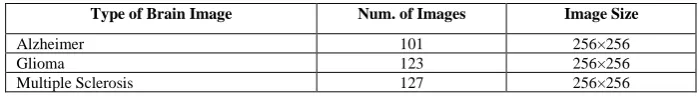
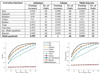
Table 2 shows the highest average training accuracy achieved for each activation function. The training accuracies obtained at different number of hidden layer nodes in these experiments are represented in Fig 1 to Fig 3. All experiments are conducted by varying the number of nodes with an increment of 5 nodes, up to 60 nodes. That acti-vation function is chosen as the most suitable actiacti-vation function which shows best accuracy at lesser number of nodes compared to other activation functions tested here. By inspecting the accuracy results obtained using different activation function it is ob-served that the Multi-quadratic activation function shows better classification accura-cies for all the three datasets. In fact, we limit the maximum number of nodes to 60 for our comparative study as in all three datasets it is found that by 60 nodes the training accuracies obtained reaches the maximum.
Table 2. Recognition of Performance of Activation Functions for the Three Datasets
Activation functions Alzheimer Glioma Multi Sclerosis
Training Accuracy
No. of Nodes
Training Accuracy
No. of Nodes
Training Accuracy
No. of Nodes
Sine 0.918 60 0.908 60 0.918 60
Sigmoid 0.99 60 0.996 60 0.975 60
Hardlim 0.99 60 0.996 60 0.974 60
Tribas 0.5 5 0.503 60 0.5 05
Radbas 0.515 25 0.518 60 0.512 60
Tanh 0.993 60 0.995 60 0.972 60
Gaussian 0.507 35 0.517 60 0.523 60
Inv. Multi-quadratic 0.979 60 0.979 60 0.942 60
Bipolar 0.989 60 0.995 60 0.979 60
Multi-quadratic 0.999 55 1 40 0.999 55
Fig. 1. Training accuracy for activation functions in Alzheimer dataset
Fig. 3. Training accuracy for activation functions in Multiple Sclerosis dataset
After choosing the activation function with best performance, the number of nodes that led to this best performance had been considered as the optimal number of nodes for the SLFN. The network structure that we choose for the ELM classifier which gives best classification performance in all three datasets is given in Table 3.
Table 3. The Network Structure of ELM with best Classification Performance
Dataset Activation Function No. of nodes
Alzheimer Multi-quadratic 55
Glioma Multi-quadratic 40
Multi Sclerosis Multi-quadratic 55
4.2 Experiment-II: Classification and performance analysis at fixed network structure
In order to study the classification performance and effectiveness of the SFLA-ELM method, it was applied on three MRI datasets, namely, Alzheimer, Glioma and Multiple Sclerosis. Two of the established hybrid ELM models such as; ABC-ELM and PSO-ELM were also implemented for classification of the MRI datasets to have a compara-tive study of the classification performances and validate the effeccompara-tiveness of the model.
Table 4. Performance Comparison of Proposed Model Based on Accuracy, Sensitivity, Specificity
Methods Alzheimer Glioma Multi Sclerosis
Acc Sen Spe Acc Sen Spe Acc Sen Spe
SFLA-ELM 0.979 1.000 0.917 0.997 0.944 1.000 0.988 0.968 1.000
PSO-ELM 0.957 0.963 0.887 0.947 0.867 0.985 0.976 0.988 0.833
ABC-ELM 0.910 0.933 0.853 0.895 0.866 0.833 0.908 0.917 0.831
*Acc: Accuracy, Sen: Sensitivity, Spe: Specificity
were evaluated in order to compare the effectiveness of the classifier. The average clas-sification accuracy, sensitivity, specificity values obtained for SFLA-ELM, PSO-ELM and ABC-ELM in case of all the three datasets is presented in Table 4. SFLA-ELM is found to achieve highest accuracy of 0.947, 0.944 and 0.988 for Alzheimer, Glioma and Multiple Sclerosis dataset respectively. it is evident that SFLA-ELM better scores for specificity and sensitivity as compared to PSO-ELM and ABC-ELM in case of all the three datasets except the case of Multiple Sclerosis dataset, where PSO-ELM scores better than SFLA-ELM in terms of the sensitivity value.
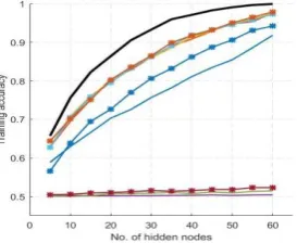
Fig. 4.Boxplots Showing the Variation in Classification Accuracy Values
Figure 4 shows the box-plots related to the distribution of the classification accuracy values. Each box in the plot corresponds to the accuracy distribution range obtained over the 10-fold cross validation performed for classification using the algorithm. As we can figure out, SFLA-ELM generates more compact boxes in all three datasets com-pared to PSO-ELM and ABC-ELM, and this confirms that SFLA-ELM gives better stability in terms of classification results compared to the other two methods.
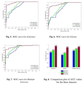
Figure 5 to Figure 7 present the ROC curves for the classification methods on the three datasets. A close observation of the ROC curves establishes that, SFLA-ELM outperforms PSO-ELM and ABC-ELM in case of all the three datasets.
Fig. 5. ROC curve for Alzheimer Fig. 6. ROC curve for Glioma
Fig. 7. ROC curve for Multiple Sclerosis
Fig. 8. Comparison plot of AUC values for the three datasets
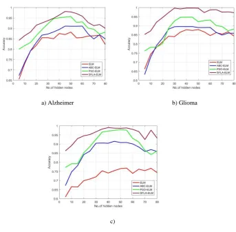
4.3 Experiment III: Classification and Performance Analysis at Varying Network Structure
a) Alzheimer b) Glioma
c)
Fig. 9. Classification Accuracy Vs. Number of Nodes for the three Datasets
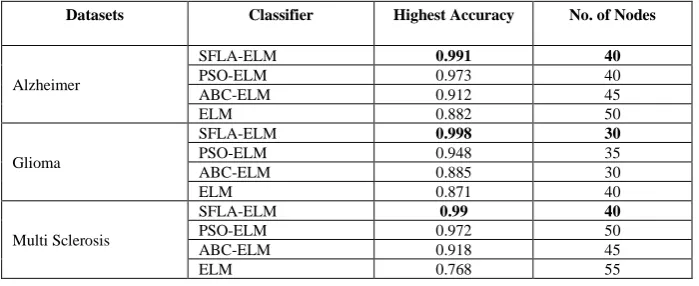
Table 5. Highest Accuracy Achieved while Varying the Number of Nodes
Datasets Classifier Highest Accuracy No. of Nodes
Alzheimer
SFLA-ELM 0.991 40
PSO-ELM 0.973 40
ABC-ELM 0.912 45
ELM 0.882 50
Glioma
SFLA-ELM 0.998 30
PSO-ELM 0.948 35
ABC-ELM 0.885 30
ELM 0.871 40
Multi Sclerosis
SFLA-ELM 0.99 40
PSO-ELM 0.972 50
ABC-ELM 0.918 45
ELM 0.768 55
5
Findings
The analysis of the of the experiments results suggested that the proposed SFLA-ELM classifier exhibits better overall performance in successfully discriminating the diseased and normal brain MRI samples with better accuracy in comparison to the ex-isting hybridized ELM methods, PSO-ELM and ABC-ELM. Some of the important findings of these experimental studies are as follows.
a) The proposed SFLA-ELM method achieves better accuracy in all three MRI datasets compared to the other two hybrid ELM based methods, i.e. ABC-ELM and PSO-ABC-ELM.
b) Analysis of the box plot results for accuracy further confirms that SFLA-ELM gives better stability in terms of classification results compared to the other two methods.
c) The ROC curve analysis of the three hybrid methods shows that the curve for SFLA-ELM is superior to ABC-ELM and PSO-ELM. The higher AUC values obtained for ELM in case of all three datasets further prove that SFLA-ELM is more successful in ranking both the positive and negative instances. d) SFLA-ELM achieves better sensitivity and specificity than the other two
hy-brid methods and thereby making it evident that, the proposed SFLA-ELM method is more appropriate for diagnosis of brain diseases from MRI data. e) While evaluating the performance of the three-classification method over
6
Conclusion and Future Scope
This paper presents a novel hybrid classification method SFLA-ELM to classify MR brain image data for diagnosis of pathological brain conditions. SFLA-ELM applies SFLA for enhancing the performance of basic ELM classifier by optimally determining the input weight and hidden bias of the SLFN. The performance validation was done under both fixed network structure as well as varying network structure of ELM. Ex-perimental results reveal that the SFLA-ELM method out performs the other two hybrid methods PSO-ELM and ABC-ELM and achieves better performance in terms of vari-ous performance measures like accuracy, ROC, AUC, sensitivity, specificity. Further-more, SFLA-ELM is found to be more successful in ranking both the positive and neg-ative instances by achieving higher AUC values and also shows better classification accuracy with a relatively compact network with better stability and thereby making it evident that, the proposed SFLA-ELM method can be more appropriate for diagnosis of brain diseases from MRI data. Future work could focus on implementing this model for multi-class classification of brain MR images and also on other type medical images for diagnosis of other diseases. Larger image dataset can also be used to enhance the training process of the classifier.
7
References
[1]A. Ortiz, J.M. Gorriz, J. Ramirez, D. Salas-Gonzalez, J.M. Llamas-Elvira, Two fully-unsu-pervised methods for MR brain image segmentation using SOM-based strategies, Appl. Soft Comput. J., 13 (2013) 2668–2682. https://doi.org/10.1016/j.asoc.2012.11.020
[2]D. Jude Hemanth, C. KeziSelva Vijila, A. Immanuel Selvakumar, J. Anitha, Performance Improved Iteration-Free Artificial Neural Networks for Abnormal Magnetic Resonance Brain Image Classification, Neurocomputing, 130 (2014) 98–107. https://doi.org/10.1016/ j.neucom.2011.12.066
[3]Geethu Mohan, M. Monica Subashini, MRI based medical image analysis: Survey on brain tumor grade classification, Biomedical Signal Processing and Control, 39 (2018) 139–161.
https://doi.org/10.1016/j.bspc.2017.07.007
[4]Amr S. Mady, Samir Abou El-Seoud, An Overview of Volume Rendering Techniques for Medical Imaging, iJOE ‒ Vol. 16, No. 6, 2020, pp. 95-106, https://doi.org/10.3991/ijoe.v1 6i06.13627
[5]Ahmed Elnakib, Hanan M. Amer, Fatma E.Z. Abou-Chadi, Early Lung Cancer Detection using Deep Learning Optimization, iJOE ‒ Vol. 16, No. 6, 2020, pp. 82-94, https://doi.org/ 10.3991/ijoe.v16i06.13657
[6]Hewawasam Puwakpitiyage C. A., Paramesura Rao V. R., Muhammad Azizi M. S. A., Tee W. J., Murugesan R. K., Hamzah M. D., A Proposed Web Based Real Time Brain Computer Interface (BCI) System for Usability Testing, iJOE ‒ Vol. 15, No. 8, 2019, pp. 108-119,.
https://doi.org/10.3991/ijoe.v15i08.10406
[8]G. Huang, Y. Qin, C.K. Siew, Extreme learning machine: theory and applications, Neuro-computing, 70(1) (2006) 489–501. https://doi.org/10.1016/j.neucom.2005.12.126
[9]G. Huang, L. Chen, C. K. Siew, Universal approximation using incremental constructive feed forward networks with random hidden nodes, IEEE Trans. Neural Networks, 17(4) (2006)870–892. https://doi.org/10.1109/TNN.2006.875977
[10]G. Huang, L. Chen, Convex Incremental extreme learning machine, Neurocomputing, 70(16) (2007) 3056–3062. https://doi.org/10.1016/j.neucom.2007.02.009
[11]G. Huang, L.Chen, Enhanced random search based incremental extreme learning machine, Neurocomputing, 71(16) (2008) 3460–3468. https://doi.org/10.1016/j.neucom.2007.10.008
[12]G. Huang, L.Chen, Optimization method based extreme learning machine for classification, Neurocomputing, 74(1) (2010) 155–163. https://doi.org/10.1016 /j.neucom.2010.02.019
[13]G. Huang, Q. Zhu, C.K. Siew, Extreme learning machine: a new learning scheme of feed forward neural networks, Proceedings of the IEEE International Joint Conference on Neural Networks, 2004.
[14]Gao Huang, Guang-Bin Huang, Shiji Song, KeyouYoua, Trends in extreme learning ma-chines: A review, Neural Networks, 61 (2015) 32–48. https://doi.org/10.1016/j.neun et.2014.10.001
[15]Bin Li, Yibin Li, Xuewen Rong, The extreme learning machine learning algorithm with tunable activation function, Neural Comput & Applic, (2013) 22:531–539. https://doi.org/ 10.1007/s00521-012-0858-9
[16]Yongshan Zhang, Jia Wu, Zhihua Cai, Peng Zhang, Ling Chen, Memetic Extreme Learning Machine, Pattern Recognition, 58(2016)135–148. https://doi.org/10.1016 /j.patcog.2016.04.003
[17]P. Mohapatra, S. Chakravarty, P. K. Dash,ann improved cuckoo search based extreme learn-ing machine for medical data classification, Swarm and Evolutionary Computation, 24 (2015) 25–49. https://doi.org/10.1016/j.swevo.2015.05.003
[18]S. Suresh, S. Saraswathi, N. Sundararajan, Performance enhancement of extreme learning machine for multi category sparse data classification problems, Engineering Applications of Artificial Intelligence, 23 (2010) 1149–1157. https://doi.org/10.1016 /j.engappai.2010.06.009
[19]J. Sánchez-Monedero, C. Hervas-Martinez, P.Gutiérrez, M. C. Ruz, M. R. Moreno, M. Cruz-Ramirez, Evaluating the performance of evolutionary extreme learning machines by a com-bination of sensitivity and accuracy measures, Neural Network World, 20 (7) (2010) 899. [20]Q.-Y. Zhu, A. K. Qin, P. N. Suganthan, G.-B. Huang, Evolutionary extreme learning
ma-chine, Pattern Recognition, 38 (10) (2005) 1759–1763. https://doi.org/10.1016/j.patcog. 2005.03.028
[21]F. Han, H.-F. Yao, Q.-H. Ling, an improved evolutionary extreme learning machine based on particle swarm optimization, Neurocomputing, 116 (2013) 87–93. https://doi.org/10. 1016/j.neucom.2011.12.062
[22]Y. Xu, Y. Shu, Evolutionary extreme learning machine-based on particle swarm optimiza-tion, Proc. of the 2006 international symposium on neural networks, 2006, pp. 644–652.
https://doi.org/10.1007/11759966_95
[23]Chao Ma, An Efficient Optimization Method for Extreme Learning Machine Using Artifi-cial Bee Colony, Journal of Digital Information Management, 15(3) (2017).
[25]Muzaffar Eusuff, Kevin Lansey, Fayzul Pasha, Shuffled frog-leaping algorithm: a memetic meta-heuristic for discrete optimization, Engineering Optimization, 38(2) (2006) 129–154.
https://doi.org/10.1080/03052150500384759
[26]Parmeet Kaur, Shikha Mehta, Resource provisioning and work flow scheduling in clouds using augmented Shuffled Frog Leaping Algorithm, J. Parallel Distrib. Comput., 101 (2017) 41–50. https://doi.org/10.1016/j.jpdc.2016.11.003
[27]Sunita Panda, Archana Sarangi, Siba Prasada Panigrahi, A new training strategy for neural network using shuffled frog-leaping algorithm and application to channel equalization, Int. J. Electron. Commun., 68 (2014) 1031–1036. https://doi.org/10.1016/j.aeue.2014.05.005
[28]Xinmin Cheng, Xiaodan Zhang, Li Zhao, Aideng Deng, Yongqiang Bao, Yong Liu, Yun-liang Jiang, The application of shuffled frog leaping algorithm to wavelet neural networks for acoustic emission source location, C. R. Mecanique, 342 (2014) 229–233.
https://doi.org/10.1016/j.crme.2013.12.006
[29]Jianping Luo, Xia Li, Min-Rong Chen, Hongwei Liu, A novel hybrid shuffled frog leaping algorithm for vehicle routing problem with time windows, Information Sciences, 316 (2015) 266–292. https://doi.org/10.1016/j.ins.2015.04.001
[30]Zhaokun Li, Jingtai Cao, Xiaohui Zhao, Wei Liu, Swarm intelligence for atmospheric com-pensation in free space optical communication—Modified shuffled frog leaping algorithm, Optics & Laser Technology, 66 (2015) 89–97. https://doi.org/10.1016 /j.optlastec.2014.08.012
[31]Jian-ping Luo, Xia Li, Min-rong Chen, Hybrid shuffled frog leaping algorithm for energy-efficient dynamic consolidation of virtual machines in cloud data centers, Expert Systems with Applications, 41 (2014) 5804–5816. https://doi.org/10.1016/j.eswa.2014.03.039
[32]Jianfu Xia, Huiling Chen, Qiang Li, Minda Zhou, Limin Chen, ZhennaoCai, Yang Fang, Hong Zhou, Ultrasound-based differentiation of malignant and benign thyroid Nodules: An extreme learning machine approach, Computer Methods and Programs in Biomedicine, 147 (2017) 37–49. https://doi.org/10.1016/j.cmpb.2017.06.005
[33]Bin Li, Yibin Li, Xuewen Rong, The extreme learning machine learning algorithm with tunable activation function, Neural Comput & Applic, 22 (2013) 531–539. https://doi.org/ 10.1007/s00521-012-0858-9
[34]Ömer Faruk Ertuğrul, A novel type of activation function in artificial neural networks: Trained activation function, Neural Networks, 99 (2018) 148–157. https://doi.org/10.1016 /j.neunet.2018.01.007
[35]E. Malar, A. Kandaswamy, D. Chakravarthy, A. GiriDharan, A novel approach for detection and classification of mammographic microcalcifications using wavelet analysis and extreme learning machine, Computers in Biology and Medicine, 42 (2012) 898–905.
https://doi.org/10.1016/j.compbiomed.2012.07.001
[36]http://www.med.harvard.edu/AANLIB
8
Authors
Sarada Prasanna Pati hascompleted hisPh.D. in Computer Science & Engineering and works as an Associate Professor in the Computer Science & Engineering Depart-ment of Siksha ‘O’ Anusandhan Deemed to be University. His research area includes medical image analysis, machine learning and soft computing.
published 165 number of research papers in reputed journals and conferences. Her re-search area includes Data Mining and soft computing techniques.
Samarjeet Borah is currently working as Professor in the Department of Computer Applications, Sikkim Manipal University (SMU), Sikkim, India. He Borah handles var-ious academics, research and administrative activities such as curriculum development, BoS, DRC, IT Infrastructure Management etc. in SMU. Dr. Borah is involved with various funded projects from AICTE (Govt. of India), DST-CSRI (Govt. of India) etc. as PI/Co-PI. Dr. Borah is involved with various journals of repute and book volumes as Guest Editor, Editor and Series Editor. [email protected]