REVIEW
Incremental value of PET and MRI in the evaluation
of cardiovascular abnormalities
Hamid Chalian1&James K. O’Donnell1&Michael Bolen2&Prabhakar Rajiah1,3
Received: 8 January 2016 / Revised: 1 April 2016 / Accepted: 22 April 2016 / Published online: 25 May 2016 #The Author(s) 2016. This article is published with open access at Springerlink.com
Abstract
The cardiovascular system is affected by a wide range of path-ological processes, including neoplastic, inflammatory, ische-mic, and congenital aetiology. Magnetic resonance imaging (MRI) and positron emission tomography (PET) are state-of-the-art imaging modalities used in the evaluation of these car-diovascular disorders. MRI has good spatial and temporal resolutions, tissue characterization and multi-planar imaging/ reconstruction capabilities, which makes it useful in the eval-uation of cardiac morphology, ventricular and valvar function, disease characterization, and evaluation of myocardial viabil-ity. FDG-PET provides valuable information on the metabolic activity of the cardiovascular diseases, including ischemia, inflammation, and neoplasm. MRI and FDG-PET can provide complementary information on the evaluation of several car-diovascular disorders. For example, in cardiac masses, FDG-PET provides the metabolic information for indeterminate cardiac masses. MRI can be used for localizing and character-izing abnormal hypermetabolic foci identified incidentally on PET scan and also for local staging. A recent advance in im-aging technology has been the development of integrated
PET/MRI systems that utilize the advantages of PET and MRI in a single examination. The goal of this manuscript is to provide a comprehensive review on the incremental value of PET and MRI in the evaluation of cardiovascular diseases.
Main Messages
•MRI has good spatial and temporal resolutions, tissue char-acterization, and multi-planar reconstruction
•FDG-PET provides valuable information on the metabolic activity of cardiovascular disorders
• PET and MRI provide complementary information on the evaluation of cardiovascular disorders
Keywords PET . MRI . Cardiac . Ischemia . Neoplasm
Introduction
Cardiovascular disease (CVD), which includes ischemia, in-flammation, neoplasia, and congenital disorders accounts for 30 % of all deaths and is a leading cause of morbidity and mortality [1]. Early and accurate diagnosis is essential for the optimal management of these disorders. Several imaging mo-dalities are used in the evaluation of cardiovascular disorders, each with their inherent advantages and disadvantages. Echocardiography is a widely available and portable modality, and there is good clinical familiarity with its application to cardiovascular disease. However, echocardiography is strong-ly operator-dependent, in some cases limited by acoustic win-dow, and has limited tissue characterization [2]. Cardiac cath-eterization is the gold standard for evaluation of coronary vascular abnormalities and for interventional procedures. Computed tomography (CT) is a noninvasive technique which offers a three dimensional data set, is relatively operator
Electronic supplementary materialThe online version of this article
(doi:10.1007/s13244-016-0494-5) contains supplementary material, which is available to authorized users.
* Prabhakar Rajiah [email protected]
1
Department of Radiology, University Hospitals Case Medical Center, Cleveland, Ohio, USA
2
Cardiovascular Imaging Laboratory, Imaging Institute, Cleveland Clinic Foundation, Cleveland, Ohio, USA
independent, and provides good spatial resolution. Functional information is typically not obtained, but can be obtained if retrospective electrocardiographic (ECG)-gated acquisitions are utilized, although they are associated with higher radiation doses. Single-photon emission computed tomography (SPECT) is widely used in the evaluation of myocardial is-chemia. Magnetic resonance imaging (MRI) and positron emission tomography (PET) are the two other commonly used imaging modalities.
The combination of PET and MRI often provides incre-mental information in the evaluation of cardiovascular dis-eases, more than each of the individual modalities separately. Based on our literature review, there is no comprehensive review on the incremental role of PET and MRI in evaluation of cardiovascular disorders, and therefore the goal of this man-uscript is to provide one.
Magnetic resonance imaging
MRI has several advantages including good spatial resolution, good temporal resolution, wide field-of-view, and multi-planar imaging/reconstruction capabilities, all without using ionizing radiation. In addition, MRI has tissue characterization capabilities due to inherent soft-tissue contrast, which can be brought out using different tissue-weighted sequences and augmented by administration of gadolinium-based contrast agents. MRI provides exquisite morphological information, and in addition, ventricular and valvular function can be quan-tified. There are numerous available MRI sequences that can be selected for a tailored approach each clinical scenario. (Table1). Limitations of MRI include the cost, availability, and long duration of scanning, as well as a relative paucity of skilled readers and technologists. MRI cannot be used in pa-tients with contraindications including those with metallic de-vices and those with claustrophobia. MRI is also avoided in patients with severe renal dysfunction due to the risk of nephrogenic systemic fibrosis [3].
Positron emission tomography
PET is based on the beta decay of radioisotopes that result in the emission of positron, a positively charged beta particle, which travels for few millimetres after emission and then col-lides with an electron resulting in annihilation of both and subsequent formation of two high-energy (511 kev) gamma rays. FDG (18F-fluoro-deoxy-glucose) is one of the most com-mon PET isotopes, used for evaluating metabolism. In the cardiovascular system, FDG is used in the evaluation of myo-cardial viability, inflammatory/infectious disorders, neo-plasms, and atherosclerosis. FDG-PET is very helpful for stag-ing malignancies, optimizstag-ing biopsy location, guidstag-ing radia-tion therapy, assessing tumour response to therapy, and detect-ing tumour recurrence [4]. 11C acetate and 11C palmitate
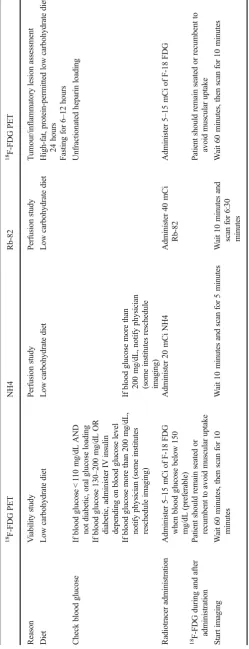
provide insight into myocardial metabolism. Rubidium 82 chloride, 13-N ammonia, and 15O water are useful in the assessment of myocardial perfusion [5,6]. While PET has limited spatial resolution and anatomical localization, this can be overcome by adding CT (PET/CT), which also helps in attenuation correction—however, it adds to the radiation dose. Cardiac PET protocols and patient preparation are sum-marized in Table2[5–8].
Cardiac PET and MRI
MRI and FDG-PET can provide complementary information on the evaluation of several cardiovascular disorders. The ex-quisite morphological information and tissue characterization capabilities of MRI can be complementary to the metabolic information of FDG-PET. The information obtained from PET and MRI acquisitions can be interpreted separately or can be fused using dedicated software algorithms. A signifi-cant challenge in fusion is achieving both spatial and temporal alignment, particularly in pulmonary and cardiac systems [9]. Cardiac motion can be improved by ECG-gating of both PET and MRI, while respiratory motion requires navigator gating for MRI or list mode for PET. Another challenge is the differ-ence in acquisition times, which is 5–6 minutes for PET and just a few seconds for each MRI sequence. Studies have shown that fusion of PET and MRI can show misalignment as much as 2.0 ± 1.6 mm [10].
Hybrid PET/MRI
significant time savings as compared with performing two separate examinations, improves throughput, and also reduces patient discomfort [9].
However, several challenges also exist in achieving wide-spread acceptance of this novel technology. This includes hard-ware issues such as photomultiplier tubes in the PET not work-ing with strong magnetic fields, PET detectors producwork-ing mag-netic field heterogeneities, and MRI surface coils causing un-wanted attenuation interfering with gamma rays. Another im-portant challenge is to develop a robust attenuation correction technique to accurately quantify the standardized uptake values (SUVs), since MRI signal does not contain information about tissue attenuation but reflects the distribution of protons. MR attenuation correction can be performed based on segmenta-tion or atlas-based techniques. The segmentasegmenta-tion approach is based on co-registration of the MR images to the PET trans-mission images using a surface matching technique, which is then segmented into several different classes. Using either a T1-weighted spoiled gradient echo, or T1-weighted 2-point mDixon, or ultra-short TE sequence, a three-segment model (air, soft tissue, lung) or a four-segment model (air, soft tissue, fat, and lung) can be used [11,12]. In the atlas-based approach, an atlas is generated using a surplus of prior scans with corres-ponding known attenuation corrections. Each individual scan can be co-registered to the atlas by comparing each voxel to its nearest neighbours in the atlas. Then, the attenuation coeffi-cients can be interpolated from the generated atlas [12].
Applications of cardiac PET and MRI
In the following sections we will discuss the utility of PET and MRI in specific clinical cardiovascular scenarios. In each sce-nario, we will begin by discussing the current role of MRI and PET individually in the evaluation of these diseases and fol-low it up with the complementary information provided by both, as well as integrated PET/MRI technology (Table3).
Cardiac masses
Cardiac masses may be non-neoplastic (thrombus, hematoma, lipomatous hypertrophy, pericardial cyst), benign neoplastic ( m y x o m a , f i b r o e l a s t o m a , l i p o m a , h e m a n g i o m a , paraganglioma, rhabdomyoma, fibroma), or malignant neo-plastic (metastasis, sarcoma, lymphoma, leukemia, mesothe-lioma). The most common cardiac mass is thrombus. Secondary tumours are significantly more common than pri-mary tumours [13]. Myxoma is the most common primary tumour in the heart [14]. The role of imaging is (1) to charac-terize the mass, since it determines treatment strategy; (2) determine local staging, including extent of the tumour and involvement of adjacent structures, which is essential for sur-gical mapping; (3) to determine if chemo- or radiotherapy is required along with surgery; and (4) to assess response to therapy [15].
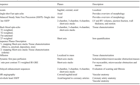
Table 1 MRI sequences and their utility
Sequence Planes Description
Scouts Sagittal, coronal, axial Localizer
Single-shot Fast spin echo Axial Provides overview of morphology Balanced Steady State Free Precession (SSFP)- Single shot Axial Provides overview of morphology Cine SSFP 2-chamber, 3-chamber, 4-chamber,
short-axis stacks
LV and RV volumes, ejection fraction, wall thickness, and motion
Black blood spin-echo - T1-weighted, - T2-weighted, - Fat saturated
2-chamber, 3-chamber, 4-chamber, short-axis stacks
Tissue characterization
T2*-weighted
Sequence Planes Description
T1- mapping Short-axis stacks Tissue characterization (fibros is, amyloid, deposition, iron)
T2- mapping Short-axis stacks Tissue characterizaton (edema)
Short axis Iron quantification
Diffusion Localized to mass Tissue characterization
Dynamic first pass perfusion Short-axis stacks Ischemia/infarct/microvascular obstruction/masses 2 min post contrast T1-weighted IR GRE Short-axis stacks For myocarditis, microvascular obstruction and
masses Delayed enhancement sequences 2-chamber, 3-chamber, 4-chamber,
short-axis stacks
Evaluation of scarring and fibrosis
MR angiography Coronal/sagittal/axial Vascular anatomy 3d-whole heart SSFP Axial/targeted to coronary arteries Coronary artery anatomy
To characterize cardiac masses, MRI is performed using multiple tissue weightings (T1-w, T2-w, fat saturated se-quences), diffusion, and various stages of contrast enhance-ment (early, dynamic perfusion, delayed enhanceenhance-ment). MRI features that suggest a benign or malignant lesion in-clude margin, size, location, calcification, and pericardial effusion. Based on these factors, MRI has shown to be ac-curate in the prediction of lesion type (area under curve for two observers, 0.88 and 0.92, with p values < 0.0003 for agreement between the observers) [15]. Using long inver-sion times (>500 milliseconds), a thrombus can be distin-guished from a neoplasm, since only a thrombus stays dark at this sequence. In addition, MRI also provides informa-tion on the extent of the mass, including involvement of adjacent structures. It also provides functional information,
such as ventricular and valvular function, particularly if there is a valvular extension.
FDG-PET is also valuable in the evaluation of cardiac neo-plasm. For evaluating cardiac mass, normal myocardial up-take is suppressed by fasting for at least 6 hours. Since malig-nant cells accumulate more glucose than normal cells do as a result of predominant glycolytic catabolism, significant up-take of F-18 FDG is indicative of malignant tissue. PET-CT has been shown to have 100 % sensitivity and 86 % specificity in differentiating benign from malignant cardiac tumours at SUV cut-off of 3.5 and 94 % sensitivity with 100 % specificity with SUV cut-off of 4.5. Benign tumours show only slight FDG uptake (2.8 + 0.9 vs 9.5 + 4.0 in malignant lesions). Extra-cardiac tumour manifestations may also be elucidated by whole-body PET/CT [16]. However, the CT component of
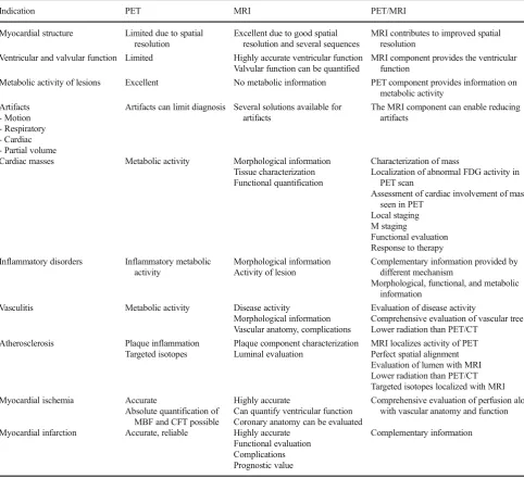
Table 3 Advantages of PET, MRI, and PET/MRI in different cardiac applications
Indication PET MRI PET/MRI
Myocardial structure Limited due to spatial resolution
Excellent due to good spatial resolution and several sequences
MRI contributes to improved spatial resolution
Ventricular and valvular function Limited Highly accurate ventricular function Valvular function can be quantified
MRI component provides the ventricular function
Metabolic activity of lesions Excellent No metabolic information PET component provides information on metabolic activity
Artifacts - Motion - Respiratory - Cardiac - Partial volume
Artifacts can limit diagnosis Several solutions available for artifacts
The MRI component can enable reducing artifacts
Cardiac masses Metabolic activity Morphological information Tissue characterization Functional quantification
Characterization of mass
Localization of abnormal FDG activity in PET scan
Assessment of cardiac involvement of mass seen in PET
Local staging M staging
Functional evaluation Response to therapy Inflammatory disorders Inflammatory metabolic
activity
Morphological information Activity of lesion
Complementary information provided by different mechanism
Morphological, functional, and metabolic information
Vasculitis Metabolic activity Disease activity
Morphological information Vascular anatomy, complications
Evaluation of disease activity
Comprehensive evaluation of vascular tree Lower radiation than PET/CT
Atherosclerosis Plaque inflammation Targeted isotopes
Plaque component characterization Luminal evaluation
MRI localizes activity of PET Perfect spatial alignment Evaluation of lumen with MRI Lower radiation than PET/CT Targeted isotopes localized with MRI Myocardial ischemia Accurate
Absolute quantification of MBF and CFT possible
Highly accurate
Can quantify ventricular function Coronary anatomy can be evaluated
Comprehensive evaluation of perfusion along with vascular anatomy and function
Myocardial infarction Accurate, reliable Highly accurate Functional evaluation Complications Prognostic value
PET is often inadequate to provide morphological informa-tion, which is essential for pre-surgical evaluation.
Fig. 1 Characterization of mass.a20-year-old female with irregular
heart rate noted on physical exam, which eventually led to a cardiac MRI. Short-axis T2-weighted image shows intermediate to low signal 9 × 4 cm mass originating from the inferolateral basal to mid left ventricle (arrow).bThere was no immediate contrast enhancement (not shown here), but there is intense delayed contrast enhancement in short-axis inversion recovery sequence (arrow).cFour-chamber FDG-PET scan shows no abnormal uptake in the mass, indicating a benign mass. The imaging findings were thought to be suggestive of fibroma. Based upon these findings, and the patient’s lack of symptoms, a choice was made to not intervene, and instead to obtain clinical follow-up as well as serial imaging. The patient has done well over six years, and the mass has shown no interval change in size
Fig. 2 Characterization of mass.aAxial FDG-PET/CT scan in a patient
with known thyroid cancer showed intensely hypermetabolic lesion (arrow) in the heart, which could not be localized clearly. b Four-chamber delayed enhancement cardiac MRI shows the mass to be entirely located within the left ventricular cavity. The mass has two distinct components, an enhancing component (red arrow) and a non-enhancing component (yellow arrow), which correspond to metastatic lesion and superimposed and bland thrombus, respectively. The patient was placed on anticoagulants in addition to chemotherapy
Fig. 3 Characterization of mass.a Contrast-enhanced 3d SSFP MR
There are several scenarios in which the combination of MRI and PET provides additive information. This includes (1) the characterization of mass; (2) localization of abnormal FDG activity seen in PET scan; (3) assessment of cardiac involvement of mass seen in PET scan; (4) local staging, mor-phological information, and aggressiveness; (5) M staging; (6) functional evaluation; and (7) response to therapy, distinguishing scar tissue from recurrence.
CharacterizationAlthough MRI and FDG-PET by
them-selves can characterize many tumours, the combination of both improves the diagnostic confidence in distinguishing malignant from benign lesions. If there is no FDG activity in a lesion, a malignancy can be excluded in most cases—an important piece of information that helps in management. False-positive FDG-uptake can be seen in inadequate patient preparation, inflam-matory conditions (e.g. sarcoidosis), infection, abscess, surgical changes, radiation changes, and brown fat. Rarely, uptake can be seen in myxoma, which is also a false-positive finding [17]
(Figs.1,2,3and4). False-negative findings are seen in small lesions and carcinoid. In addition, specific isotopes such as 18 F-FDOPA, 18 F-FDA (fluorodopamine), 11C-hyroxyephedrine (11C-HED), and 68 Ga labelled may be use-ful in the evaluation of paragangliomas.
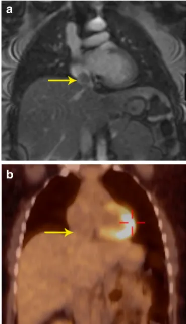
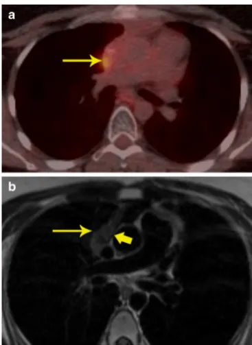
Localization of abnormal activity seen in PET scanPET
scans performed for evaluation of tumours or other purposes occasionally show abnormal focus of hypermetabolism in the chest, either in or adjacent to the heart. It is often not possible to accurately localize this lesion using PET or the correspond-ing low-dose CT scan. In such situations, MRI can provide additional information in localizing and characterizing these masses [3,15] (Figs.5and6).
Assessment of cardiac involvement of mass seen in PET scanFurther to the above utility, occasionally there are masses identified in PET imaging, which are in or adjacent to the heart (Fig.7). It is important to identify the extent of involvement of the heart, particularly the involvement of cardiac chambers, myo-cardium, or pericardium. Correlative MRI images will allow for greater precision in cardiac mass anatomic localization [3].
Fig. 4 Characterization of mass.a Coronal first-pass perfusion MR
image in a patient who presented with chest pain and IVC mass on echocardiography shows a mass that demonstrates contrast enhancement similar to the liver parenchyma. The mass also showed similar signal to liver in all other MRI sequences (not shown here).b
Coronal FDG-PET/CT image shows no uptake in the lesion, indicating it is a benign process. This was proven to be a rare case of aberrant liver, which extended into the IVC
Fig. 5 Localization of abnormal activity.aAxial FDG-PET/CT image
Evaluation of aggressiveness of lesionAs discussed above, morphological information on the extent of tumour and in-volvement of adjacent structures is essential for surgical plan-ning (Fig.8). Neither PET nor the low-dose CT of PET/CT routinely provide this information and MRI is well suited for this purpose [3]. However, FDG-PET provides information on distal spread throughout the body [16].
M staging Additional information obtained from PET can
help in identifying non-cardiac primary or secondary neoplas-tic processes (Fig.9). For example, paraganglioma is a tumour that is most often benign, but can be malignant in 10 % of cases. There are no specific imaging, or histological features to distinguish a benign or malignant paraganglioma. The only way to establish malignancy is to evaluate the presence of distal spread. Using FDG-PET, this can be determined and this helps in deciding the nature of the neoplasm (Fig.10). Probst et al. reported the beneficial use of PET/MR in a patient with squamous cell carcinoma of lung with metastasis to the cardiovascular system [18]. Recently, whole-body MR
Fig. 6 Localization of abnormal activity.aFDG-PET/CT image in a
patient with breast cancer shows intense uptake in the anterior mediastinum abutting the heart. The exact location and extent of this lesion is not evident.bAxial delayed enhancement image shows that there is a heterogeneously enhancing mass in the right ventricle (arrow) that is extending to the right ventricular apex and also invading the right ventricular free wall
Fig. 7 Evaluation of cardiac infiltration.aFDG PET/CT in a patient with
lymphoma shows a focus of hypermetabolism adjacent to the SVC. The exact extent of this is not evident in the PET or the concomitant low-dose CT scan.bMRI was performed for evaluating cardiac extension. T1-weighted axial image shows that the mass (thin arrow) has a clear fat plane (thick arrow) with the SVC and cardiac structures, indicating that it is not infiltrating the cardiac structures
Fig. 8 Local extension.aAxial FDG-PET/CT image in a patient with
diffusion-weighted imaging has been used in oncology. This technique has a high contrast between tumour and background tissues and is used for detection of tumour (primary, recur-rence, secondaries), tumour grading, and therapy monitoring, particularly for bone metastases [19].
Functional informationThe MRI component also provides
valuable functional information such as the involvement of crucial structures e.g., valve and papillary muscles. In
addition, accurate quantification of ventricular and valvular function can be made.
Response to therapyTumour response to therapy can also be
evaluated using the metabolic activity of lesions with PET. PET and MRI are also useful in distinguishing scar tissue from recurrence [20].
A recent study showed that while FDG-PET and MRI sep-arately had a sensitivity of 100 % and specificity of 92 % in determining cardiac malignancy, PET/MR achieved sensitiv-ity and specificsensitiv-ity of 100 % (Fig.10) [21]. In summary, PET/ MR provides valuable information on local and distant staging
Fig. 9 Staging.aShort-axis delayed enhancement MRI in a 72-year-old
patient shows a mass in the ventricular septum (arrow) that has peripheral enhancement and central non-enhancement.bFDG-PET/CT image shows intense uptake in the mass in the ventricular septum, indicating that this is malignant.cCoronal FDG PET/CT image in the same patient shows the septal lesion (thin arrow). In addition, there was also a hypermetabolic right apical lung mass (thick arrow), consistent with lung cancer. The septal lesion is a metastasis. There were also metastatic lesions in the spine
Fig. 10 Characterization and staging.a2-chamber STIR image obtained
and also evaluates treatment response. With a hybrid PET/MR technique, this information can be obtained in a single study.
Inflammatory disorders
The combination of PET and MRI is valuable in several in-flammatory disorders, such as myocarditis, sarcoidosis, peri-carditis, and valvar infections.
Myocarditis
Myocarditis is characterized by an inflammatory reaction of the myocardium. The aetiology of myocarditis can be endog-enous (toxins, autoimmune processes, etc.) or exogendog-enous (bacteria, viruses, fungal, or parasites) [22]. MRI and PET provide complementary information for the assessment of myocardial inflammatory processes, including myocarditis [23]. MRI sequences used for myocarditis include the follow-ing: (1) T2-weighted (T2-W) images, which show myocardial oedema, an important hallmark of reversible inflammatory injury. Typically, myocardial oedema is seen locally, in a non-vascular distribution in subepicardial or mid-myocardial location. Occasionally, the oedema is diffuse and can only be detected by quantification of the T2 value, particularly when it is two standard deviations above that of skeletal muscle [24]; (2) early gadolinium enhancement, i.e. T1-weighted (T1-W) fast-spin echo images taken 1–2 mins after the injection of contrast agent, which shows enhancement in acute myocardi-tis due to hyperemia and capillary leak; and (3) delayed en-hanced images taken 10–15 minutes after the injection of con-trast agent, which shows enhancement due to fibrosis and necrosis [24–26], thus representing irreversible myocardial injury. The overall sensitivity, specific, accuracy, positive pre-dictive value, and negative prepre-dictive value of MRI for myo-carditis is 59 %, 86 %, 68 %, 89 %, and 53 %, respectively (24). On18F-FDG PET scans, myocarditis presents as diffuse increased metabolic activity with areas of heterogeneity. Increased metabolic activity in myocarditis is secondary to microvascular and myocyte damage as well as changes in fatty acid metabolism [23]. Increased metabolic activity in myocar-dium is, however, nonspecific and can also be seen in conges-tive heart failure, right ventricular strain, and hypertrophy due to pulmonary hypertension [26].
The abnormalities in PET and MRI are based on differ-ent mechanisms, and hence they may provide complemen-tary information in myocarditis. Other than functional in-formation on the myocardial activity from MRI, PET can demonstrate activity of the inflammatory process in the myocardium (Fig.11). MRI also provides functional infor-mation of the ventricles and valves. Since myocarditis usu-ally affects younger patients in comparison to neoplastic cardiac masses, an argument could be made for the use of
PET/MR as compared to PET/CT due to lower radiation exposure. PET and MRI can also be used for assessment of the response to medical treatment. However, other than short case reports, there is still lack of published reports on use of PET/MR in identification and follow-up of pa-tients with myocarditis [27].
Sarcoidosis
Sarcoidosis is a systemic granulomatous disease of unknown aetiology, likely due to an alteration in the immune response after exposure to an environmental, occupational, or infectious agent in genetically susceptible individuals [28]. Pathologic hallmark of sarcoidosis is non-caseating granuloma [29]. On MRI, acute phase of sarcoidosis manifests with myocardial thickening and oedema. Patchy myocardial oedema is seen due to active inflammation and is more commonly seen in the subepicardial region and less commonly in the mid-myocardial region. The oedema corresponds to areas of active inflammation, may present with ventricular arrhythmia or conduction disturbance, and usually responds to steroid ther-apy [30]. In the chronic phase, there may be wall thinning. Delayed enhancement is seen in up to 50 % of patients, in a mid-myocardial or subepicardial distribution due to replace-ment fibrosis [31]. Sarcoidosis often involves the septum (par-ticularly basal portion) and left ventricular wall with rare in-volvement of the right ventricle and papillary muscles [32]. Wall motion abnormalities are also seen.
Fig. 11 Myocarditis.aShort-axis delayed enhancement MRI in a patient
18
F-FDG PET is also useful in the identification of cardiac sarcoidosis, with a sensitivity and specificity of 89 % and 78 %, respectively [29]. Recently, published data support the growing role of18F-FDG PET-CT in the diagnosis, risk strat-ification, and assessment of response to treatment of patients with cardiac sarcoidosis [4].
With PET/MR, the MR component evaluates for active inflammation, wall motion abnormalities, and fibrosis. FDG uptake indicates inflammation (Fig.12). Since the disease detection is based on different mechanisms, the information obtained may be complementary, resulting in possible improved sensitivity in disease detection. PET/MRI also has the advantage of providing valuable information on the exact anatomical extension of sarcoidosis in the cardiovascular sys-tem, functional assessment of the myocardium as well as
assessment of the disease activity and response to treatment (Fig.12). This can help to improve the selection of available treatment options including medical management and cardiac resynchronization therapy [33].
PET/MR is also useful in other inflammatory disorders, such as Erdheim Chester disease (Fig.13), which is a rare disorder characterized by abnormal proliferation of non-Langerhans his-tiocytes, resulting in fibrosis that affects multiple organs.
Pericarditis
Inflammation of the pericardium can have diverse aetiologies including idiopathic, autoimmune diseases, infections, post myocardial infarction, uremia, and radiation. Pericarditis can be of acute, chronic inflammatory, or chronic fibrosing types. MRI has become an important modality in the evaluation of pericardial disease. In acute pericarditis, there is pericardial thickening, pericardial effusion (Fig.14a), and pericardial in-flammation, which is manifested as delayed enhancement. In the chronic phase, pericardial thickening is present, but with lower amount of effusion and inflammation than acute type. In chronic fibrosing type, there is pericardial thickening with/ without calcification and features of pericardial constriction may be seen. MRI is valuable in the evaluation of pericardial constriction, since it shows features of ventricular interdepen-dence such as exaggerated septal flattening in real-time imag-ing (Movie1), diastolic septal bounce, and abrupt cessation of diastolic filling. MRI is increasingly being used in the evalu-ation of pericardial inflammevalu-ation, particularly in the context of transient pericardial constriction, which may be seen in the acute or subacute phase of pericarditis due to impaired cardial distensibility. Although the standard treatment of peri-cardial constriction is aggressive periperi-cardial stripping, in tran-sient pericardial constriction, anti-inflammatory therapy (NSAIDs, colchicine, steroids) may be beneficial [34]. The presence of inflammation on MRI, even in the absence of clinical and serologic evidence of inflammation warrants con-tinued therapy [34].
Inflammatory processes involving the pericardium demon-strate a mild-to-moderate FDG uptake (Fig.14b), but occasion-ally no FDG uptake can be seen. In contrast, markedly in-creased FDG uptake is seen in proliferative neoplastic disor-ders that often corresponds with a localized mass [32]. A pre-liminary study showed that the FDG uptake is strong in tuber-culous pericarditis compared to mild to moderate uptake in idiopathic pericarditis. In addition, hypermetabolic lymph nodes were also seen in tuberculous pericarditis [35]. Other causes of pericarditis can be distinguished based on history and FDG uptake patterns. Following surgery, there is diffuse and mild uptake and following radiation, there is diffuse uptake corresponding to the radiation port. Chemotherapy pericarditis is homogeneous and diffuse. Chronic pericarditis shows thick-ening and diffuse mild uptake with small or no effusion [35].
Fig. 12 Sarcoidosis.a32-year-old male admitted after a syncopal
Although most cases of pericarditis do not require PET/MR, unique information provided by PET can be superimposed to MRI or CT and thus help to improve identification of inflam-matory processes or masses in uncertain cases, to exclude other infectious foci, and aid in evaluation of response to therapy.
Cardiac valve abscess
Valve abscess is considered a complication of infective endo-carditis (IE), either in a native or prosthetic valve, and requires urgent surgery with debridement of all infected and necrotic tissues. Echocardiography identifies only 40 % of surgically proven myocardial abscesses [35]. On MRI, morphological and functional evaluation of the valve can be performed along with quantification of valvular lesions such as regurgitation or stenosis. Inflammation or abscess is seen as soft tissue sur-rounding the valve. Inflammatory tissue has high signal on T2-weighted sequences (Fig.15a) and abscesses show fluid signal with peripheral enhancement.
PET-CT has been shown to have higher sensitivity than echocardiography in the detection of valve abscess, showing intense uptake in the affected valves (Fig.15b) [7]. Hybrid PET/MR imaging can aid in the characterization and loca-lization of the high uptake zone on PET due to the excellent spatial resolution capabilities of MRI. The tissue
characterization of MRI can also help to improve assessment of post-surgical cases in which high uptake can be expected due to post-surgical changes or residual/recurrent abscess. Information obtained from hybrid PET/MR can also achieve better differentiation of valve abscess from infective valve vegetations. Although there are some case series on the diag-nostic value of PET-CT [36], there is no published report on the additional value of PET/MR in identification and manage-ment of cardiac valve abscesses [36]. MR or PET/MRI in patients with prosthetic valves can be challenging due to sus-ceptibility artefacts. Sequences with lower sussus-ceptibility arte-facts such as a conventional gradient-echo than a steady-state free precession (SSFP) should be used.
Vascular disease
Vasculitis
Vasculitis is inflammation of the vascular wall and can be classified based on the size of the affected vessels, location of vessels, and underlying cause. Aetiologies for vasculitis include autoimmune diseases, infection, and post chemother-apy [37]. Signs and symptoms are primarily dependent on the affected vessels and severity of disease. Although definitive
Fig. 13 Inflammatory disease.a46-year-old male with history notable
for hypertension, bilateral orbital xanthogranulomas, and chronic low-grade fevers. As part of work-up, the patient had an aortic MRI that demonstrated diffuse confluent soft tissue within the mediastinum (arrow).b There were additional soft tissue changes as well as stranding surrounding both kidneys (arrows).cFDG PET/CT showed only minimal FDG avidity in the paraaortic soft tissue.dFDG-PET/CT
diagnosis is established using biopsy, inflammatory markers and imaging are also helpful in diagnosis.
On MRI, active large vessel vasculitis presents with wall thickening, which may show oedema on STIR and enhance-ment with contrast. The oedema seen on STIR images, how-ever does not show a consistent relationship with disease ac-tivity [38]. MR angiography provides comprehensive evalua-tion of the vasculature, able to identify abnormalities such as aneurysms and stenosis, and can be used to screen high-risk patients without any radiation exposure. MRI has been shown to have a sensitivity of 80 % and specificity of 90 % in the diagnosis of giant cell arteritis [39]. Since18F-FDG accumu-lates in the macrophage-rich areas, vasculitis is seen in PET with high uptake in the vascular wall in a linear pattern. However, the tracer uptake for inflammatory disease is lower than for oncologic disease [40]. Also, PET/CT lacks the capa-bility to show the vessel wall in the low-dose non-contrast CT.
FDG-PET.CT has been shown to have an overall sensitivity and specificity of 80 % and 89 % for the diagnosis of giant cell arteritis when compared to reference clinical criteria [39].
PET/MR combines the anatomic information provided with MRI with the metabolic activity of PET, providing a reliable diagnosis of vasculitis with lower radiation dose than PET/CT, also at the same time enabling assessment of aneu-rysms and stenosis in the vasculature. In a pilot study on 14 consecutive examinations, PET/MR was found to be efficient in the evaluation of large vessel vasculitis and that only a combination of PET and MRI revealed the whole disease ex-tent (Fig.16). In MRI, abnormal segments were seen in 32/ 149, and in FDG 78/149, for an overall number of 83/149. Also, there was a strong and significant correlation between CRP and the number of vessel segments affected by inflam-mation in PET/MR (r = 0.86, p = 0.01) in contrast to PET or MRI only. Thus, PET and MRI are complementary for di-agnosis and likely to be more accurate to assess the true extent of disease than either modality alone [41]. While
Fig. 14 Pericarditis.aCoronal FDG PET/CT in a patient showed intense
uptake in the pericardium.bShort-axis delayed enhancement MRI shows diffuse circumferential delayed pericardial enhancement (arrows), which is consistent with pericardial inflammation. There was also pericardial thickening in black blood images (not shown here)
Fig. 15 Perivalvular abscess.aThree-chamber Cine SSFP MRI image in
the disease activity can be evaluated more accurately with PET, MRA provides a comprehensive evaluation of the vascular tree without the need for ionizing radiation [42].
Atherosclerotic disease
Atherosclerosis is a diffuse, chronic inflammatory disorder characterized by deposition of lipid and fibrous products in the vessel wall, following complex biological processes. Current imaging techniques focuses on the detection of steno-sis, grading of stenosis and evaluation of perfusion abnormal-ities distal to the stenosis. However, major events such as myocardial infarction (MI) and stroke are not produced by plaques with associated stenosis, but by non-stenotic plaques that get disrupted, the so-called vulnerable plaque. Hence, it is vital to be able to detect, quantify, and characterize the biolog-ical activity and stability of plaque at a subclinbiolog-ical stage. Identification of these high-risk features along with plaque burden will significantly help in the diagnosis and manage-ment of these patients [43].
High-resolution multi-contrast imaging of the vessel wall (T1-w, T2-w, PD, TOF) and dynamic contrast-enhanced angi-ography can characterize the specific tissue components of complex plaques, which helps to delineate biologic activity (Table4) [43]. High-risk features of an atherosclerotic plaque include a large lipid-rich necrotic core, thin fibrous cap, endo-thelial denudation with superficial platelet aggregation, fissured/ injured plaque and active inflammation. Stenosis > 90 %, super-ficial calcified nodule, intraplaque haemorrhage, outward posi-tive remodelling and adventitial inflammation/neovascularity are the other features of vulnerability. Flow can be evaluated using time-of-flight sequence. Due to the smaller size, evalua-tion of coronary arteries is challenging, but this technique has been well established in the evaluation of carotid plaque [43].
FDG-PET has also been shown to be useful for assessment of inflammatory activity within plaque since macrophages in an inflamed plaque show avid FDG uptake and accumulation. High uptake has been shown both in symptomatic and asymp-tomatic plaques [44]. It has also been shown that there is a correlation between FDG uptake and extent of macrophage infiltration in a vulnerable carotid plaque [45]. However, low spatial resolution of clinical PET machines limits identifica-tion of small plaques and localizaidentifica-tion of high FDG uptake zones.
With PET/MR, the high spatial resolution of MRI helps in localizing the metabolic activity obtained from PET (Fig.17)
Fig. 16 Vasculitis.aAxial STIR MRI image performed in a patient with
suspected vasculitis following lung cancer chemotherapy shows an intense high signal in the wall of the ascending and descending aorta (arrows), which is indicative of active vasculitis.bSagittal FDG PET image in the same patient shows the intense uptake along the wall of the thoracic aorta (arrow).cThis high uptake is localized better to the aortic wall (arrow) using the fused PET/MR image (bottom)
Table 4 Characteristics of plaque
components in high-resolution multi-contrast MRI
Component T1-w T2-w PD Contrast enhancement
Lipid High Intermediate Intermediate No
Fibrocellular High High High No
Calcium Low Low Low No
Necrotic core Low High High No
Fibrotic cap Low Low Low Yes
[46]. Simultaneous acquisition also results in perfect spatial alignment. In addition, MRI also provides additional informa-tion on plaque components as described above [43]. Thus, the biological features of plaque obtained by PET provides addi-tional information with multi-parametric plaque morphology assessment with MRI. The lumen can also be assessed at the same time for stenosis/occlusion. There is also potential for using targeted PET isotopes, which hone in on specific mo-lecular targets of atherosclerosis, such as macrophage, or re-ceptors of angiogenesis, vascular adhesion, and apoptosis. Some potential agents include 68 Ga-DOTATATE and 68 Ga- NOTA-RGD [46]. A recent study in HIV patients found congruence between PET/MR and PET/CT SUV values and improved delineation of outer and inner wall of carotid artery from MRI [47,48]. MRI can be used for mor-phological follow-up and the combination can be used in drug trials.
Coronary artery disease (CAD)
Myocardial ischemia
Myocardial ischemia occurs when the oxygen requirement of part of myocardium exceeds the oxygen supply derived by coronary arteries, typically caused by coronary atherosclerotic disease. Identification of myocardial ischemia is essential in patients with coronary artery disease for management and for prognosis.
MRI is very useful in the evaluation of myocardial ische-mia. Dynamic first-pass perfusion MRI performed at rest and stress has been shown to have high accuracy in the evaluation of myocardial ischemia. The CE-MARC trial showed sensi-tivity (86.5 %) and negative predictive value (90.5 %) of MRI to be statistically superior to that of SPECT (66.5 % and 79.1 %, respectively), while the specificity (83.4 % vs 82.6 %) and PPV (77.2 % vs 71.4 %) were non-statistically significantly superior [49]. Myocardial ischemia is seen as a hypointense perfusion defect at first pass perfusion imaging only at stress, with normal perfusion at rest.
Rubidium-82, Nitrogen-13 ammonia and 15 O labelled water are the PET isotopes used for perfusion imaging. PET has high accuracy, with sensitivity and specificity of 90 % in detection of obstructive coronary disease [50]. In addition, PET imaging is also valuable in quantification of myocardial blood flow (MBF) and coronary flow reserve (CFR), which is useful in multi-vessel disease, early changes of vasoreactivity and follow-up [9]. Also, PET findings have prognostic value [51].
With an integrated PET/MR system, simultaneous 13 N ammonia PET-MR myocardial perfusion has the advantage of increasing the accuracy of MR perfusion by the PET com-ponent, since the N-13 ammonia perfusion imaging has more coverage than stress perfusion MR. 13 N-ammonia PET has been shown to be an important cost-effective strategy in clin-ical decision making [52]. Utilizing MR attenuation correction data eliminates the radiation associated with CT attenuation data. Also, the examination time is shortened, since rest-phase 13 N-ammonia images are not required and are replaced by delayed enhancement images. In addition, MRI can also help address artefacts caused by motion or partial volume averag-ing. Ventricular function, global and regional, can be evaluat-ed in the same study. A study which comparevaluat-ed MRI with PET showed high accuracy of MRI in evaluating pathology with sensitivity of 91 % and specificity of 94 % compared to PET scan, but with significant underestimation of CFR [53]. Challenges of perfusion PET include lack of a good flow tracer for PET MPI, and short half-life of the isotopes, which necessitates having a cyclotron on site.
Myocardial infarction
Myocardial infarction results from death of myocardial tissue due to blockage of coronary arteries. Imaging is useful in the diagnosis, prognosis, and evaluation of complications. An im-portant contribution of imaging is detection of viable myocar-dium, which is dysfunctional but not dead, and hence can recover its function following revascularization.
Delayed enhancement imaging is highly sensitive and spe-cific in the evaluation of myocardial infarct, both in acute (sensitivity of 99 %) and chronic phase (sensitivity 94 %) [53]. MI is manifest as a subendocardial or transmural pattern
Fig. 17 Carotid atherosclerosis.aAxial proton density-weighted MRI
of enhancement confined to a vascular territorial distribution [54]. MRI is accurate in the quantification of myocardial func-tion and scar, which has prognostic value. Dysfuncfunc-tional seg-ments with scarring > 50 % of myocardial thickness have been shown to have lower probability of functional recovery after revascularization [55]. MRI can also evaluate several compli-cations of MI including thrombus, rupture, ventricular septal defect, pericarditis, etc. MRI has prognostic value and can also provide functional information [56]. The difference between oedema seen in T2-weighted images and the scar seen in de-layed enhancement is considered the salvageable area by myocardial reperfusion [57].
PET is also very reliable in the evaluation of myocardial viability, quantifying the degree of viability [58]. With PET, myocardial viability is detected by using a combination of
perfusion imaging and FDG study [58]. Infarcted scarred tis-sue does not have18F-FDG uptake (Fig.18), while a viable hibernating myocardium has 18F-FDG uptake (Fig. 19). Hence, a matched defect, i.e. a defect in perfusion imaging
Fig. 18 Myocardial infarction.aShort-axis delayed enhancement MRI
in a patient with myocardial infarction shows a large transmural scar of the anterior wall (yellow arrow) and partial thickness scarring of the inferolateral segment (red arrow). Viability of this segment is borderline.bFused PET/MR image shows no uptake in the anterior as well as inferolateral segments, indicating that these are not viable. Thus, the combination of these imaging modalities improves the diagnostic confidence
Fig. 19 Hibernating myocardium.a 57-year-old male with coronary
and FDG scan, is indicative of a scar, while a mismatched defect, i.e. a defect in perfusion imaging, but normal FDG uptake, is indicative of hibernating myocardium [59].
A study using FDG and 13 NH3 PET and MRI showed that MRI can depict areas of non-transmural enhancement in via-ble areas of PET due to higher spatial resolution [60]. There was good correlation between the modalities for location and extent of infarct. Both modalities have comparable PPV for functional recovery after revascularization using 50 % thick-ness enhancement and 50 % FDG activity as cut off [58]. Both these techniques have high sensitivity but low specificity (63 %) in predicting functional recovery after revasculariza-tion, indicating that segments called viable do not always im-prove [61].
PET-MRI can also evaluate and characterize the heteroge-neity of the scar tissue, since this is a marker for inducible monomorphic ventricular tachycardia [53]. The combination of PET SUV maps, metabolic function, delayed enhancement and T1 mapping may be better predictor of arrhythmia sus-ceptibility than MR alone. The combination of PET and MRI can also assess other features such as decreased wall thicken-ing, regional functionional abnormalities, lack of contractile response to low-dose dobutamine, preserved sub-epicardial myocardium, and preserved perfusion [9]. Both modalities provide prognostic information, although FDG-PET depends on mismatch between flow and glucose uptake, and MRI de-pends on infarct size.
Ventricular function
Cardiac MRI is considered the gold standard in evaluation of myocardial function [60]. PET scan has significantly lower spatial and temporal resolution than MRI, limiting its accuracy in evaluation of cardiac function. Left ventricular function is calculated by algorithm-dependent assumptions on endocar-dial and epicarendocar-dial border. Hence, the MRI component of PET/MR provides more accurate quantification of ventricular function.
Coronary angiography
MRI also provides morphological information on the coro-nary arterial anatomy, including luminal stenosis [62]. Although inferior to CT, MRI can also be used for the evaluation of the anatomy of coronary arteries and evaluate coronary artery disease, with reasonably good accuracy. A recent multicenter trial showed sensitivity of 88 %, specificity of 72 %, PPVof 71 %, and NPVof 79 % compared to coronary angiography [62]. MR coronary angiography is performed using a 3d- SSFP sequence with navigator gating, images acquired during end-expiration and in diastole, with myocar-dial suppression by T2 prepared sequence. Thus, MRI pro-vides concomitant anatomic information.
Emerging and future applications
There are several emerging potential areas where hybrid PET/ MRI is valuable in cardiovascular disorders. The most impor-tant application would be to utilize the high spatial resolution of MRI to localize the uptake of novel PET isotopes that are targeted towards specific pathophysiological process. This combines the anatomical detail of MRI with the highly sensi-tive metabolic information of PET. For example, 11C-labelled metahydroxyephedtrine (HED) and β-adrenoreceptor tracers can image defects of sympathetic innervation in cardiac disor-ders such as CAD, heart failure, and arrhythmias, thus helping in diagnosis, prognosis and selecting the appropriate therapy. 19 F- galacto RGE enables imaging ofαvβ3 integrin, which is a marker of angiogenesis and also accumulates in the injured myocardium and thus can monitor effectiveness of therapies that aim to heal myocardium. Stem cell therapy can be directly visualized by reporter gene PET imaging [9].
Conclusion
MRI and PET are clinically established imaging modalities, with complementary strengths which can be invaluable in the evaluation of a range of cardiovascular disorders. A combina-tion of PET and MRI can provide synergistic informacombina-tion in several scenarios for the enhanced characterization of cardio-vascular disorders. With use of hybrid PET/MRI scanners, this information can be obtained in a single scan.
Compliance with ethical standards
Financial support None
Conflict of Interest None
Open AccessThis article is distributed under the terms of the Creative
C o m m o n s A t t r i b u t i o n 4 . 0 I n t e r n a t i o n a l L i c e n s e ( h t t p : / / creativecommons.org/licenses/by/4.0/), which permits unrestricted use, distribution, and reproduction in any medium, provided you give appropriate credit to the original author(s) and the source, provide a link to the Creative Commons license, and indicate if changes were made.
References
1. Nichols M, Townsend N, Scarborough P et al (2015) Cardiovascular disease in Europe 2014: epidemiological update. European Heart Journal 36(40):2673–4
3. Hundley WG, Bluemke DA, Finn JP et al (2010) ACCF/ACR/ AHA/NASCI/SCMR 2010 expert consensus document on cardio-vascular magnetic resonance: a report of the American college of cardiology foundation task force on expert consensus documents. J Am Coll Cardiol 55(23):2614–62
4. Skali H, Schulman AR, Dorbala S (2013) (18)F-FDG PET/CT for the assessment of myocardial sarcoidosis. Curr Cardiol Rep 15(5): 352
5. Delbeke D, Coleman RE, Guiberteau MJ et al (2006) Procedure guideline for tumor imaging with 18F-FDG PET/CT 1.0. J Nucl Med 47(5):885–95
6. Dorbala S, Di Carli MF, Delbeke D et al (2013) SNMMI/ASNC/ SCCT guideline for cardiac SPECT/CT and PET/CT 1.0. J Nucl Med 54(8):1485–507
7. Cardiac F-18 FDG PET. December 10, 2014]; Available from: http://www.asnc.org/media/PDFs/PPPETFDG081511.pdf. 8. Surasi DS, Bhambhvani P, Baldwin JA et al (2014) 18F-FDG PET
and PET/CT patient preparation: a review of the literature. J Nucl Med Technol 42(1):5–13
9. Nekolla SG, Martinez-Moeller A, Saraste S (2009) PET and MRI in cardiac imaging: from validation studies to integrated applications. Eur J Nucl Med Mol Imaging 36(Suppl 1):S121–30
10. Sinha S, Sinha U, Czernin J et al (1995) Noninvasive assessment of myocardial perfusion and metabolism: feasibility of registering gat-ed MR and PET images. AJR Am J Roentgenol 164(2):301–7 11. Partovi S, Kohan A, Rubbert C et al (2014) Clinical oncologic
applications of PET/MRI: a new horizon. Am J Nucl Med Mol Imaging 4(2):202–12
12. Hofmann M, Pichler B, Scholkopf B et al (2009) Towards quanti-tative PET/MRI: a review of MR-based attenuation correction tech-niques. Eur J Nucl Med Mol Imaging 36(Suppl 1):S93–104 13. Yusuf SW, Reardon MJ, Banchs J (2014) Cardiac tumors.
Cardiology 129(3):197–8
14. Anvari MS, Naderan M, Eslami Shahr Babaki A et al (2014) Clinicopathologic review of non-myxoma cardiac tumors: a 10-year single-center experience. Cardiology 129(3):199–202 15. Hoffmann U, Globits S, Schima W et al (2003) Usefulness of
mag-netic resonance imaging of cardiac and paracardiac masses. Am J Cardiol 92(7):890–5
16. Rahbar K, Seifarth H, Schafers M et al (2012) Differentiation of malignant and benign cardiac tumors using 18F-FDG PET/CT. J Nucl Med 53(6):856–63
17. Agostinhi D, Babatasi G, Galateau F et al (1999) Detection of cardiac myxoma by F-18 FDG PET. Clin Nucl Med 24(3):159–60 18. Probst S, Seltzer A, Spieler B et al (2011) The appearance of cardiac metastasis from squamous cell carcinoma of the lung on F-18 FDG PET/CT and post hoc PET/MRI. Clin Nucl Med 36(4):311–2 19. Wilhem T, Stieltjes B, Schlemmer HP (2013) Whole-body- MR
diffusion weighted imaging in oncology. Rofo 185(10):950–8 20. Tokmak H, Demir N, Demirkol MO (2014) Cardiac angiosarcoma:
utility of [(18)F]fluorodeoxyglucose positron emission tomography-computed tomography in evaluation of residue, metas-tases, and treatment response. Vasc Health Risk Manag 10:399–401 21. Nensa F, Tezgah E, Poeppel TD et al (2015) Integrated 18F-FDG PET/MR imaging in the assessment of cardiac masses: a pilot study. J Nucl Med 56(2):255–60
22. Liu PP, Mason JW (2001) Advances in the understanding of myo-carditis. Circulation 104(9):1076–1082
23. Miyagawa M, Yokoyama R, Nishiyama Y et al (2014) Positron emission tomography-computed tomography for imaging of in-flammatory cardiovascular diseases. Circulation Journal 78(6): 1302–1310
24. Friedrich MG, Sechtem U, Schulz-Menger J et al (2009) Cardiovascular magnetic resonance in myocarditis: A JACC White Paper. J Am Coll Cardiol 53(17):1475–87
25. Mayroeni S (2012) Myocarditis in systemic diseases and the role of MRI. Hellenic J Cardiol 53(2):142–7
26. Erba PA, Sollini M, Lazzeri E et al (2013) FDG-PET in cardiac infections. Semin Nucl Med 43(5):377–95
27. Nensa F, Schlosser T (2014) Cardiovascular hybrid imaging using PET/MRI. Rofo 186(12):1094–101
28. Rossman MD, Kreider ME (2007) Lesson learned from ACCESS (a case controlled etiologic study of sarcoidosis). Proc Am Thorac Soc 4(5):453–6
29. Youssef G, Leung E, Mylonas I et al (2012) The use of 18F-FDG PET in the diagnosis of cardiac sarcoidosis: a systematic review and metaanalysis including the Ontario experience. J Nucl Med 53(2): 241–8
30. Amano Y, Tachi M, Tani H, et al. (2012) T2-weighted cardiac magnetic resonance imaging of edema in myocardial diseases. Sci World J. 194069.
31. Tadamura E, Yamamuro M, Kubo S et al (2005) Effectiveness of delayed enhanced MRI for identification of cardiac sarcoidosis: comparison with radionuclide imaging. AJR Am J Roentgenol 185(1):110–5
32. Vignaux O (2005) Cardiac sarcoidosis: spectrum of MRI features. AJR Am J Roentgenol 184(1):249–54
33. Schneider S, Batrice A, Rischpler C et al (2014) Utility of multi-modal cardiac imaging with PET/MRI in cardiac sarcoidosis: im-plications for diagnosis, monitoring and treatment. Eur Heart J 35(5):312
34. Gentry J, Klein AL, Jellis CL (2016) Transient constrictive pericar-ditis: current diagnostic and therapeutic strategies. Current Cardiology Reports 18(5):41
35. Dong A, Dong H, Wang Y et al (2013) 18F-FDG PET/CT in dif-ferentiating acute tuberculous from idiopathic pericarditis: prelimi-nary study. Clin Nucl Med 2013 38(4):e160–5
36. Saby L, Le Dolley Y, Lass O et al (2012) Early diagnosis of abscess in aortic bioprosthetic valve by 18F-fluorodeoxyglucose positron emission tomography-computed tomography. Circulation 126(14): e217–20
37. Belli L, Magistretti G, Puricelli GP et al (1997) Arteritis following intra-arterial chemotherapy for liver tumors. Eur Radiol 7(3):323–6 38. Tso E, Flamm SD, White RD et al (2002) Takayasu arteritis: utility and limitations of magnetic resonance imaging in diagnosis and treatment. Arthritis Rheum 46(6):1634–42
39. Khan A, Dasgupta B (2015) Imaging in giant cell arteritis. Curr Rheumatol Rep 17:52
40. Blockmans D (2003) The use of (18F)fluoro-deoxyglucose positron emission tomography in the assessment of large vessel vasculitis. Clin Exp Rheumatol 21(6 Suppl 32):S15–22
41. Einspieler I, Thurmel K, Eiber M et al (2014) Imaging large vessel vasculitis with fully integrated PET/MR: a pilot study. J Nucl Med 55(Supplement 1):185
42. Balink H, Bennink RJ, van Eck-Smit BL et al (2014) The role of 18F-FDG PET/CT in large-vessel vasculitis: appropriateness of cur-rent classification criteria? Biomed Res Int 2014:687608 43. Chu B, Ferguson MS, Chen H et al (2009) Magnetic resonance
imaging features of the disruption-prone and the disrupted carotid plaque. JACC Cardiovasc Imaging 2(7):883–96
44. Davies JR, Rudd JH, Weissberg P (2004) Molecular and metabolic imaging of atherosclerosis. J Nucl Med 45(11):1898–907 45. Gaebe M, Pederson SF, Borgwardt L et al (2009) Molecular
pathol-ogy in vulnerable carotid plaques: correlation with [18]-fluorodeoxyglucose positron emission tomography (FDG-PET). Eur J Vasc Endovasc Surg 37(6):714–21
46. Lee JS, Paeing JC (2015) Nuclear molecular imaging for vulnerable atherosclerotic plaques. Korean J Radiol 16(5):955–966
18F-FDG PET scanning exploring plaque vulnerability. J Nucl Cardiol 18(6):1066–75
48. Ripa RS, Mnudsen A, Hag AM et al (2013) Feasibility of simulta-neous PET/MR of the carotid artery: first clinical experience and comparison to PET/CT. Am J Nucl Med Mol Imaging 3(4):361–71 49. Greenwood JP, Maredia N, Younger JF et al (2012) Cardiovascular magnetic resonance and single-photon emission computed tomog-raphy for diagnosis of coronary heart disease (CE-MARC): a pro-spective trial. Lancet 379(9814):453–60
50. Klocke FN, Baird MG, Lorell BJ et al (2003) ACC/AHA/ASNC guidelines for the clinical use of cardiac radionuclide imaging– executive summary: a report of the american college of cardiology/American heart association task force on practice guide-lines (ACC/AHA/ASNC committee to revise the 1995 guideguide-lines for the clinical use of cardiac radionuclide imaging). J Am Coll Cardiol 42(7):1318–33
51. Yoshinaga K, Chow BJ, Williams K et al (2006) What is the prog-nostic value of myocardial perfusion imaging using rubidium-82 positron emission tomography? J Am Coll Cardiol 48(5):1029–39 52. Siegrist PT, Husmann L, Knabenhans M et al (2008) (13)N-ammo-nia myocardial perfusion imaging with a PET/CT scanner: impact on clinical decision making and cost-effectiveness. Eur J Nucl Med Mol Imaging 35(5):889–95
53. Schmidt A, Azevedo CF, Cheng A et al (2007) Infarct tissue het-erogeneity by magnetic resonance imaging identifies enhanced car-diac arrhythmia susceptibility in patients with left ventricular dys-function. Circulation 115(15):2006–14
54. Kim RJ, Albert TS, Wible JH et al (2008) Performance of delayed-enhancement magnetic resonance imaging with gadoversetamide contrast for the detection and assessment of myocardial infarction: an international, multicenter, double-blinded, randomized trial. Circulation 117(5):629–37
55. Kim RJ, Wu E, Rafael A et al (2000) The use of contrast-enhanced magnetic resonance imaging to identify reversible myocardial dys-function. N Engl J Med 343(20):1445–53
56. Rajiah P, Desai MY, Kwon D, Flamm SD (2013) MR imaging of myocardial infartion. Radiographics 33(5):1383–412
57. Aletras AH, Tilak GS, Natanzon A et al (2006) Retrospective de-termination of the area at risk for reperfused acute myocardial in-farction with T2-weighted cardiac magnetic resonance imaging: histopathological and displacement encoding with stimulated ech-oes (DENSE) functional validations. Circulation 113(15):1865–70 58. Schinkel AF, Pldermans D, Elhendy A et al (2007) Assessment of myocardial viability in patients with heart failure. J Nucl Med 48(7):1135–46
59. Underwood SR, Bax JJ, vom Dahl J et al (2004) Imaging tech-niques for the assessment of myocardial hibernation. Report of a Study Group of the European Society of Cardiology. Eur Heart J 25(10):815–36
60. Kuhl HP, Lipke CS, Krombach GA et al (2006) Assessment of reversible myocardial dysfunction in chronic ischaemic heart dis-ease: comparison of contrast-enhanced cardiovascular magnetic resonance and a combined positron emission tomography-single photon emission computed tomography imaging protocol. Eur Heart J 27(7):846–53
61. Bellenger NG, Davies LC, Francis JM et al (2000) Reduction in sample size for studies of remodeling in heart failure by the use of cardiovascular magnetic resonance. J Cardiovasc Magn Reson 2(4): 271–8