R E S E A R C H
Open Access
SNHG15
is a bifunctional MYC-regulated
noncoding locus encoding a lncRNA that
promotes cell proliferation, invasion and
drug resistance in colorectal cancer by
interacting with AIF
Morvarid Saeinasab
1,2, Ahmad Reza Bahrami
1,2, Jovanna González
3,4, Francesco P. Marchese
3,4, Dannys Martinez
3,4,
Seyed Javad Mowla
5, Maryam M. Matin
1,2*and Maite Huarte
3,4*Abstract
Background:Thousands of long noncoding RNAs (lncRNAs) are aberrantly expressed in various types of cancers, however our understanding of their role in the disease is still very limited.
Methods:We applied RNAseq analysis from patient-derived data with validation in independent cohort of patients. We followed these studies with gene regulation analysis as well as experimental dissection of the role of the identified lncRNA by multiple in vitro and in vivo methods.
Results:We analyzed RNA-seq data from tumors of 456 CRC patients compared to normal samples, and identified SNHG15as a potentially oncogenic lncRNA that encodes a snoRNA in one of its introns. The processedSNHG15is overexpressed in CRC tumors and its expression is highly correlated with poor survival of patients. Interestingly, SNHG15is more highly expressed in tumors with high levels ofMYCexpression, while MYC protein binds to two E-box motifs onSNHG15sequence, indicating thatSNHG15transcription is directly regulated by the oncogene MYC. The depletion ofSNHG15by siRNA or CRISPR-Cas9 inhibits cell proliferation and invasion, decreases colony formation as well as the tumorigenic capacity of CRC cells, whereas its overexpression leads to opposite effects. Gene expression analysis performed uponSNHG15inhibition showed changes in multiple relevant genes implicated in cancer progression, includingMYC,NRAS,BAG3orERBB3. Several of these genes are functionally related to AIF, a protein that we found to specifically interact withSNHG15, suggesting that theSNHG15acts, at least in part, by regulating the activity of AIF. Interestingly, ROS levels, which are directly regulated by AIF, show a significant reduction in SNHG15-depleted cells. Moreover, knockdown ofSNHG15increases the sensitiveness of the cells to 5-FU, while its overexpression renders them more resistant to the chemotherapeutic drug.
Conclusion:Altogether, these results describe an important role ofSNHG15in promoting colon cancer and mediating drug resistance, suggesting its potential as prognostic marker and target for RNA-based therapies.
Keywords:SNHG15, Colorectal cancer, lncRNA, Survival, Drug resistance, AIF
© The Author(s). 2019Open AccessThis article is distributed under the terms of the Creative Commons Attribution 4.0 International License (http://creativecommons.org/licenses/by/4.0/), which permits unrestricted use, distribution, and reproduction in any medium, provided you give appropriate credit to the original author(s) and the source, provide a link to the Creative Commons license, and indicate if changes were made. The Creative Commons Public Domain Dedication waiver (http://creativecommons.org/publicdomain/zero/1.0/) applies to the data made available in this article, unless otherwise stated.
* Correspondence:[email protected];[email protected]
1
Department of Biology, Faculty of Science, Ferdowsi University of Mashhad, Mashhad, Iran
3Department of Gene Therapy and Regulation of Gene Expression, Center for
Background
In the past years, advances in sequencing revealed that only 1–2% of the human genome encodes for proteins while the majority of it is transcribed into non-coding RNAs (ncRNAs) [1]. Different classes of ncRNAs are expressed in diverse biological processes and cellular pathways, including small ncRNAs (miRNAs, piRNAs and siRNAs) and long ncRNAs, which have at least 200 nucleotides of length and can be spliced [2–5]. LncRNAs can be classified into various categories based on their position relative to protein-coding genes: (1) intronic, when they are located within genes; (2) intergenic, when they are mapped between different genes and (3) anti-sense, when they are overlapping with exons of other transcript on the opposite strand [6]. The functional roles of these molecules remain mostly unclear, but re-cent studies have revealed that they contribute to vari-ous cellular processes such as transcription regulation, nuclear architecture, epigenetic regulation, enhancer as-sociation (in nucleus), maintenance of mRNA stability, sponging microRNA or regulation of protein translation (in cytoplasm) [7]. Recently, by next-generation sequen-cing, thousands of lncRNAs have been found to be aber-rantly expressed in various types of cancers [8]. While some of them may play oncogenic roles promoting pro-liferation, invasion and metastasis, others may have a tumor suppressor function, by modulating growth arrest pathways [9–11].
According to global cancer statistics, colorectal cancer (CRC) is the third most common human malignancy and the fourth leading cause of cancer-associated mor-tality [12, 13]. Therefore, its early diagnosis is an essen-tial requirement. Several studies have reported that some lncRNAs are associated with different stages of CRC [14, 15], indicating that lncRNAs could be a bio-marker or target for diagnostic, prognostic and thera-peutic applications. However, the contribution of long noncoding RNAs to this type of cancer is still poorly studied.
In the present study, we identify and characterize the oncogenic lncRNASNHG15, which is correlated to sur-vival of CRC patients. Our results describe an important effect of SNHG15 in cancerous phenotype of CRC cells and its role in drug sensitivity. Moreover, several genes deregulated after SNHG15 depletion are implicated in cancer initiation, progression and also survival pathways. Altogether, these findings suggest the potential of
SNHG15 as prognostic marker and target for
RNA-based therapies.
Methods TCGA analysis
The RNA-seq data of 456 tumor and 41 normal sam-ples were downloaded from TCGA database (https://
cancergenome.nih.gov/). The expression of lncRNAs was quantified by Cufflinks v.2.2.1 and lncRNA ex-pression levels were compared between normal tissue and tumor tissue samples.
Patients
Fresh CRC specimens and their adjacent normal tissues were obtained from 36 CRC patients who underwent sur-geries between 2014 and 2016 in Imam-Reza Hospital, Mashhad, Iran. None of patients had received preopera-tive treatment including radiotherapy or chemotherapy and all of samples were confirmed as colorectal cancer after histopathological examination. The study protocol was approved by the Ethics Committee of Ferdowsi Uni-versity of Mashhad and all patients were informed with a written for using their tissues. All clinicopathological char-acteristics of patients are presented in Table1.
Cell lines and cell culture
Human colorectal cancer cell lines DLD1, HCT 116, HT-29, LoVo, LS513, SW620 and T84 were cultured in Roswell Park Memorial Institute (RPMI-1640) medium (Gibco) supplemented with 10% fetal bovine serum (FBS, Gibco) and 1% penicillin/streptomycin (Lonza). RKO, SW480 and Caco-2 cells were cultured in Dulbec-co’s Modified Eagle Medium (DMEM, Gibco) supple-mented with 10% FBS and 1% penicillin/streptomycin. Primary dermal fibroblasts (HDFa) were cultured in High glucose Dulbecco’s Modified Eagle Medium (DMEM, Gibco) supplemented with 15% FBS and 1%
Table 1Clinicopathological characteristics of CRC patients
Clinical parameter Number Percentage
Age
≥60 13 36.1
< 60 23 63.9
Gender
Male 17 47.2
Female 19 52.8
Invasion depth
T1 2 5.6
T2 4 11.1
T3 29 80.6
T4 1 2.7
TNM stages
I 3 8.3
II 33 91.7
II & IV 0 0
Lymphatic metastasis
Yes 18 50
penicillin/streptomycin. All cell lines were obtained from American Type Culture Collection (ATCC). Cells were maintained in 5% CO2 humidified-air at 37 °C.
RNA extraction and qRT-PCR
Total RNAs were isolated from patient specimens and cultured cells using TRIzol Reagent (Invitrogen) follow-ing the manufacturer’s instructions. After DNase I (Invi-trogen) treatment, cDNAs were synthesized using High-Capacity cDNA Reverse Transcription Kit (Applied Biosystem) with random hexamer primers. qRT-PCRs were performed using SYBR Green reagent (Applied Biosystem). Expression levels of genes were calculated with the comparative cycle threshold (CT) (2-ΔCT and 2-ΔΔCT) method using glyceraldehyde 3-phosphate dehydrogenase (GAPDH) as an endogenous control. All primers used in the study are listed in Additional file1: Table S1.
Cell transient transfection
All siRNAs for targeting SNHG15 and MYC and nega-tive control siRNA were purchased from Sigma-Aldrich (USA). LoVo and SW620 cells were plated into 6 well plate (150 × 103cells per well) and transfected with siR-NAs at a final concentration of 25 nM for 48 h, using Li-pofectamine 2000 (Invitrogen, USA) according to manufacturer’s protocol. The sequence of siRNAs are as follows: SNHG15#1 (Sense: 5′-CCUUGAGUCUCAUG UUCAA-3′, Anti-sense: 5′- UUGAACAUGAGACU CAAGG-3′), SNHG15#2 (Sense: 5′- GAGCUUACU GUCACAGCAA-3′, Anti-sense: 5′- UUGCUGUGA CAGUAAGCUC-3′), MYC (Sense: 5′- GGUCAGAGU CUGGAUCACC-3′, Anti-sense: 5′- GGUGAUCCA GACUCUGACC-3′), Ctrl (Sense: 5′- CAGUCGCGU UUGCGACUGGC-3′, Anti-sense: 5′- GCCAGUCGC AAACGCGACUG-3′).
For overexpression ofSNHG15, we purchasedSNHG15 cDNA sequence (837 bp) was cloned in pDNR-LIB (BC092459; Source Bioscience-UK) and subcloned it into pcDNA3.1 plasmid. Then pcDNA3.1 vectors (empty vec-tor and SNHG15) were transfected into HCT 116 and SW480 cells (300 × 103cells per well) at final concentra-tion 250 ng/mL using Lipofectamine 2000 and subsequent studies were done after 48 h.
Polysome fractionation
LoVo cells were cultured in 15 cm dishes one day before experiment to reach 80% confluency. The day after, one plate was treated with cycloheximide (100μg/mL) and another one treated with EDTA (25 mM) to disassemble the polysomes as negative control followed by incuba-tion at 37 °C for 5 min. After removing media and wash-ing 3 times with PBS, cells were harvested by scrappwash-ing and transferred to 15-mL tubes for centrifugation
(200×g for 5 min). Cell pellets were resuspended in 425μL of a hypotonic buffer [5 mM Tris-HCl (pH 7.5), 1.5 mM KCl, 2.5 mM MgCl2 and 1X protease inhibitor
cocktail], followed by adding 5μL of 10 mg/mL CHX or EDTA, 1μL of 1 M DTT and 100 units of RNase inhibi-tor and vortexed for 5 s. Then 25μL of 10% Triton X-100 and 25 μLof 10% sodium deoxycholate were added and vortexed for 5 s again. Cell lysates were cen-trifuged at 16000×g for 7 min at 4 °C and supernatants (~ 500μl) were loaded onto sucrose gradient. Ultracen-trifuge was performed at 33000 rpm for 150 min at 4 °C using Optima L-100 XP Ultracentrifuge (BECKMAN) with SW41Ti rotor. 12 fractions were separated carefully and transferred into 2 mL tubes. 1 mL TRIzol Reagent was added to each fraction and RNA extraction was per-formed according to manufacturer’s instructions. Ex-pression levels of SNHG15 in each fraction were quantified by qRT-PCR and normalized relative to the first fraction collected. Also GAPDH expression was evaluated as a translated mRNA (positive control).
Cell proliferation and Colony formation assay
Transfected cells were plated in 96-well plates at a dens-ity of 1 × 10³ cells per well. Then cell proliferation was evaluated using CellTiter96 Aqueous Non-Radioactive Cell Proliferation Assay (MTS) kit (Promega) every 24 h.
For colony formation assay, transfected cells (0.5 × 10³ cells per well) were seeded in a six-well plate. After 10 days, colonies were fixed with 0.5% Glutaraldehyde (Sigma) for 20 min and subsequently washed with PBS for 3 times. Then stained for 30 min with 0.5% crystal violet (Sigma) and the number of colonies was counted in each well.
Cell-cycle and apoptosis assays
For cell-cycle analysis, transfected cells were harvested after 48 h and stained with propidium iodide. Cell cycle assay was performed in a FACSCalibur flow cytometer (BD Biosciences) and data were analyzed by BD Cell-Quest and Flow Jo software. For time-line studies, G1/S synchronized cells were generated by double thymidine block procedure. Briefly, cells were grown in medium containing 2 mM thymidine for 16 h. Then cultured in normal medium for 9 h followed by 16 h incubation in presence of 2 mM thymidine again.
Apoptosis assay was performed by Annexin V and 7-AAD staining using Apoptosis Detection Kit I (BD Biosciences) according to manufacturer’s instructions and detected by FACSCalibur flowcytometer.
Transwell invasion assay
was diluted with PBS (3 mg/mL) and polymerized at 37 °C for 2 h. 48 h after transfection, 105 CRC cells (LoVo or HCT116) in 100μL of medium containing 1% FBS, were plated onto the upper side of inserts and after 4 h, 300μL of medium containing 10% FBS was added to the lower chamber to induce cell attraction. Plates were incubated at 37 °C for 48. Then cells were fixed in 4% formaldehyde for 20 min and non-invading cells on the upper side of the insert were removed with cotton swabs. The lower part of the insert was stained with 0.1% crystal violet (Sigma). Im-ages were captured from four fields in each well using the Leica DMIL LED inverted microscope (Leica Microsys-tems) and the numbers of invasive cells were counted from five random fields in each image. Experiments were performed independently at least three times.
Mouse xenograft experiments
Female BALB/c-Rag2/−IL2cc/immunodeficient mice aged 6–7 weeks were used in this study. The study was per-formed under specific pathogen-free conditions at Center of Medical Application (CIMA) University of Navarra, Spain. For each mice, 1 × 10⁶ SNHG15 overexpressing HCT 116 and HCT116 transfected with an empty vector were resuspended in 100μL PBS and mixed with the same amount of Matrigel to inject subcutaneously into the hind limb. Tumor size was measured externally every 4 days using a precision caliper for a total period of 28 days. Tumor volume (V) was estimated using the following for-mula: V =π/6 × width2 × length. The tumor weight was measured on the last day after removal.
For LoVo cells, after CRISPR-Cas9 editing, 4× 10⁶cells were injected per mice and tumor size was measured every 4 days. After 48 days, tumors were dissected out and their weight were measured.
CRISPR-Cas9 editing
Two single-guide RNAs (sgRNAs) were designed to delete the region between exon 3 to 5 of SNHG15 using a tool from the Zhang Lab (http://crispr.mit.edu/). Oligonucleo-tides to clone the guide RNA (Additional file2: Table S2) were then annealed and cloned into pX330 vector contain-ing CAS9 [16] and subsequently co-transfected with GFP expressing plasmid (pmax-GFP) into LoVo cells. GFP posi-tive cells were sorted in 96 well plate by BD FACSAria IIu cytometer. After single cells reached confluency, genomic DNA was extracted using QuickExtract reagent (Epicentre) and PCR was performed using a pair of primers flanking the depleted region and positive clones were identified by PCR product length. Furthermore, RNA was extracted to perform qRT-PCR by specific primer (Primer pair 1 and 2) to validate deletion of aimed region. After selecting clones (WT3, CL10 and CL83), subsequent experiment (cell pro-liferation, colony formation, cell cycle, apoptosis assay and tumor formation) were performed as described before.
Nuclear-cytoplasmic fractionation
Cells were cultured in 10 cm dishes and harvested into two tubes for subcellular fractions and whole-cell extrac-tion. After centrifugation at 1000×g for 5 min at 4 °C, cell pellets were resuspended in 500μL of lysis buffer (10 mM Tris-HCl, pH 7.5, 140 mM NaCl, 1.5 mM MgCl2, 0.05% IGEPAL supplemented with RNasin 10 U/
mL and protease inhibitor cocktail) and incubated on ice for 10 min. Then TRIzol Reagent was added to one tube containing cell lysate to extract total RNA. For obtaining nuclear and cytoplasmic fractions, 500μL of lysis buffer containing sucrose was added into a clean tube and cell lysate was added to this tube without mixing the two phases. After 10 min centrifugation at 12000×g and 4 °C, around 500μL of the upper phase was collected as cyto-plasmic fraction. Remaining pellet was resuspended in 500μL of lysis buffer as nuclear fraction and finally cyto-plasmic and nuclear RNAs were extracted using TRIzol Reagent.
RNA pull-down
RNA pull-down was performed as described previously [17]. Briefly, biotinylated RNA of SNHG15 was gener-ated in vitro and incubgener-ated with protein lysate of LoVo cells and then streptavidin magnetic beads. Interacting proteins were loaded in a NuPAGE Novex 4–12% bis-Tris gel (Invitrogen) and stained with SilverQuest Silver Staining Kit (ThermoFisher). One differential band was seen and sent for mass spectrometry to Taplin Mass Spectrometry Facility (Harvard Medical School; USA).
Western blot
RNA immunoprecipitation (RIP)
LoVo cells were cultured in 10 cm dishes and then har-vested to prepare cell lysate by RIPA buffer. After centri-fugation, cytoplasmic fraction was collected and pre-cleared with Protein G beads (Invitrogen). Five per-cent of samples were used as input and remaining were divided to two parts and incubated with AIF monoclonal antibody or IgG overnight at 4 °C. RNAs bound to spe-cific proteins, were separated by Dynabeads protein G beads (Invitrogen) and extracted by TRIzol. SNHG15 RNA levels were quantified by qRT-PCR using 3 pairs of
SNHG15 primers. MALAT-1, U6, GAPDH and HPRT
were used as negative controls and the data were pre-sented to the value obtained from IgG.
Immunofluorescence
A density of 75 × 103LoVo cells were seeded on coverslips placed in the bottom of each well in 12 well plates. 48 h after inhibition by siRNAs, cells were fixed in 4% PFA solu-tion for 30 min at RT and then washed with washing buffer. Non-specific binding was blocked using block solution con-taining 10% FBS. Cells were incubated in anti-AIF prepared in block solution (1:250; sc-13,116; Santa Cruz) for 30 min at RT. Then washed two times with washing buffer, followed by 30 min incubation in secondary Alexa fluor 488 donkey anti-mouse IgG (A-21202, Thermo Fisher). Cover-slips were mounted on slides using mounting solution. All images were captured using LSM 800 (Zeiss, Jena, Germany) inverted confocal microscope equipped with a 63x Plan-Apochromat objective (NA1.4 oil).
ROS assay
LoVo cells were transfected with combination of two si-SNHG15 and si-control. After 48 h, cells were col-lected and cell lysates were prepared. Cellular ROS level were measured by OxiSelect in vitro ROS/RNS assay kit (Cell biolabs) according to the manufacturer’s instruc-tions, using fluorescence plate reader. Results are shown as change in relative fluorescence unit (RFU).
Chemotherapy sensitivity assay
24 h after transfection of LoVo or HCT 116 cells, 5 × 10³ cells were plated in each well of 96-plates. Plates were incubated for another 24 h and then treated with 0 to 50μg/mL of 5-FU (Sigma). Cells proliferation was deter-mined using CellTiter96 Aqueous Non-Radioactive Cell Proliferation Assay (MTS) kit (Promega®) every 24 h for upto 3 days. Viability of the cells was determined by the following equation: (cells treated with 5-FU Abs/ Un-treated cells Abs) × 100.
RNA sequencing
LoVo cells were transfected with combination of two siRNA and control si-RNA. RNA was extracted by
Maxwell 16 LEV simply RNA kit (Promega) from three biological replicates and the quality of them, were assessed by High sensitivity RNA ScreenTape system (Agilent Technologies). Library preparation was per-formed with the TruSeq Stranded mRNA kit (Illumina). Libraries were then sequenced on an Illumina NextSeq (75 bp paired-end). Sequenced reads were aligned using bowtie2 (against hg19) and the differential gene expres-sion analysis was carried out with DeSeq2. Biological knowledge extracted by using Ingenuty Pathway Analysis (QIAGEN Inc., https://www.qiagenbioinformatics.com/ products/ingeuity-pathway-analysis).
Statistical analysis
All data were analyzed using GraphPad Prism software (GraphPad Software, version 5.01, CA, USA). Two-tailed student’s t-test was used to analyze normal distributed data and differences were considered significant atp< 0.05.
Results
SNHG15is upregulated in colorectal cancer and is highly correlated with poor survival
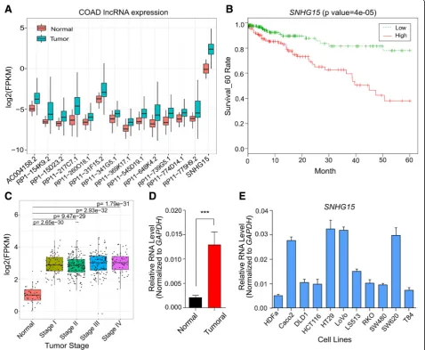
To characterize lncRNAs deregulated in CRC and corre-lated with survival, we profiled their expression in Tumor Cancer Genome Atlas (TCGA) cohort of 456 tu-moral and 41 normal tissue samples. We found 14 lncRNAs as the most significantly deregulated tran-scripts (Fig. 1a, Additional file 3: Table S3), for which their upregulation was related to a significant decrease in survival of CRC patients (Table S3). Among them, we focused onSNHG15due to its highly significant upregu-lation in tumors (pvalue =7.5e-23) and also the highest correlation with poor survival of patients (p value = 4e-5) (Fig. 1b). More investigations in this cohort of tu-mors containing different stages of CRC revealed that al-though there is a significant upregulation of SNHG15 expression in tumors versus normal samples, there is no obvious difference among CRC patients at various stages (Fig. 1c). These results suggest that SNHG15 upregula-tion is an early event in colorectal cancer promoupregula-tion and its expression is maintained at high levels until last stage.
To confirm this observation, we obtained 36 fresh colorectal cancer tumors and their adjacent normal tis-sues immediately after surgery from Iranian CRC pa-tients (Table 1). SNHG15 expression was examined by qRT-PCR and its upregulation was observed in tumoral samples (Fig. 1d, p < 0.001). Moreover, we profiled
SNHG15 expression in ten CRC cell lines (Caco-2,
SNHG15expression is regulated in the cell cycle and transcriptionally controlled by MYC
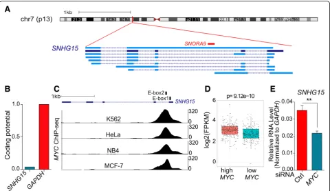
SNHG15 (small nucleolar RNA host gene 15) is located
on chromosome 7 and composed of 5 exons. According to GENCODE V29 annotation, it can be spliced into dif-ferent isoforms, although isoform 2 is the most abundant [18]. Interestingly, SNORA9 (small nucleolar RNA, H/ ACA box 9) is encoded in intron 2 ofSNHG15(Fig.2a).
SNORA9also named ACA9 is a member of H/ACA
pseu-douridylation guide RNA machinery, which contributes to pseudouridine synthesis in snRNAs and rRNAs [19]. Cod-ing potential analysis revealed that SNHG15 sequence does not have ability to code for proteins (Fig. 2b). In agreement with this, polysome fractionation methods
revealed that SNHG15 is not associated with polysomes (Additional file4: Figure S1).
Previous studies revealed that there are two E-box (CACGTG) binding motifs for transcription factor MYC on the first exon and first intron of SNHG15 [20]. We therefore analyzed ChIP-seq data from ENCODE and confirmed that MYC is bound to these boxes in different cancerous cell lines (Fig.2c).
In order to investigate the transcriptional regulation of
SNHG15 by MYC, we analyzed colorectal
adenocarcin-oma RNA-seq data from TCGA, finding thatSNHG15is significantly upregulated in the tumors with high level of
MYCexpression (Fig.2d). In agreement with this obser-vation, the depletion of MYC in LoVo CRC cell line
Fig. 1aExpression of candidate lncRNAs deregulated in CRC tumors compared to normal samples and with higher expression significantly correlated with decreased survival of patients analyzed by RNA-seq from The Cancer Genome Atlas (TCGA).Pvalues were calculated using Wilcoxon signed rank test.bKaplan–Meier analyses of the correlations betweenSNHG15expression level and overall survival of 450 patients with CRC (TCGA).cSNHG15expression levels in different stages of CRC compared with normal tissues (TCGA).dRelative expression level ofSNHG15in 36 Iranian CRC patients compared with corresponding adjacent tissue.P-value is calculated by two-tailed Student’s t-test.eExpression levels of
resulted in a significant decrease in the level ofSNHG15 (Fig. 2e). These results indicate thatSNHG15 transcrip-tion is controlled by MYC.
Since MYC is well known to regulate cell cycle, we in-vestigated expression of SNHG15 during cell cycle. To do this, we obtained G1/S synchronized cells by double thymidine block procedure and collected the cells at dif-ferent time points after block release. Results showed
that SNHG15 expression is regulated during the cell
cycle, with an increased expression ofSNHG15in G2/M phase (Additional file5: Figure S2). Together, these data suggest that SNHG15 is transcriptionally regulated by MYC in a cell-cycle dependent manner.
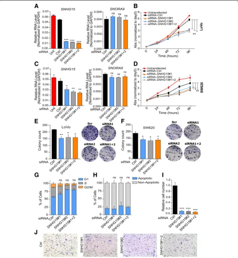
SNHG15deregulation has strong effects on proliferation, invasion and tumor formation abilities of CRC cells To investigate SNHG15 function in CRC, we designed two siRNAs to knock it down, and transfected LoVo and SW620 cells with each one of them individually or a combination of both. qRT-PCR analysis showed that 48 h after transfection, SNHG15 transcript was significantly reduced, while the expression level of
SNORA9, which is located in one of its introns, was
not changed (Fig. 3a and c). These results confirmed
that the designed siRNAs target SNHG15 exons after splicing, leaving intact SNORA9, and allowing
study-ing SNHG15 function independently of SNORA9.
Fur-ther investigation showed that the knockdown of
SNHG15 significantly inhibited cell proliferation (Fig.
3b and d) and colony formation capacity of these cells (Fig. 3e-f ). However, downregulation of SNHG15 did not significantly influence the cell cycle profile or the percentage of apoptotic cells (Fig. 3g-h). On the other hand, the invasion capacity of the cells was sig-nificantly decreased after SNHG15 inhibition, as quan-tified by transwell assays (Fig. 3i-j).
We subsequently investigated the effects of SNHG15 overexpression on cell growth, invasion and tumor formation capacity. To do so, SNHG15 cDNA se-quence (837 bp) was cloned and expressed transiently in HCT 116 and SW480 cells, which express SNHG15 at lower levels compared to LoVo and SW620 cells. qRT-PCR analysis indicated a significant increase in
SNHG15 RNA levels relative to the control cells
transfected with the empty vector (Fig. 4a and d). MTS assay showed that the enforced expression of
SNHG15 led to a significant increase in cell prolifera-tion (Fig. 4b and e). Colony formation assay also
indicated that SNHG15-overexpressing cells not only could form more colonies but also of larger size (Fig.
4c and f ). Consistently with the phenotype observed upon depletion, SNHG15 overexpression did not in-fluence cell cycle or apoptosis in HCT 116 cells (Fig.
4g-h). In addition, the overexpression of SNHG15 in HCT 116 cells increased their invasion capacity (Fig.4i).
To further explore the role of SNHG15 in the tumorigenicity of CRC cells, SNHG15-overexpressing and control HCT 116 cells were injected into
immunodeficient mice, and the tumor size was mea-sured every 4 days. As shown in Fig. 4j, tumors grew faster in cells overexpressed SNHG15, and larger and heavier tumors were formed by these cells after 4 weeks.
In order to confirm our results and exclude possible off target effects of the siRNAs, we knocked-outSNHG15by CRISPR-Cas9 system in LoVo cells. Two guide RNAs (gRNAs) were designed to delete a region ofSNHG15of around 1400 bp without affecting SNORA9 sequence
Fig. 4aLevel ofSNHG15after overexpression in HCT116 cells determined by qRT-PCR (b) Change in proliferation ability of HCT116 cells after overexpression ofSNHG15in comparison with control.cColony formation ability in overexpressing cells comparing to control cells.dSNHG15
(Fig. 5a). Several clones were obtained with homozygote deletion. After screening of the clones, we chose two independent clones with deletion in the aimed region confirmed by qRT-PCR with two pairs of primer de-signed specifically for deleted and non-deleted region. Of note, they no changes in SNORA9 expression were observed (Fig. 5b). The experimental characterization
of these two clones showed their low proliferation and colony formation capacity (Fig. 5c, d), while didn’t show significant changes in cell cycle profile and percentage of apoptotic cells (Fig. 5e-f ). More-over, xenograft mice model experiments confirmed our previous data and revealed that the tumors formed by knock-out cells were smaller and lighter
than those formed by the wild type cells (Fig. 5g). Together, these results suggest that SNHG15 pro-motes the oncogenic capacity of CRC cells.
SNHG15depletion in CRC cells affects the expression of genes with roles in cell proliferation, migration and survival
To determine the biological processes and pathways reg-ulated by SNHG15, a gene expression analysis by RNA-seq was performed in LoVo cells after transfection with combination of two siRNAs or a negative control siRNA. Our results revealed an elevated number of genes significantly deregulated upon SNHG15 depletion (Fig. 6a–Additional file 6: Table S4). Among 766 genes with a significant change of expression (FDR < 0.05), 372 genes were upregulated and 394 genes were downregu-lated. In order to obtain more information about
SNHG15function, Ingenuity Pathway Analysis (IPA) was performed, showing that the genes affected by SNHG15 knockdown are preferentially associated to cancer as well as cell death and survival (Fig. 6b). Interestingly, more detailed canonical pathway analysis showed that these genes contribute to some important molecular pathways and mechanisms of cancer, including poly-amine regulation in colon cancer, GADD45 signaling, chromosomal replication during cell cycle and role of CHK protein in cell cycle checkpoint control (Fig. 6c). We chose 20 candidates among the genes detected by the RNA-seq and pathway analyses to validate the changes in their expression caused by SNHG15 deple-tion. Results showed thatCTGF,GADD45A,GADD45B,
HAS2,LAMC3,NRAS,BAG3,ERBB3, MYCandCASP3
were deregulated afterSNHG15inhibition with each in-dividual siRNA or by the combination of them (Fig.6c), confirming the effect of SNHG15 in the regulation of these relevant genes.
SNHG15interacts with AIF in the cytoplasm and contributes to the resistance to stress
The gene expression analysis performed reflected the physiological changes caused bySNHG15downregulation in CRC cells in terms of proliferation and tumorigenicity. However the analysis did not provide insights into the mechanism by which SNHG15affects the biology of the tumor cells. To investigate the mechanism by which
SNHG15regulates CRC proliferation, we first set to deter-mine its subcellular localization. Nuclear-cytoplasmic frac-tionation in LoVo cells showed that this lncRNA is mainly cytoplasmic (Fig.7a). We hypothesized that SNHG15 in-teracts with cytoplasmic proteins to carry out its func-tions, so we focused on identifying specific physical interactions with SNHG15. To that end, we performed RNA pull-down using in vitro transcribedSNHG15RNA or an unrelated RNA of similar length (murine linc-p21)
as control. The RNAs were incubated with cytoplasmic extract of LoVo cells and mass spectrometry (MS) was performed on the differential band found on the retained proteins (Fig.7b, upper panel). Apoptosis Induced Factor (AIF) was identified as a protein bound toSNHG15with 8 unique peptides but absent in the control RNA pull-down. AIF is a bifunctional protein that exhibits distinct roles based on its subcellular localization. After translation in the cytosol AIF is transported to the mitochondria, where it acts as an NADH oxidase to generate O2− and
subse-quently H2O2[21]. In consequence, AIF has an effect on
reactive oxygen species (ROS) levels with a strong impact in various cellular stress and survival pathways [22, 23]. On the other hand, upon apoptotic stimulus AIF can be cleaved and translocated to the nucleus, where it binds to DNA and promotes chromatin condensation and DNA fragmentation, which are necessary for the apoptosis pro-gram [24]. Interestingly, the molecular weight of the iso-form that we found as interacting withSNHG15is around 67 kDa (full length protein), indicating thatSNHG15binds to the precursor AIF.
The interaction betweenSNHG15and AIF was further validated by western blot using specific antibody against AIF (Fig. 7b). Moreover, it was confirmed by RNA im-munoprecipitation (RIP) with an antibody able to specif-ically immunoprecipitate the endogenous AIF (Fig. 7c). To further investigate the functional relationship be-tween SNHG15 and AIF, immunofluorescence studies were performed showing that inhibition of SNHG15did not change AIF subcellular localization from cytoplasm to nucleus (Fig. 7d), suggesting that SNHG15 is not im-plicated in the pro-apoptotic function of AIF.
Based on these observations and the phenotype ob-served in the cells uponSNHG15inhibiton, we hypothe-sized thatSNHG15binding to AIF does not influence AIF role in apoptosis, but may affect the other mechanism in which the protein has been involved, i.e. respiratory chain and stress response [22, 23]. To test this hypothesis, we measured ROS levels in SNHG15-depleted cells in com-parison to controls. As shown in Fig.7e, after depletion of
SNHG15, ROS levels resulted in a significant reduction. Since enhancement of ROS is known to prevent cellular toxicity by neutralizing chemical stresses [22,23], we ex-amined whether the expression ofSNHG15had an influ-ence on survival of CRC cells after treatment with 5-FU as chemical stress. To do this, we depleted or overexpressed
SNHG15in CRC cells (LoVo or HCT 116) and exposed
cells showed more resistance and higher survival to the drug treatment than control cells (Fig.7f ). These results suggest that the increased levels of SNHG15 are related
with the capacity of CRC cells to cope with the cytotoxic stress caused by 5-FU, which could be mediated by its interaction with AIF. In agreement with these findings,
pathway analysis on the genes affected bySNHG15 knock-down revealed that many of them are functionally related to AIF. Moreover, the role of this group of genes in many important cellular mechanisms such as tissue morphology, cell death and survival, cellular growth and proliferation, cell cycle, organismal development and cellular movement (migration) was confirmed. It was also revealed that these genes contributed in some disorder like cancer, gastro-intestinal disease, organismal injury and abnormalities (Fig.7g).
Discussion
Since colorectal cancer is the third most common hu-man malignancy worldwide [13], many researchers have focused on the characterization of CRC-related lncRNAs as new biomarkers for diagnosis or targeted therapy of this disease. Some of these important lncRNAs include
CCAT1, H19, HOTAIR, MALAT1, UCA1 and PTENP1
[14,25]. In this study, we searched for lncRNAs strongly associated with poor prognosis of CRC. SNHG15 was identified as an oncogenic lncRNA whose upregulation
was related to poor survival of CRC patients. Interest-ingly, the classification of colorectal adenocarcinoma TCGA samples relative to MYCexpression showed that
SNHG15 is upregulated in the samples with high levels of MYC expression. More investigation confirmed that MYC has two binding sites (E-box) on SNHG15 se-quence and bound them in different cancerous cell lines. Furthermore, its inhibition by siRNA, led to decrease
SNHG15 level in CRC cell line and confirmed that
SNHG15 is transcriptionally regulated by MYC. On the
other hand, RNA-seq results showed significant reduc-tion of MYC transcript after depletion of SNHG15. These finding suggest a feedback loop betweenSNHG15 and MYCexpression that introduceSNHG15as an add-itional component of the pro-proliferative network acti-vated by this oncogenic transcription factor. Consistently with this notion, the depletion of SNHG15 by different experimental methods leads to decreased cell proliferation, while its enforced expression promotes cell proliferation and clonogenicity.
Long non-coding RNAs have been involved in a variety of mechanisms that can take place in different cellular compartments. Although some studies have addressed the role of SNHG15 in the nucleus [18], in LoVo cells
SNHG15is mainly localized in the cytoplasm, suggesting that it has a role in this cellular compartment. To eluci-date the role of SNHG15, we searched for proteins with specific physical interactions with SNHG15, identifying AIF as associated withSNHG15. AIF mRNA is translated in the cytoplasm into a 613-amino acid precursor (67 kDa) that is transported to mitochondria via mitochon-drial localization signal (MLS). After being imported into the intermembrane space, the first 34 amino acids are re-moved and the 62 kDa mature AIF (AIFmit) is generated to contribute to the respiratory chain as an NADH oxi-dase [21,26]. If cells receive an apoptotic stimulus, a dif-ferent cleavage occurs in AIFmit by calpain or cathepsin, and a 57 kDa AIF is formed to induce DNA condensation and DNA fragmentation in the nucleus [27–29].
It has been shown that AIF maintains the transformed state of CRC cells via its NADH oxidase activity and cells show more apoptosis sensitivity as a result of AIF knockout and decreases in ROS level. In other words, AIF helps to neutralize chemical stress, and increased protein level of AIF leads to enhancement of ROS to prevent cellular toxicity [22, 23]. Interestingly, we did not observe significant changes in the number of apop-totic cells after dysregulation of SNHG15. In addition, inhibition of SNHG15 did not induce translocation of AIF into nucleus after 48 h. These data suggest that under these experimental conditions, i.e. in the absence of apoptotic stimulus, SNHG15 mainly affects the oxidation-related function of AIF. Given the full-length size of the AIF protein interacting with SNNH15, and
the subcellular localization of the lncRNA, we speculate
that SNHG15 could interact with AIF upon the protein
translation, coupling it to its correct translocation to the mitochondria. Interestingly, many studies have demon-strated that several members of the HSP70 family could bind to AIF and neutralize this protein [30]. Gurbuxani et al. showed that fragment between amino acids 150 to 228 of AIF is necessary for binding to HSP70 and this interaction block AIF nuclear localization [31]. As this fragment exists in both isoform of AIF, this process may occur for mitochondrial localization, so it is possible that binding SNHG15 to AIF could prevent AIF neutralization by HSP70 family and help it to translocate to intermembrane space of mitochondria for contribut-ing in respiratory chain activities and stress response. In agreement with our observations, previous studies have shown that SNHG15contribute to the molecular mech-anism of cellular stress response. This lncRNA is a short-lived lncRNA (t1/2 < 4 h) and its expression level is increased significantly after 24 h cycloheximide used as a stressor, when its half-life was increased from 3.4 to more than 24 h after this treatment [32]. We showed that after depletion of SNHG15, ROS levels are signifi-cantly decreased, and drug sensitivity experiments showed that inhibition of SNHG15 could sensitize CRC cells to 5-FU, which is a basic chemotherapeutic drug for CRC. Based on these results, we propose that the interaction between AIF andSNHG15may be help to in-corporate this protein in ROS formation pathway.
Gene expression analysis after depletion of SNHG15 revealed significant deregulation of multiple genes in-cluding CTGF, GADD45A, GADD45B, HAS2, LAMC3,
NRAS, BAG3, ERBB3, MYC and CASP3. Most of these
genes are known to play an important role in CRC tumor development and response to treatment. For ex-ample, CTGF decreases cell apoptosis and enhances CRC chemoresistance to 5-FU [33]. The inhibition of
remains to be shown if the combination of other mecha-nisms with the AIF-dependent here described confers
SNHG15its full pro-oncogenic activity.
Beyond CRC, the dysregulation and oncogenic role of
SNHG15 has been indicated in various types of cancer, including gastric [47] and hepatocellular carcinoma [48], osteosarcoma [44], as well as pancreatic [49] and breast cancers [46]. Our study, together with this body of work, including a recent study relating this lncRNA with in-creased liver metastasis of CRC tumors [50], indicate that SNHG15 possesses a broad oncogenic activity, and suggests that the development of tools to target the lncRNA could have therapeutic value across multiple cancer types.
Conclusion
Our results describe a role for the MYC-regulated
SNHG15 locus in colorectal cancer, role dependent on
the lncRNA encoded by this bifunctional gene. The lncRNA SNHG15 is able to promote colon cancer and mediating drug resistance, suggesting its potential as prognostic marker and target for RNA-based therapies.
Additional files
Additional file 1:Table S1.Primers used for qRT-PCR. (DOCX 12 kb)
Additional file 2:Table S2.Guide RNAs used for CRISPR-Cas9 editing. (DOCX 11 kb)
Additional file 3:Table S3.The most significantly deregulated lncRNAs with high expression correlated with low survival of patients. (DOCX 13 kb)
Additional file 4:Figure S1.Association possibility ofSNHG15to polysomes,GAPDHexpression was evaluated as a positive control. (PNG 1830 kb)
Additional file 5:Figure S2.(A) Cell cycle analysis of CRC cells after synchronization with double thymidine block procedure. (B)SNHG15 expression level of synchronized cells after each time point. Graphs shows mean ± SEM of values. (PNG 604 kb)
Additional file 6:Table S4.RNA seq analysis of LoVo cells after SNHG15 depletion.(XLSX 163 kb)
Abbreviations
5-FU:Fluorouracil; AIF: Apoptosis Induced Factor; ATCC: American Type Culture Collection; BAG3: BCL2 Associated Athanogene 3; CASP3: Caspase 3; CCAT1: Colon Cancer Associated Transcript 1; ChIP: Chromatin
immunoprecipitation; CHX: Cycloheximide; CL: Clone; CRC: Colorectal cancer; CRISPR: clustered regularly interspaced short palindromic repeats;
CTGF: Connective Tissue Growth Factor; Ctrl: Control; DAPI: 4′ ,6-diamidino-2-phenylindole; DMEM: Dulbecco’s modified Eagle’s medium;
EDTA: Ethylenediaminetetraacetic acid; ERBB3: Erb-B2 Receptor Tyrosine Kinase 3; FBS: Fetal bovine serum; FDR: False Discovery Rate;
GADD45A: Growth Arrest and DNA Damage Inducible Alpha; GADD45B: Growth Arrest and DNA Damage Inducible Beta; GAPDH: Glyceraldehyde 3-phosphate dehydrogenase;
GENCODE: Encyclopedia of DNA Elements; GFP: Green Fluorescent Protein; gRNAs: guide RNAs; HAS2: Hyaluronan Synthase 2; HOTAIR: HOX Transcript Antisense RNA; HPRT: Hypoxanthine Phosphoribosyltransferase; IPA: Ingenuty Pathway Analysis; LAMC3: Laminin Subunit Gamma 3; lncRNA: Long Noncoding RNA; MALAT1: Metastasis associated lung adenocarcinoma transcript 1; miRNAs: microRNA; MLS: Mitochondrial Localization Signal; MS: Mass Spectrometry; NC: Negative Control; ncRNAs: non-coding RNAs; PBS: Phosphate Buffered Saline; PFA: Paraformaldehyde; piRNAs:
Piwi-interacting RNA; PTENP1: Phosphatase and Tensin Homolog Pseudogene 1; qRT-PCR: Quantitative Reverse Transcription Polymerase Chain Reaction; RFU: Relative Fluorescence Unit; RIP: RNA Immunoprecipitation; ROS: Reactive Oxygen Species; RPMI-1640: Roswell Park Memorial Institute; RT: Room Temperature; SD: Standard Deviation; SEM: Standard Error of the Mean; sgRNAs: single-guide RNAs; siRNA: Small interfering RNA; SNHG15: Small Nucleolar RNA Host Gene 15; SNORA9: Small Nucleolar RNA, H/ACA box 9; snoRNAs: Small Nucleolar RNA; TCGA: The Cancer Genome Atlas; TNM: Tumor, lymph node, metastasis; UCA1: Urothelial Cancer Associated 1; WT: Wild Type
Acknowledgements
We thank Dr. Tomás Aragón for his assistance with experimental work, and all patients involved in this study. Authors appreciate the ENCODE project and The Cancer Genome Atlas database for their valuable datasets.
Funding
This work has been supported by BFU2014–58027-R. This study was part of a PhD dissertation was supported by Ferdowsi University of Mashhad (No. 41615).
Availability of data and materials
All data analyzed during current study are included in this published article. Further details could be available upon reasonable request. The RNA-seq data from LoVo cells with knockdown ofSNHG15generated during this study are available at the Gene Expression Omnibus: GSE128998.
Authors’contributions
MS, MMM and MH designed the experiments and MS mainly did the experiments and wrote the paper. DM performed bioinformatics analyses and JG helped in flowcytometry and in vivo experiments. ARB and SJM contributed in sample processing and management and also helped in data analysis. MH and MMM provided the financial support and supervised laboratorial processes and contributed equally to this work. All authors read and approved the final manuscript.
Ethics approval and consent to participate
This study was authorized by the Ethics Committee of Ferdowsi University of Mashhad and all CRC patients signed informed consent forms. All the animal studies were performed according to the Research Ethics Committee (CEI) of Navarra University.
Consent for publication
Not applicable.
Competing interests
The authors declare that they have no competing interests.
Publisher’s Note
Springer Nature remains neutral with regard to jurisdictional claims in published maps and institutional affiliations.
Author details
1Department of Biology, Faculty of Science, Ferdowsi University of Mashhad,
Mashhad, Iran.2Industrial Biotechnology Reasearch Group, Institute of
Biotechnology, Ferdowsi University of Mashhad, Mashhad, Iran.3Department
of Gene Therapy and Regulation of Gene Expression, Center for Applied Medical Research, University of Navarra, Pamplona, Spain.4Institute of Health
Research of Navarra (IdiSNA), Pamplona, Spain.5Department of Molecular
Genetics, Faculty of Biological Sciences, Tarbiat Modares University, Tehran, Iran.
Received: 29 December 2018 Accepted: 7 April 2019
References
1. Djebali S, Davis CA, Merkel A, Dobin A, Lassmann T, et al. Landscape of transcription in human cells. Nature. 2012;489:101–8.
2. Mattick JS, Makunin IV. Non-coding RNA. Hum Mol Genet. 2006;15:17–29. 3. Jia H, Osak M, Bogu GK, Stanton LW, Johnson R, et al. Genome-wide
4. Cabili MN, Trapnell C, Goff L, Koziol M, Tazon-Vega B, et al. Integrative annotation of human large intergenic noncoding RNAs reveals global properties and specific subclasses. Genes Dev. 2011;25:1915–27.
5. Harrow J, Frankish A, Gonzalez JM, Tapanari E, Diekhans M, et al. GENCODE: the reference human genome annotation for the ENCODE project. Genome Res. 2012;22:1760–74.
6. Mercer TR, Dinger ME, Mattick JS. Long non-coding RNAs: insights into functions. Nat Rev Genet. 2009;10:155–9.
7. Hombach S, Kretz M. Non-coding RNAs: classification, biology and functioning. Adv Exp Med Biol. 2016;937:3–17.
8. Bhan A, Mandal SS. Long noncoding RNAs: emerging stars in gene regulation, epigenetics and human disease. Chem Med Chem. 2014;9: 1932–56.
9. Vitiello M, Tuccoli A, Poliseno L. Long non-coding RNAs in cancer: implications for personalized therapy. Cell Oncol. 2015;38:17–28. 10. Bartonicek N, Maag JL, Dinger ME. Long noncoding RNAs in cancer:
mechanisms of action and technological advancements. Mol Cancer. 2016; 15:43–52.
11. Kornfeld JW, Bruning JC. Regulation of metabolism by long, non-coding RNAs. Front Genet. 2014;5:57–64.
12. Brenner H, Kloor M, Pox CP. Colorectal cancer. Lancet. 2014;383:1490–502. 13. Torre LA, Bray F, Siegel RL, Ferlay J, Lortet-Tieulent J, et al. Global cancer
statistics, 2012. CA Cancer J Clin. 2015;65:87–108.
14. Xie X, Tang B, Xiao YF, Xie R, Li BS, et al. Long non-coding RNAs in colorectal cancer. Oncotarget. 2016;7:5226–39.
15. Luo J, Qu J, Wu D-K, Lu ZL, Sun YS, et al. Long non-coding RNAs: a rising biotarget in colorectal cancer. Oncotarget. 2017;8:22187–202.
16. Cong L, Ran FA, Cox D, Lin S, Barretto R, et al. Multiplex genome engineering using CRISPR/Cas systems. Science. 2013;339:819–23. 17. Mari’n-Be’jar O, Huarte M. RNA pulldown protocol for invitrodetection and
identification of RNA-associated proteins. Methods Mol Biol. 2015;1206: 87–95.
18. Jiang H, Li T, Qu Y, Wang X, Li B, et al. Long non-coding RNASNHG15 interacts with and stabilizes transcription factor slug and promotes colon cancer progression. Cancer Lett. 2018;425:78–87.
19. Kiss AM, Jády BE, Bertrand E, Kiss T. Human box H/ACA pseudouridylation guide RNA machinery. Mol Cell Biol. 2004;24:5797–807.
20. Thomas LR, Wang Q, Grieb BC, Phan J, Foshage AM, et al. Interaction with WDR5 promotes target gene recognition and tumorigenesis by MYC. Mol Cell. 2015;58:440–52.
21. Miramar MD, Costantini P, Ravagnan L, Saraiva LM, Haouzi D, et al. NADH oxidase activity of mitochondrial apoptosis-inducing factor. J Biol Chem. 2001;276:16391–8.
22. Urbano A, Lakshmanan U, Choo PH, Kwan JC, Ng PY, et al. AIF suppresses chemical stress-induced apoptosis and maintains the transformed state of tumor cells. EMBO J. 2005;24:2815–26.
23. Liou G-Y, Storz P. Reactive oxygen species in cancer. Free Radic Res. 2010; 44:479–96.
24. Vahsen N, Cande C, Dupaigne P, Giordanetto F, Kroemer RT, et al. Physical interaction of apoptosis-inducing factor with DNA and RNA. Oncogene. 2006;25:1763–74.
25. Yang Y, Junjie P, Sanjun C, Ma Y. Long non-coding RNAs in colorectal cancer: progression and future directions. J Cancer. 2017;8:3212–25. 26. Daugas E, Nochy D, Ravagnan L, Loeffler M, Susin SA, et al. Apoptosis
inducing factor (AIF): a ubiquitous mitochondrial oxidoreductase involved in apoptosis. FEBS Lett. 2000;476:118–23.
27. Otera H, Ohsakaya S, Nagaura Z, Ishihara N, Mihara K. Export of mitochondrial AIF in response to pro-apoptotic stimuli depends on processing at the intermembrane space. EMBO J. 2005;24:1375–86. 28. Yuste VJ, Moubarak RS, Delettre C, Bras M, Sancho P, et al. Cysteine protease
inhibition prevents mitochondrial apoptosis-inducing factor (AIF) release. Cell Death Differ. 2005;12:1445–8.
29. Polster BM, Basañez G, Etxebarria A, Hardwick JM, Nicholls DG. Calpain I induces cleavage and release of apoptosis-inducing factor from isolated mitochondria. J Biol Chem. 2005;280:6447–54.
30. Ravagnan L, Gurbuxani S, Susin SA, Maisse C, Daugas E, et al. Heat- shock protein 70 antagonizes apoptosis-inducing factor. Nat Cell Biol. 2001;3:839–43. 31. Gurbuxani S, Schmitt E, Cande C, Parcellier A, Hammann A, et al. Heat shock
protein 70 binding inhibits the nuclear import of apoptosis-inducing factor. Oncogene. 2003;22:6669–78.
32. Tani H, Torimura M. Identification of short-lived long non-coding RNAs as surrogate indicators for chemical stress response. Biochem Biophys Res Commun. 2013;439:547–51.
33. Yang K, Gao K, Hu G, Wen Y, Lin C, et al. CTGF enhances resistance to 5-FU-mediating cell apoptosis through FAK/MEK/ERK signal pathway in colorectal cancer. Onco Targets Ther. 2016;9:7285–95.
34. Smith ML, Kontny HU, Zhan Q, Sreenath A, Pm O’C, et al. Antisense GADD45 expression results in decreased DNA repair and sensitizes cells to UV-irradiation or cisplatin. Oncogene. 1996;13:2255–63.
35. Moreira MP, Silva LM, Martins WK. The role of GADD45A in resistance to oxidative stress-mediated cell death in human Colon tumor cell lines. Applied Cancer Research. 2009;29:179–84.
36. Wang L, Xiao X, Li D, Chi Y, Wei P, et al. Abnormal expression of GADD45B in human colorectal carcinoma. J Transl Med. 2012;10:215.
37. Kim HR, Wheeler MA, Wilson CM, Iida J, Eng D, et al. Hyaluronan facilitates invasion of colon carcinoma cells in vitro via interaction with CD44. Cancer Res. 2004;64:4569–76.
38. Misra S, Obeid LM, Hannun YA, Minamisawa S, Berger FG, et al. Hyaluronan constitutively regulates activation of COX-2-mediated cell survival activity in intestinal epithelial and colon carcinoma cells. J Biol Chem. 2008:28314335–44. 39. Fernandez-Medarde A, Santos E. Ras in cancer and developmental diseases.
Genes & cancer. 2011;2:344–58.
40. Haigis KM, Kendall KR, Wang Y, Cheung A, Haigis MC, et al. Differential effects of oncogenic K-Ras and N-Ras on proliferation, differentiation and tumor progression in the colon. Nat Genet. 2008;40:600–8.
41. Shi H, Xu H, Li Z, Zhen Y, Wang B, et al. BAG3 regulates cell proliferation, migration, and invasion in human colorectal cancer. Tumour Biol. 2016;37: 5591–7.
42. Li N, Chen M, Cao Y, Li H, Zhao J, et al. Bcl-2- associated athanogene 3 (BAG3) is associated with tumor cell proliferation, migration, invasion and chemoresistance in colorectal cancer. BMC Cancer. 2018;18:793. 43. Rosati A, Ammirante M, Gentilella A, Basile A, Festa M, et al. Apoptosis
inhibition in cancer cells: a novel molecular pathway that involves BAG3 protein. Int J Biochem Cell Biol. 2007;39:1337–42.
44. Liu K, Hou Y, Liu Y, Zheng J. LncRNASNHG15contributes to proliferation, invasion and autophagy in osteosarcoma cells by spongingmiR-141. J Biomed Sci. 2017;24:46–56.
45. Ma Y, Xue Y, Liu X, Qu C, Cia H, et al.SNHG15affects the growth of glioma microvascular endothelial cells by negatively regulatingmiR-153. Oncol Rep. 2017;38:3265–77.
46. Kong Q, Qiu M. Long noncoding RNASNHG15promotes human breast cancer proliferation, migration and invasion by spongingmiR-211-3p. Biochem Biophys Res Commun. 2018;495:1594–600.
47. Chen SX, Yin JF, Lin BC, Su HF, Zheng Z, et al. Upregulated expression of long noncoding RNASNHG15promotes cell proliferation and invasion through regulatesMMP2/MMP9in patients with GC. Tumour Biol. 2016;37: 6801–12.
48. Zhang JH, Wei HW, Yang HG. Long noncoding RNASNHG15, a potential prognostic biomarker for hepatocellular carcinoma. Eur Rev Med Pharmacol Sci. 2016;20:1720–4.
49. Ma Z, Huang H, Wang J, Zhou Y, Pu F, et al. Long non-coding RNASNHG15 inhibitsP15andKLF2expression to promote pancreatic cancer proliferation through EZH2-mediated H3K27me3. Oncotarget. 2017;8:84153–67. 50. Huang L, Lin H, Kang L, Huang P, Huang J, et al. Aberrant expression of long