O R I G I N A L A R T I C L E
Open Access
A new method for estimating patient body
weight using CT dose modulation data
Dominic Gascho
1*, Lucia Ganzoni
1, Philippe Kolly
2, Niklaus Zoelch
1,3, Gary M. Hatch
4, Michael J. Thali
1,
and Thomas D. Ruder
1,5Abstract
Background:Body weight (BW) is a relevant metric in emergency care. However, visual/physical methods to estimate BW are unreliable. We have developed a method for estimating BW based on effective mAs (mAseff) from computed
tomography (CT) dose modulation.
Methods:The mAseffof CT examinations was correlated with the BW of 329 decedents. Linear regression analysis was
used to calculate an equation for BW estimation based on the results of decedents with a postmortem interval (PMI) < 4 days (n = 240). The equation was applied to a validation group of 125 decedents. Pearson correlation andt-test statistics were used.
Results:We found an overall strong correlation between mAseffand BW (r= 0.931);rvalues ranged from 0.854
for decedents with PMI≥4 days to 0.966 for those with PMI < 4 days; among the latter group,rwas 0.974 for females and 0.960 for males and 0.969 in the presence and 0.966 in the absence of metallic implants (all correlations withpvalues < 0.001). The estimated BW was equal to 3.732 + (0.422 × mAseff)–(3.108 × sex index), where the sex
index is 0 for males and 1 for females. The validation group showed a strong correlation (r= 0.969) between measured BW and the predicted BW, without significant differences overall (p= 0.119) as well as in female (p= 0.394) and in male decedents (p= 0.196). No outliers were observed.
Conclusions:CT dose modulation is a rapid and reliable method for BW estimation with potential use in clinical practice, in particular in emergency settings.
Keywords:Body weight, Computed tomography, Dose modulation, Emergency radiology, Virtopsy
Key points
CT using dose modulation can be used to estimate BW
Effective mAs values showed strong correlation with measured BW
An equation can be calculated to estimate BW This method has potential use in emergency settings
Background
The estimation of body weight (BW) is a relevant issue in emergency care as accurate drug dosing [1, 2], such as in thrombolysis of acute ischaemic stroke [3, 4] or the dosage
of contrast media [5, 6], is related to BW. Patients in emergency care may be unresponsive and thus unable to state their BW, and visual estimates of BW are unreliable [1, 7, 8]. A few methods to estimate BW (beyond a simple visual estimate), applicable to both the living and the dead, are mentioned in the literature [2, 9]. Recording BW of a decedent prior to autopsy is a standard procedure in forensic medicine [10, 11]. However, these methods yield moderate accuracy [2] or are at least technically challen-ging and time consuming [9]. Therefore, developing a new approach for BW estimation is a relevant issue.
At our institute of forensic medicine, we use a cali-brated floor scale to measure BW accurately. Addition-ally, each decedent undergoes computed tomography (CT) as a supplement to autopsy. Postmortem CT exams utilize tube current modulation [12]. A main pur-pose of tube current modulation is the adjustment of
* Correspondence:[email protected]
1Department of Forensic Medicine and Imaging, Institute of Forensic Medicine, University of Zurich, 8057 Zurich, Switzerland
Full list of author information is available at the end of the article
dose exposure to body anatomy, yielding almost con-stant image noise along the scan [13, 14]. By measuring beam attenuation during the localizer scan, automated dose modulation calculates a dose distribution based on a reference value of mAs, i.e. a user-selected reference mAs value (mAsref), and on body anatomy. The shape
and size of a typical adult person with a BW of 70– 80 kg served as reference for this technique. Thus, in-creased tube current is applied for overweight people (higher attenuation detected in the localizer) and decreased tube current for underweight people (lower attenuation detected in the localizer) [15]. Since dose modulation adjusts dose exposure according to individ-ual deviations from the ideal patient and the reference standard of 70–80 kg [13, 14], we assumed that adjusted mAs values over the whole body (effective mAs, mAseff)
may correlate with BW of adults.
The aim of this study was to evaluate the correlation between mAseff values and measured BW to develop a
linear regression equation for BW estimation in adults.
Methods Study population
Scan data were acquired as part of a forensic judicial in-vestigation. Data usage is conformant with Swiss laws and ethical standards as approved by the Ethics Com-mittee of the Canton of Zurich (written approval, KEK ZH-Nr. 2015-0686).
We reviewed all cases that underwent postmortem whole body CT between September 2015 and June 2016 (n = 459). Exclusion criteria were: decedents with an age < 17 years (n = 15), use of non-standard scan parameters in the context of research purposes (n = 20), and dis-membered corpses (n = 95). Thus, the final study popu-lation consisted of 329 decedents (105 females and 224 males) with a mean age of 59.0 years (standard deviation [SD] 59.0 ± 18.0 years; range 18–95 years). Taking into consideration that decomposition- or putrefaction-related changes usually start to appear after 72 h after demise [16], the study population was divided into two groups with dif-ferent postmortem interval (PMI): 240 decedents with a PMI < 4 days (78 females and 162 males) and 89 decedents with a PMI≥4 days (27 females and 62 males). The former group was further subdivided into subgroups according to gender (78 female and 162 males) as well as according to the presence of metallic medical implants (38 with and 202 without). After evaluation of the data distribution using the Kolmogorov–Smirnov test, Pearson’s correlation coeffi-cient (r) was used to assess the correlation between mea-sured BW and mAsefffor each group and subgroup. Thep
values of the correlations were also calculated.
Linear regression analyses were used to create a model to be used to estimate BW based on mAseff,
taking into consideration sex and implants. The group
with PMI < 4 days was used for the calculation of an equation for BW estimation; therefore, the calculated constant and the unstandardized coefficients (B) were used to develop the equation. According to the multi-variate linear regression analysis, sex and/or implants were taken into account for the equation. Further, the standard error of the estimates (SEE) was calculated. The final equation was applied on a validation group, which included all cases between December 2016 and March 2017 (n = 204). Exclusion criteria were the same as mentioned above with the addition of a PMI≥4 days. The final validation group consisted of 125 decedents (43 females and 82 males) with a mean age of 56.4 years (SD 56.4 ± 18.3 years; range 18–96 years). After evaluation of data distribution using the Kolmogorov–Smirnov test, the Student t-test was applied to reveal significant differences between actual BW and BW predicted by the linear regression equation.
All CT exams utilized automated dose modulation and were performed at the request of local legal authorities.
Imaging protocol
Postmortem CT was performed on a 128-slice scanner (SOMATOM Definition Flash, Siemens Healthcare, For-chheim, Germany) using the dose modulation technique (CARE Dose 4D™, Siemens Healthcare, Forchheim, Germany). The CT scan protocol included frontal and lateral localizer topogram or scout view using 120 kVp and 35 mA. Dose modulation was based on attenuation measurements automatically taken during the lateral localizer. The whole body scan was performed according to the calculated dose distribution and the initial refer-ence mAs value (mAsref). The scan parameters of the
whole body CT were as follows: reference tube current 400 mAsref; tube voltage 120 kVp; rotation time 0.5 s;
pitch 0.35; acquisition 128 × 0.6 mm. The actual tube current levels were based on dose modulation with an average adaptation to patient size. After each scan, the effective mAs values (mAseff) according to the effective
dose exposure of the whole body scan was documented in an automated dose report.
Descriptive data
The actual mAseff values and the CT examination
9.0, Armonk, NY, USA). PMI in days was calculated according to the time period between the estimated time of death and the CT examination date. The presence or absence of metallic medical implants (orthopaedic implants and pacemakers) was noted by reviewing all image data. Actual BW measurements (kg) on the readout of the calibrated floor scale (Mul-tiRange ID5, Mettler-Toledo International Inc., Ohio, Columbus, US) were documented during body intake at our institution, according to our routine protocol.
Statistical analysis
All statistical analyses were computed using dedicated software (R version 3.3.2., R Core Team, R Foundation for Statistical Computing, Vienna, Austria). Apvalue of < 0.05 was considered to be statistically significant.
Results
The study population yielded a mean actual BW of 73.8 kg (SD 73.8 ± 20.1 kg, range 18–137 kg) and a mean value of 165.8 mAseff (SD 165.8 ± 46.4 mAseff, range 30–
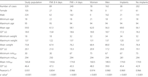
Table 1Descriptive data and statistical analyses of the study population and of subgroups
Study population PMI≥4 days PMI < 4 days Women Men Implants No implants
Number of cases 329 89 240 78 162 38 202
Female 105 27 78 78 0 17 61
Male 224 62 162 0 162 21 141
Minimum age 18 22 18 21 18 21 18
Maximum age 95 95 94 94 94 94 94
Mean age 59.0 61.3 58.1 62.0 56.3 69.0 56.1
SDa(±) 18.0 15.8 18.6 18.0 18.7 17.2 18.2
Minimum weight 18 18 32 32 34 34 32
Maximum weight 137 122 137 131 137 120 137
Mean weight 73.8 67.4 76.2 68.4 80.0 75.0 76.4
SDa(±) 20.1 21.0 19.3 20.9 17.2 20.0 19.1
Minimum mAseff 30 30 67 75 67 67 75
Maximum mAseff 294 250 294 294 294 281 294
Mean mAseff 165.8 143.6 174.0 160.5 180.5 174.0 174.0
SDa(±) 46.4 47.3 43.3 48.3 39.0 45.4 42.9
Pearson’srb 0.931 0.854 0.966 0.974 0.960 0.969 0.966
pvaluec <0.001 < 0.001 < 0.001 < 0.001 < 0.001 < 0.001 < 0.001 The study population indicated a strong correlation between measured BW and mAseffvalues (r= 0.931). The Pearson coefficient was higher for PMI < 4 days (r= 0.966) than for PMI≥4 days (r= 0.854);rwas 0.974 for females with PMI < 4 days and 0.960 for males with PMI < 4 days. Further subgroups with PMI < 4 days for implants (r= 0.969) and no implants (r= 0.966) revealed both strong and nearly equal correlations. All correlation coefficients were statistically significant (p< 0.001)
a
Standard deviation b
Pearson correlation coefficient between mAseff
and body weight c
pvalue of the correlation
294 mAseff). The correlation between the measured BW
and mAseff was stronger for PMI < 4 days (r= 0.966)
than for PMI≥4 days ( r= 0.854). The descriptive data and statistical analyses of the study group and of all sub-groups are listed in Table 1. The Kolmogorov–Smirnov test showed normal data distributions for all groups and subgroups except females (mAseff, p= 0.002; weight, p=
0.001) and males (weight,p= 0.032) with a PMI < 4 days. The correlation was found to be strong for both females (r= 0.974) and males (r= 0.960). The same applied to the subgroups for implants (r= 0.969) and no implants (r= 0.966). All correlation coefficients were statistically significant (p< 0.001). Correlations between mAseff and
measured BW for the study population, for PMI < 4 days and for PMI≥4 days are illustrated in Fig. 1.
Multivariate linear regression analysis for PMI < 4 days taking into account the mAseff(p< 0.001), sex (p< 0.001)
and implants (p= 0.271) revealed that sex was a signifi-cant factor, whereas implants were not. Therefore, the implants variable was not included in the equation. Based on the results of the multivariate linear regression analysis for PMI < 4 days (constant = 3.732, p= 0.007) taking into account mAseff(B = 0.422,p<0.001) and sex
(B =−3.108, p <0.001), we propose the following linear regression equation to estimate BW:
Estimated BW¼3:732þð0:422mAseffÞ−ð3:108sex indexÞ
where the sex index is 0 for males and 1 for females. The SEE was 4.82.
The validation group yielded a mean actual BW of 74.8 kg (SD 74.8 ± 16.7 kg, range 32–128 kg) and a mean mAseff of 169.5 (SD 168.5 ± 38.3 mAseff, range
65–274 mAseff). The mean predicted BW calculated by
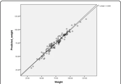
the equation was 74.2 kg (SD 74.2 ± 16.6 kg, range 28.1–119.4 kg). Descriptive data of the validation group and of all subgroups are listed in Table 2. The statistical evaluation of data distribution showed normal distribu-tions for the main group and both subgroups. The actual BW and BW predicted by the equation were strongly correlated (r= 0.969; women,r= 0.972; men,r= 0.960). The coefficient of determination (R2) was 0.938. The val-idation group showed no outliers (maximum deviation ±9 kg; mean deviation−0.6 kg; Fig. 2). The Studentt-test revealed no statistically significant difference between actual BW and predicted BW for the validation group (p = 0.119; females,p = 0.394; males, p = 0.196).
Discussion
This study presents a reliable method to estimate BW using CT dose modulation through a simple equation. We found a strong correlation between BW, measured with the standard scale, and mAseff values based on CT dose
modulation. The proposed equation, taking into account
mAseffand sex, fits 93.8% (R2= 0.938) of the data
regard-ing decedents with PMI < 4 days, without any outliers in the validation group. Thus, a rapid and robust method to determine BW of non-decomposed human decedents is now available. In the forensic setting, this could have value in situations of equipment failure, data loss, or if images were evaluated in isolation. Moreover, this method may have potential in clinical radiology as whole body CT has gained increasing importance in emergency settings such as polytrauma [17–20] or other conditions. Notably, our equation was derived from data obtained with the CT scanner and the protocol we used and may not provide the same results when different CT models from other vendors and other protocols are used. However, this study clearly describes how institutes can calculate an equation for their own whole body CT unit and protocol.
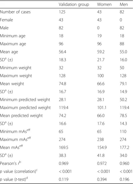
Table 2Descriptive data and statistical analyses for the validation group
Validation group Women Men
Number of cases 125 43 82
Female 43 43 0
Male 82 0 82
Minimum age 18 19 18
Maximum age 96 96 88
Mean age 56.4 59.2 55.0
SDa(±) 18.3 21.7 16.0
Minimum weight 32 32 50
Maximum weight 128 100 128
Mean weight 74.8 66.6 79.1
SDa(±) 16.7 16.9 14.9
Minimum predicted weight 28.1 28.1 50.2
Maximum predicted weight 119.4 101.1 119.4
Mean predicted weight 74.2 66.0 78.5
SDa(±) 16.6 17.6 14.3
Minimum mAseff 65 65 110
Maximum mAseff 274 238 274
Mean mAseff 169.5 154.9 177.2
SDa(±) 38.3 41.8 34.0
Pearson’srb 0.969 0.972 0.960
pvalue (correlation)c < 0.001 < 0.001 < 0.001
pvalue (t-test)d 0.119 0.394 0.196 Applying the equation on the validation group revealed a strong correlation between measured BW and predicted BW (r= 0.969). The Pearson coefficientr
was 0.972 for females and 0.960 for males. All correlation coefficients were statistically significant (p< 0.001). The Studentt-test revealed no significant difference between actual BW and predicted BW for the validation group (p= 0.119; females,p= 0.394; males,p= 0.196)
a
Standard deviation b
Pearson correlation coefficient between actual weight and predicted weight (calculated by the equation)
c
pvalue of the correlation d
The study population was divided into cases with PMI < 4 days and cases with PMI≥4 days, because of decom-position- or putrefaction-related changes. This temporal separation was chosen based on the experiences of our forensic pathologists. Although decomposition is dependent on several factors [21], in our temperate cli-mate, generalized bloating usually starts to appear after 72 h after demise [16]. For this study the chosen point of time for temporal separation seemed appropriate. All decedents with PMI < 4 days showed an excellent correl-ation between mAseff and BW (r = 0.966). By contrast,
the correlation was weaker in decedents with PMI ≥ 4 days (r = 0.854), probably due to decomposition- or putrefaction-related changes (e.g. gaseous distention or putrefaction fluid). It is conceivable that decedents with a shorter PMI (or living patients) may show even higher correlation between mAseffand BW. In the field of
post-mortem imaging, Jackowski et al. [9] presented a method using postmortem CT. The method was derived from a study by Abe et al. [22], who calculated a soft tissue multiplication factor for detecting whole body skeletal muscle mass in the living. Based on 50 cases (30 adults and 20 paediatrics) with a short PMI (not described more accurately), Jackowski et al. [9] calculated a multi-plication factor to estimate BW of decedents based on whole body segmentation. However, whole body seg-mentation requires specialized skills and software and additional imaging processing steps and can be time consuming. Conversely, the use of dose-modulated mAs and an equation enable rapid BW estimation.
Rapid BW calculation based on dose modulation for adult patients may show potential in emergency radiology with respect to drug dosage or dosage of contrast media, which are usually based on patient BW. Fernandes et al.
[1] demonstrated that 33% of estimates from physicians and nurses deviate by more than 10% from actual BW of ambulatory patients (indicated with a 95% confidence interval). As mentioned by the authors, BW estimates for patients in the supine position may be even less accurate. An equation by Buckley et al. [2] yielded greater accuracy compared to visual BW estimates made by physicians and nurses. Deviations greater than ±10 kg from measured BW still occurred in 15% of male patients and 27% of female patients. Thus, the authors recommended the lin-ear regression equation only for male patients when patients are not able to state their BW. By contrast, the present study revealed strong correlations for both fe-males and fe-males with a PMI < 4 days. However, the data of each of these two subgroups were not normal distrib-uted; therefore, the results are less robust. The mean BW of males (80.0 kg) was in the range of the standard refer-ence patient BW of 70–80 kg used in dose modulation software and revealed a strong correlation (r = 0.960). Despite the fact that the mean BW of females (68.4 kg) was below the range of the standard reference patient BW of 70–80 kg, the correlation was also strong (r= 0.974). Al-though, metallic implants affect x-ray attenuation [23], the correlation between decedents with implants (r = 0.969) was nearly equal to decedents without implants (r= 0.966). In contrast to sex, taking implants into account was not statistically significant in the multivariate linear regression analysis; thus, implants were not considered as a factor to consider. Therefore, the presence or absence of metallic im-plants was not taken into account in the linear regression equation. We hypothesize that small medical devices may also have little influence on the correlation.
In our study, the applied dose modulation (CARE Dose 4D™) was used with an average adaptation to patient BW. CARE Dose 4D™ also allows for different adaptation options regarding patient size (very strong, strong, weak, and very weak), which can be selected for adult slim or adult obese patients. Different adap-tation settings result in different mAseff values [15].
Therefore, changes in adaptation options would result in different correlations between mAseff and patient
BW. We hypothesize that separate equations for slim or obese patients using weak or strong adaptations, respectively, will result in more precise BW estima-tions. Further, dose modulation was based on a lateral whole body localizer. Our postmortem CT protocol included at first a frontal localizer and afterwards a lateral localizer. The correlation between dose modu-lations based on attenuation measurements during the frontal localizer and BW was not evaluated in this study. However, this study clearly describes the calcu-lation of an equation for BW estimation based on mAseff values, which can be easily calculated for any
clinical CT protocol using dose modulation.
Admittedly, this study has several limitations when considering the clinical perspective. First, our results are based on a standardized postmortem CT proto-col according to the literature [12]. Radiation dose to the decedent can be neglected in postmortem im-aging; therefore, a high mAsref value of 400 is
stand-ard for whole body scans. Further studies are required regarding mAseff values from clinical
proto-cols. Second, the estimation of BW based on CT using dose modulation requires a whole body scan. Therefore, this approach is limited to polytrauma patients who undergo whole body CT scans. Third, automatic exposure control systems are available from several CT vendors [13, 14] but dose modula-tion strategies vary between vendors. The results of this study are based on the dose modulation strategy of a single vendor. However, we hypothesize that other vendors provide similar correlations, which can be investigated in the same way as the present study.
To summarize, this study demonstrates a rapid and reliable method for BW estimation. Given the lack of re-liable methods for practitioners to estimate patient BW based on visual parameters or physical exam, BW esti-mation based on CT dose modulation may have poten-tial use in clinical radiology and polytrauma patients. Certainly, further studies are required.
Authors’contributions
DG drafted the manuscript, performed the computed tomography scans and participated in performing the statistical analysis. DG and TR conceived of the study and coordinated it. LG carried out the data collection. PK performed the statistical analysis. LG, PK, NZ, GH and TR reviewed the manuscript. MT provided technical equipment. All authors read and approved the final manuscript.
Publisher’s Note
Springer Nature remains neutral with regard to jurisdictional claims in published maps and institutional affiliations.
Author details
1Department of Forensic Medicine and Imaging, Institute of Forensic Medicine, University of Zurich, 8057 Zurich, Switzerland.2Department of Clinical Research, University of Bern, 3008 Bern, Switzerland.3Hospital of Psychiatry, Department of Psychiatry, Psychotherapy and Psychosomatics, University of Zurich, 8032 Zurich, Switzerland.4Center for Forensic Imaging, Departments of Radiology and Pathology, University of New Mexico School of Medicine, Albuquerque, NM 87102, USA.5Institute of Diagnostic, Interventional, and Pediatric Radiology, University Hospital Bern, 3010 Bern, Switzerland.
Received: 16 August 2017 Accepted: 20 October 2017
References
1. Fernandes CM, Clark S, Price A, Innes G (1999) How accurately do we estimate patients’weight in emergency departments? Can Fam Physician 45:2373–2376
2. Buckley RG, Stehman CR, Dos Santos FL et al (2012) Bedside method to estimate actual body weight in the emergency department. J Emerg Med 42:100–104
3. Lorenz MW, Graf M, Henke C et al (2007) Anthropometric approximation of body weight in unresponsive stroke patients. J Neurol Neurosurg Psychiatry 78:1331–1336
4. Breuer L, Nowe T, Huttner HB et al (2010) Weight approximation in stroke before thrombolysis: the WAIST-Study: a prospective observational “dose-finding”study. Stroke 41:2867–2871
5. Bae KT, Tao C, Gürel S et al (2007) Effect of patient weight and scanning duration on contrast enhancement during pulmonary multidetector CT angiography. Radiology 242:582–589
6. Bae KT (2010) Intravenous contrast medium administration and scan timing at CT: considerations and approaches. Radiology 256:32–61
7. Hall WL II, Larkin GL, Trujillo MJ et al (2004) Errors in weight estimation in the emergency department: Comparing performance by providers and patients. J Emerg Med 27:219–224
8. Menon S, Kelly A-M (2005) How accurate is weight estimation in the emergency department? Emerg Med Australas 17:113–116 9. Jackowski C, Schwendener N, Zeyer-Brunner J, Schyma C (2015) Body
weight estimation based on postmortem CT data—validation of a multiplication factor. Int J Legal Med 129:1121–1125
10. Saukko P, Knight P (2015) Chapter 1. The forensic autopsy. In: Saukko P, Knight B (eds) Knight’s forensic pathology, 4th edn. CRC Press–Taylor & Francis, Boca Raton, FL, United States, pp 1–54
11. Di Maio V, Di Maio D (2001) Appendix. The autopsy report. In: Di Maio VJ, Di Maio D (eds) Forensic pathology, 2nd edn. CRC Press–Taylor & Francis, Boca Raton, FL, United States, pp 549–551
12. Flach PM, Gascho D, Schweitzer W et al (2014) Imaging in forensic radiology: an illustrated guide for postmortem computed tomography technique and protocols. Forensic Sci Med Pathol 10:583–606 13. Kalra MK, Maher MM, Toth TL et al (2004) Techniques and applications of
automatic tube current modulation for CT. Radiology 233:649–657 14. McCollough CH, Bruesewitz MR, Kofler JM (2006) CT Dose reduction and
dose management tools: overview of available options. RadioGraphics 26:503–512
15. Söderberg M, Gunnarsson M (2010) The effect of different adaptation strengths on image quality and radiation dose using Siemens Care Dose 4D. Radiat Prot Dosimetry 139:173–179
16. Di Maio V, Di Maio D (2001) Chapter 2: Time of death. In: Di Maio VJ, Di Maio D (eds) Forensic pathology, 2nd edn. CRC Press–Taylor & Francis, Boca Raton, FL, United States, pp 21–41
17. Poletti P-A, Wintermark M, Schnyder P, Becker CD (2002) Traumatic injuries: role of imaging in the management of the polytrauma victim (conservative expectation). Eur Radiol 12:969–978
18. Linsenmaier U, Krötz M, Häuser H et al (2002) Whole-body computed tomography in polytrauma: techniques and management. Eur Radiol 12:1728–1740
19. Huber-Wagner S, Lefering R, Qvick L-M et al (2009) Effect of whole-body CT during trauma resuscitation on survival: a retrospective, multicentre study. Lancet 373:1455–1461
20. Wurmb TE, Quaisser C, Balling H et al (2011) Whole-body multislice computed tomography (MSCT) improves trauma care in patients requiring surgery after multiple trauma. Emerg Med J 28:300–304
21. Zhou C, Byard RW (2011) Factors and processes causing accelerated decomposition in human cadavers–an overview. J Forensic Leg Med 18:6–9
22. Abe T, Kearns CF, Fukunaga T (2003) Sex differences in whole body skeletal muscle mass measured by magnetic resonance imaging and its distribution in young Japanese adults. Br J Sports Med 37:436–440