UDC: 636.5.09:[616.98:578.8.083.3
Original Scienti
fi
c Article
ABSTRACT
Available online at www.macvetrev.mk
Corresponding author: Ass. Dodovski Aleksandar, MsC
e-mail address: [email protected]
Present address: Faculty for Veterinary Medicine Veterinary Institute, Lazar Pop Trajkov, 5-7, 1000 Skopje Macedonia
tel: +389 2 3240 752; fax: +389 2 3114 619
INTRODUCTION
Newcastle disease (ND) virus may be present in natural or experimental hosts in 241 species from 27 to 50 orders of birds and it is very likely that all birds are susceptible to infection, but the severity of the disease would depend on the type of the bird (19). The ND virus (NDV) belongs to order Mononegavirales, family Paramyxoviridae, subfamily Paramyxovirinae, genus Avulavirus (22). In this genus there are 11 serotypes of APMV, labeled APMV-1 to APMV-11 (9, 24). Newcastle
CLASSICAL AND MOLECULAR CHARACTERIZATION OF
PIGEON PARAMYXOVIRUS TYPE 1 (PPMV-1) ISOLATED
FROM BACKYARD POULTRY – FIRST REPORT IN MACEDONIA
Dodovski Aleksandar
1, Krstevski Kiril
1, Naletoski Ivancho
21Veterinary Institute, Faculty of Veterinary Medicine,
University “Ss. Cyril and Methodius” Skopje, Macedonia
2Joint FAO/IAEA Division of Nuclear Techniques in Food and Agriculture, Vienna, Austria
Received 22 May 2013; Received in revised form 26 June 2013; Accepted 9 July 2013
Aim of this study was to characterize pigeon variant of Newcastle disease virus (NDV) isolated from backyard poultry using classical and molecular methods. In standard hemagglutination inhibition (HI) test both polyclonal NDV antiserum and monoclonal antibodies 161/617 specifi c for pigeon variants of NDV showed inhibition of heamagglutination of the isolated virus. Intracerebral pathogenicity index (ICPI) has shown that the isolate is mesogenic virus (ICPI = 0.81). One-step RT-qPCR for detection of M gene was performed indicating a presence of NDV and RT-qPCR for discrimination between lentogenic and velogenic strains based on F gene was also performed indicating a presence of virulent NDV. A portion of the F gene was amplifi ed and sequenced for determination of virulence and phylogenetic characterization. The F protein cleavage site sequence of the isolate had multiple basic amino acids at residues 112–116 and a phenyl alanine at residue 117 (112RRQKR*F117) which is typical for velogenic strains. The nucleotide sequence of 374 bp was aligned to begin at nt 47 and fi nish at 420 immediately after the cleavage site and compared with other reference strains from the region and worldwide. In the phylogenetic tree, the isolate clustered into genotype VIb, typical for PPMV-1. This strain is phylogenetically very similar to other PPMV-1 isolated from pigeons in Macedonia. Poultry infected with PPMV-1 can spread the virus in the absence of clinical signs, thus PPMV-1’s are constant threat to domestic poultry. This is the fi rst report of evidenced spillover of PPMV-1 into poultry in Macedonia.
Key words: PPMV-1, backyard poultry, RT-qPCR, nucleotide sequencing
disease virus is APMV-1, for which there are two different classifi cations based on genomic structure (2, 7, 12).
epizootics in pigeons (29). Virus is characterized as an antigenic variant of NDV with use of monoclonal antibodies (mAb’s) (6). Because of the antigenic difference and for pragmatic purposes these viruses are termed pigeon paramyxovirus type 1 (PPMV-1) viruses beside the fact being able to infect poultry but with decreased virulence (23). These viruses are also able to infect wild pigeons, doves and ornamental birds (17, 20). The disease in pigeons has an enzootic character with occasional spread to wild pigeons and doves and represents a constant threat to poultry (4).
Amino acid sequence of cleavage site (CS) of fusion (F) protein is a major determinant of virulence (25, 26), although other proteins have a role in virulence depending on the strain of the virus (14). In order for the virus to be virulent a basic amino acid at residue 113, a pair of basic amino acid at residues 115 and 116 and phenylalanine at residue 117 of the F gene is required (10).
For proper diagnosis it is not enough only to detect NDV or prove infection with the virus, but it is also necessary to distinguish whether the virus is virulent or not by recommended classical and molecular methods (30). Nucleotide sequencing can determine virulence and can assess genetic makeup, genotype and phylogenetic characteristics of NDV (11, 21, 27). These techniques allow only 250 nucleotides to be suffi cient for reliable phylogenetic analysis (2, 21, 27).
The role of pigeons in epizootiology of NDV in Macedonia has not been studied previously. The aim of this study was to characterize PPMV-1 isolated from backyard poultry by classical and molecular methods and to assess its phylogenetic relationship with other NDV isolates.
MATERIAL AND METHODS
Virus and virus isolation
Pool of internal organs obtained from dead chickens were homogenized in antibiotic medium using sterile sand, checked for sterility and inoculated in to the 9-11 days old embryonated chicken eggs (ECE). All dying embryos and remaining embryos after the incubation period of fi ve days were checked for haemagglutination activity according to recommended protocols (29).
country of origin/laboratory identifi cation number (reference number)/year of sampling.
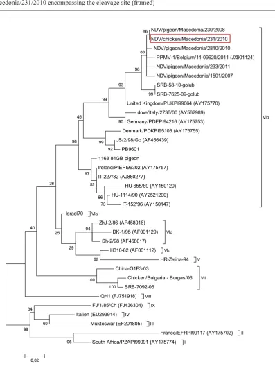
GenBank accession numbers of the viruses used for construction of the phylogenetic tree are shown in parentheses in Fig. 2. Several sequences without accession numbers were obtained from colleagues from the region. Accession number of the Macedonian strain is given in the results section.
Strain under investigation was isolated from backyard chicken on 10.02.2010 from village Rankovce in the northeastern part of Macedonia (N 42o10’03. E 22o07’03.) Isolation was donefrom the pool of visceral organs. Strain was labeled as NDV/chicken/Macedonia/231/2010 according to serotype/host/location/laboratory number/sampling date.
Hemagglutination inhibition test (HI test)
The supernatant of allantoic fl uid was collected and used in standard HI test using polyclonal NDV antiserum and monoclonal antibodies (mAb): U85 for detection of classical strains, 161/617 for detection of PPMV-1 strains and 7D4 for detection of F and La Sota vaccine strains.
In vivo pathogenicity test
The pathogenicity of PPMV-1 isolate was assessed by intracerebral pathogenecity index (ICPI) test. One-day-old specifi c pathogen free (SPF) chickens were inoculated intracerebrally with 0.1 ml of a 1:10 dilution of infective allantoic fl uid. Chicks were monitored during an 8-day observation period and scored daily as normal (score 0), sick (score 1), and dead (score 2). Total scores were determined and the mean daily scores were calculated to obtain the ICPI.
Preparation of viral RNA, real-time reverse transcription-polymerase chain reaction (RT-qPCR) and reverse transcription-polymerase chain reaction (RT-PCR)
Viral RNA was extracted from allantoic fl uid of ECE using Invisorb Spin Virus RNA Mini Kit (Invitek, Germany) following manufacturers protocol.
5’-AGTGATGTGCTCGGACCTTC-3’, reverse primer
М-4220 5’-CCTGAGGAGAGGCATTTGCTA-3’ and hydrolysis probe М+4169 5’- (FAM) TTCTCTAGCAGTGGGACAGCCTGC(TAMRA) - 3’. Thermal protocol was as follows: 50 0C 10 min,
95 0C 5 min and 40 cycles on 95 0C 10 seconds and
55 0C 30 seconds.
The procedure for detection of fusion (F) gene is the same as described above except for primers and hydrolysis probe used: forward primer F+4839 5’-TCCGGAGGATACAAGGGTCT-3’, reverse primer F-4939 5’-AGCTGTTGCAACCCCAAG-3’ and hydrolysis probe F+4894 (VFP-1) 5’-(FAM) AAGCGTTTCTGTCTCCTTCCTCCA(TAM RA)-3’ and the temperature of annealing of primers which was 58 0C instead of 55 0C, according to Wise
et al. (31).
Two-step RT-PCR for the F gene was performed according to methods described by Collins et al., (10) with primers according to Aldous et al., (2) where forward primer was used instead of random primers in the RT step. In the PCR step forward primer MSF1 5’-GACCGCTGACCACGAGGTTA-3’ and reverse primer #2 5’-AGTCGGAGGATGTTGGCAGC-3’ were used with the thermal protocol of 94 0C for 3
min, 42 cycles of 94 0C for 1 min, 50 0C for 1 min
and 72 0C for 3 min, and fi nal extension at 720C for10
min. The PCR product of 700 base pairs (bp) was synthesized, subjected to electrophoresis in 1.5% agarose gel and the DNA band was excised from the gel and purifi ed using QIAquick Gel Extraction Kit (Qiagen, Valencia, CA). The purifi ed PCR product was used for sequencing.
Nucleotide sequencing and analysis of sequence data
The sequence of the amplifi ed 374 bp region of the F-gene was obtained using Big Dye Terminator v3.1 kit (Applied Biosystems, USA) and F-gene-specifi c primers, forward primer #7 5’-GACCGCTGACCACGAGGTTA-3’ and reverse primer #2 5’-TTAGAAAAAACACGGGTAGAA-3’ according to Aldous et al., (2). All sequencing reactions were performed with fl uorescent dideoxynucleotide terminators in the ABI 310 automated sequencer (Applied Biosystems Inc., Foster City, CA) and sequencing product was purifi ed with 50 μl of ethanol (96-100%), 2 μl of 3М sodium acetate and 2 μl of 125 mМ EDTA. Sequence editing
was performed with BioEdit Sequence Alignment Editor version 7.0.9.0 while alignment with Clustal V method was done in MEGA5 software (MEGA, version 5). The same region of the F-gene was used to construct phylogenetic trees and to classify PPMV-1 isolate among other class II genotype reference sequences. Phylogenetic analysis was performed using MEGA5 software (MEGA, version 5) (28). The evolutionary distances were inferred using un-rooted maximum-likelihood method with 1,000 bootstrap replicates to give credibility to the grouping and included the fi rst, second, and third coding and noncoding positions.
RESULTS
Virus was isolated from a holding of 40 chickens, out of which fi ve have died. Clinical signs involved inapetence, stretched wings and dispnoea. Gross lesions were located predominantly in the respiratory system and involved congestion and edema of the lungs. Haemagglutination activity of allantoic fl uid was detected after the second passage in inoculated ECE. Infective allantoic fl uid has demonstrated inhibition of haemagglutination with polyclonal NDV serum with titer of 8 log2 while it
was negative for H5N1 and H7N1 antisera. When tested with mAb’s it demonstrated inhibition of haemagglutination with mAb 617/161 with titre of 5 log2 while it was negative for mAb U85 and mAb
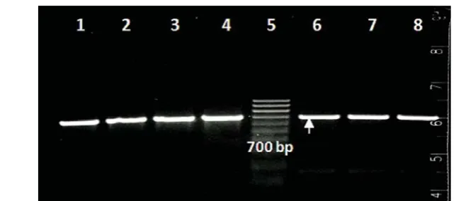
7D4. Value of ICPI was 0.81 classifying the virus as mesogenic strain regarding pathogenicity. On the basis of RT-qPCR for detection of the M and the F gene, the virus proved to be virulent strain of class II NDV with Ct values of 21.3 and 27.5, respectively. These results were confi rmed with RT-PCR for the F gene with visible band on the gel with expected size of 700 bp.
NDV/pigeon/Macedonia/230/2008 NDV/chicken/Macedonia/231/2010
NDV/pigeon/Macedonia/2810/2010
PPMV-1/Belgium/11-09620/2011 (JX901124) NDV/pigeon/Macedonia/233/2011
NDV/pigeon/Macedonia/1501/2007 SRB-58-10-golub
SRB-7625-09-golub United Kingdom/PUKPI99064 (AY175770)
dove/Italy/2736/00 (AY562989) Germany/PDEPI94216 (AY175753) Denmark/PDKPI95103 (AY175755) JS/2/98/Go (AF456439)
PB9601 1168 84GB pigeon
Ireland/PIEPI96302 (AY175757) IT-227/82 (AJ880277)
HU-655/89 (AY150120) HU-1114/90 (AY2521200)
IT-152/96 (AY150147)
VIb
VIa Israel70
ZhJ-2/86 (AF458016) DK-1/95 (AF001129) Sh-2/98 (AF458017)
VId
VIc H310-82 (AF001112)
V HR-Zelina-94 China-G1F3-03
Chicken/Bulgaria - Burgas/06 SRB-7092-06
VII
VIII QH1 (FJ751918)
IX FJ/1/85/Ch (FJ436304)
IV Italien (EU293914)
III Mukteswar (EF201805)
II France/EFRPI99117 (AY175702)
I South Africa/PZAPI99091 (AY175774)
100
96
100
34
60 99
94
62
99
92
40
29 25
99
38
73 86 52 97 96
45
95 99
93 98
86
83
0,02
Figure 1. Electropherogram of part of the nucleotide sequence of the F gene of NDV/chicken/ Macedonia/231/2010 encompassing the cleavage site (framed)
DISCUSSION
Despite vaccination PPMV-1 is still enzootic in many countries, and birds other than domestic pigeons, such as doves and wild birds and birds in ZOOs, get infected (1, 23). Most PPMV-1 strains have reduced virulence for chickens, but because of the presence of virulent cleavage site motif of the F gene they belong to the group of virulent strains (1, 23, 29). Simultaneous presence of virulent CS of the F gene with lower ICPI values is not uncommon for PPMV-1 and it is previously reported (11, 23). This phenomenon is not associated with the F protein (13) but with replication complex consisting of nucleoprotein, phosphoprotein and polimerase protein (15). It is reported that PPMV-1 isolates from dead racing pigeons with nervous signs possessing amino acid motif of the CS of the F gene 112RRQKR*FIG119 have highly variable but low ICPI values (average 0,69) while PPMV-1 with 112GRQKR*FIG117 have high values of ICPI (average 1,44) (23). These results support recommendation that in vivo pathogenecity test should always be accompanied by sequencing (13).
Based on the partial nucleotide sequence of the F gene segment, strain NDV/chicken/ Macedonia/231/2010 belongs to genotype VI, subgenotype VIb according to classifi cation by Czegledi et al., (12). This subgenotype is further divided into two groups (28) and according to this classifi cation Macedonian strain belongs to the group VIb/1 of recent European strains (EU/re). This group of strains (VIb/1) probably originated in North-East Africa (29). Strain NDV/chicken/ Macedonia/231/2010 possess amino acid CS motif
Figure 3. Results of PCR visualizationof strain NDV/chicken/Macedonia/231/2010 using primers ‛ MSF1 и #2 (Aldous et al., 2003), Size 700 bp; (arrow)1-4, 7-8 Different PPMV-1 strains isolated in Macedonia; 5- Marker 100-1000 bp
of the F gene 112RRQKR*FIG119 that classifi es it in virulent viruses (10) which is characteristic for PPMV-1 isolated from the 1990s onwards (23).
Pigeon variants are very contagious and can spread from infected pigeons to other pigeons or other poultry when inadequate biosecurity measures are present. Poultry infected with PPMV-1 can spread the virus in the absence of clinical signs. Thus they are a constant threat to domestic poultry. In the EU during the period 2000-2009, 14 outbreaks of NDV caused by PPMV-1 in poultry are reported with most of the outbreaks occurring in small backyard and ornamental fl ocks (4). Based on the nucleotide sequence, strain NDV/ chicken/Macedonia/231/2010 even though isolated from chickens belongs to PPMV-1 . This strain is phylogenetically very similar to other PPMV-1 isolated from pigeons in Macedonia in 2007, 2008, 2010 and 2011. Most probably chickens contracted the virus from infected pigeons carrying the virus. It is possible that other introductions of the PPMV-1 into poultry have occurred but have gone unnoticed. If there is a large reservoir of PPMV-1 in domestic and wild pigeons including other wild birds there is a large possibility for spread of the virus to poultry (4).
ACKNOWLEDGEMENT
We would like to acknowledge:
1. Dr. Vladimir Savić from the Croatian Veterinary Institute, Poultry Centre, Zagreb, Croatia for the initial training in techniques performed in this study. 2. Dr. Dejan Vidanović from the Specialized Veterinary Institute Kraljevo, Serbia for provision of Serbian sequences.
3. Dr. Christian Grund from the National Reference Laboratory for Newcastle Disease, FLI-Riems, Germany for his generous help in performing the ICPI test.
4. Dr. Ruth Manvell from the Animal Health and Veterinary Laboratories Agency, Weybridge UK for supplying monoclonal antibodies used in this study.
REFERENCES
1. Aldous, E.W., Fuller, C.M., Mynn, J.K., Alexander, D.J., (2004). A molecular epidemiological investigation of isolates of the variant avian paramyxovirus type 1 virus (PPMV-1) responsible for the 1978 to present panzootic in pigeons. Avian Pathol 33, 258– 269.
2. Aldous, E.W., Mynn, J.K., Banks, J., Alexander, D.J., (2003). A molecular epidemiological study of avian paramyxovirus type 1 (Newcastle disease virus) isolates by phylogenetic analysis of a partial nucleotide sequence of the fusion protein gene. Avian Pathol 32, 239–257. 3. Alexander, D.J, (2001). Newcastle disease The
Gordon Memorial Lecture. Br Poult Sci 42, 5-22.
4. Alexander, D.J., (2011). Newcastle disease in the European Union 2000 to 2009. Avian Pathol 40, 547-558.
5. Alexander, D.J., Manvell, R.J., Lowings, J.P., Frost, K.M., Collins, M.S., Russell, P.H. and Smith, J.E., (1997). Antigenic diversity and similarities detected in avian paramyxovirus type 1 (Newcastle disease virus) isolates using monoclonal antibodies. Avian Pathol 26, 399-418.
6. Alexander, D.J., Russell, P.H., Parsons, G.,
Engstrom, B., Fevereiro, M., Fleury, H.J.A., Guittet, M., Kaleta, E.F., Kihm, U., Kosters, J., Lomniczi, B., Meister, J., Meulemans, G., Nerome, K., Petek, M., Pokomunski, S., Polten, B., Prip, M., Richter, R., Saghy, E., Samberg, Y., Spanoghe, L., and Tumova, B, (1985). Antigenic and biological characterisation of avian paramyxovirus type 1 isolates from pigeons-an international collaborative study. Avian Pathol 14, 365-376.
7. Ballagi-Prоdany, A., Wehmann, E., Herczeg, J., Belak, S., Lomniczi, B., (1996). Identifi cation and grouping of Newcastle disease virus strains by restriction site analysis of a region from the F gene. Arch Virol 141, 243–261.
8. Biancifi ori, F., and Fioroni, A., (1983). An occurrence of Newcastle disease in pigeons: virological and serological studies on the isolates. Comp Immunol Microbiol Infect Dis 6, 247–252.
9. Briand, F-X., Henry, A., Massin, P., and Jestin, V., (2012). Complete Genome Sequence of a Novel Avian Paramyxovirus. J Virol 86, 7710. 10. Collins, M.S., Bashiruddin, J.B., Alexander,
D.J., (1993). Deduced amino acid sequences at the fusion protein cleavage site of Newcastle disease viruses showing variation in anti- genicity and pathogenicity. Arch Virol 128, 363-70.
11. Collins, M.S., Franklin, S., Strong, I, Meulemans, G., and Alexander, D.J., (1998). Antigenic and phylogenetic studies on a variant Newcastle disease virus using anti-fusion protein monoclonal antibodies and partial sequencing of the fusion protein gene. Avian Pathol 27, 90-96.
12. Czegledi, A., Ujvari, D., Somogyi, E., Wehmann, E., Werner, O. and Lomniczi, B, (2006). Third genome size category of avian paramyxovirus serotype 1 (Newcastle disease virus) and evolutionary implications. Virus Research 120, 36-48.
14. Dortmans, J.C.F.M., Koch, G., Rottier, P.J.M., Peeters, B.P.H., (2011). Virulence of Newcastle disease virus: what is known so far? Vet Res 42, 122.
15. Dortmans, J.C.F.M., Rottier, P.J.M., Koch, G., Peeters, B.P.H., (2010). The viral replication complex is associated with the virulence of Newcastle disease virus. J Virol 84, 10113-10120.
16. Doyle, T.M., (1927). A hitherto unrecorded disease of fowls due to a fi lter-passing virus. Journal of Comparative Pathology and Therapeutics, 40, 144-169.
17. Kaleta, E.F., (1992). Paramyxoviruses in free-living and captive birds, a brief account. In: Kaleta, E.F., Heffels-Redmann, U. (Eds.), Workshop on Avian Paramyxoviruses, vol. 27-29. Rauischholzhausen, Germany, 262-271.
18. Kaleta, E.F., Alexander, D.J. and Russell, P.H., 1985. The fi rst isolation of the avian PMV-1 virus responsible for the current panzootic in pigeons? Avian Pathol 14, 553–557.
19. Kaleta, E.F., and Baldauf, C., (1988). Newcastle disease in free-living and pet birds. In D.J. Alexander (Ed.). Newcastle Disease (pp. 197_246). Boston: Kluwer Academic Publishers.
20. Lister, S.A., Alexander, D.J., Hogg, R.A., (1986). Evidence for the presence of avian paramyxovirus type 1 in feral pigeons in England and Wales. Vet. Rec. 118, 476-479.
21. Lomniczi, B., Wehmann, E., Herczeg, J., Ballagi-Pordany, A., Kaleta, E.F., Werner, O., Meulemans, G., Jorgensen, P.H., Mante, A.P., Gielkens, A.L., et al., (1998). Newcastle disease outbreaks in recent years in western Europe were caused by an old (VI) and a novel genotype (VII). Arch Virol 143(1), 49-64.
22. Mayo, M.A., (2002). Virus Taxonomy - Houston 2002. Arch Virol 147, 1071–1076.
23. Meulemans, G., Van den Berg, T.P., Decaesstecker, M., Boschmans, M., (2002). Evolution of pigeon Newcastle disease virus strains. Avian Pathol 31, 515-519.
24. Miller, P.J., Afonso,C.L., Spackman, E., Scott, M.A., Pedersen, J.C., Senne, D.D., Brown, J.D., Fuller, C.M., Uhart, M.M., Karesh, W.B., Brown, I.H., Alexander, D.J., and Swayne, D.E., (2010a). Evidence for a new avian Paramyxovirus serotype 10 detected in Rockhopper Penguins from the Falkland Islands. J Virol 84, 11496–11504.
25. Nagai, Y., Klenk, H.D., Rott, R., (1976). Proteolytic cleavage of the viral glycoproteins and its signifi cance for the virulence of Newcastle disease virus. Virology 72, 494-508.
26. Ogasawara, T., Gotoh, B., Suzuki, H., Asaka, J., Shimokata, K., Rott, R., Nagai, Y., (1992). Expression of factor X and its signifi cance for the determination of paramyxovirus tropism in the chick embryo. EMBO J 11, 467-472. 27. Seal, B.S., King, D.J., and Bennett, J.D., (1995).
Characterization of Newcastle disease virus isolates by reverse transcription PCR coupled to direct nucleotide sequencing and development of sequence database for pathotype prediction and molecular epidemiological analysis. J Clin Microbiol 33, 2624-2630.
28. Tamura, K, Dudley, J, Nei, M, Kumar, S., (2007). MEGA 4: Molecular Evolutionary Genetics Analysis (MEGA) software version 40. Mol Biol Evol 24, 1596 – 1599.
29. Ujvari, D., Wehmann, E., Kaleta, E.F., Werner, O., Savic, V., Nagy, E., Czifra, G., Lomniczi, B., (2003). Phylogenetic analysis reveals extensive evolution of avian paramyxovirus type 1 strains of pigeons (Columba livia) and suggests multiple species transmission. Virus Res 96, 63-73.
30. World Organization for Animal Health (OIE), (2012). Chapter 2.3.14 Newcastle Disease. In: Manual of diagnostic tests and vaccines for terrestrial animals, version adopted May 2012. OIE, Paris, France.