Application of lens capsules tension reducing capsulorhexis and
conventional capsulorhexis in the white cataract phacoemulsification
Rong-Feng Liu
, Gui-Qiu Zhao
Department of Ophthalmology, The Affiliated Hospital of Qingdao University Medical College, Qingdao 266003, China
Journal of Hainan Medical University
http://www.hnykdxxb.com
ARTICLE INFO ABSTRACT
Article history:
Received 8 Sep 2017
Received in revised form 12 Sep 2017 Accepted 19 Sep 2017
Available online 28 Sep 2017
Keywords: White cataract
Tension reducing capsulorhexis Phacoemulsification
Corneal endothelial cells
Corresponding author: Rong-Feng Liu, Department of Ophthalmology, The Affiliated Hospital of Qingdao University Medical College, Qingdao 266003, China. Tel: 18953533891
E-mail: [email protected]
Fund Project: The study was supported by the Health Bureau Project of Qingdao City with the number of 2014-WJZD150.
1. Introduction
Cataract is a common eye disease in the clinic, is also a main factor for developing blindness, and currently has been the public health issue which can severely affect the human health and living quality[1,2]. The white cataract is characterized by the continuous development of cataract in the muturation period, liquidation of lens cortex, and capsule shrinking. Phacoemulsification is currently one of the most effective method in the treatment of cataract; however, it is found that in the clinic due to the liquidation of lens cortex and
degenerative change of ligment, the intraoperative capsulorhexis is difficult, and the anterior capsule laceration and posterior capsule rupture are easy to occur, causing lens and artificial lens dislocation[3]. The study is aimed to explore the clinical effect of lens capsules tension reducing capsulorhexis and conventional capsulorhexis in the white cataract phacoemulsification.
2. Materials and methods
2.1. General materials
A total of 68 patients (76 eyes) with white cataract who were admitted in our hospital from June, 2011 to June, 2016 for cataract phacoemulsification in combined with intraocular lens implantation
were included in the study. Exclusion criteria: (1) those who were merged with eye traumas; (2) those who were accompanied by keratopathy, glaucoma, or other eye diseases; (3) those who had inside and outside eyelids and blepharal dysraphism; (4) those who were accompanied by severe autoimmune disease; (5) those who had poor treatment compliance. The study was approved by the Medical Ethical Committee of our hospital. The informed consents were obtained from all the patients and their relatives. The patients who were given tension reducing capsulorhexis were served as the experiment group (n=34, 36 eyes), among which 13 were male, and 21 were female; aged from 65 to 82 years old, with an average age
of (71±5) years old; 11 eyes in grade II, 17 eyes in grade III, and 8 eyes in grade IV according to Emery graing. The patients who were
given the conventional capsulorhexis were served as the control group (n=34, 40 eyes), among which 14 were male, and 20 were
female; aged from 66 to 81 years old, with an average age of (68±5) years old; 12 eyes in grade II, 17 eyes in grade III, and 1 eye in grade IV according to Emery graing. The comparison of general materials
between the two groups was not statistically significant (P>0.05), and it was comparable.
2.2. Methods
Preoperative examination: after admission, all the patients were performed with light sense, color sense, distant and near naked-eye vision, ocular surface, corneal endothelium, corneal curvature, intraocular pressure, eye axis, artificial lens degree, and other vision function examinations, ECG, chest X-ray, and routine biochemical examinations. The patients were given antibiotics 3 d before operation and 1% compound topinkine before operation.
Surgical methods: the operation was performed by the same skilled physician. Alcaine (0.5%) was used for the surface anethesia. A transparent corneal tunnel incision with a length of 3.2 mm was made in 10 point of the affected eye. The anterior chamber was injected with viscoelastics. A transparent corneal side incision was made in 2 point. The anterior capsule was dyed for patients in the two groups. A small amount of viscoelastics was injected adjacent to the corneal incision of 10 point. The anterior chamber was injected
with sterilized bubble. Indocyanine green (0.5%) was slowly injected
into the anterior capsule surface. After even dying of anterior capsule membrane, indocyanine green in the anterior chamber was replaced by the sodium hyaluronate. The patients in the control group were given the conventional capsulorhexis. The anterior chamber was reinjected with viscoelastics. The anterior capsule membrane laceration site and direction were observed. The capsule membrane scissors was used for repairing. The starting point was cut off again. The circular capsulorhexis was completed in an opposite direction. The patients in the experiment group were performed with lens capsules tension reducing capsulorhexis. Before capsulorhexis, 1 mL syringe was used to extract the central liquid cortex to promote the collapse of anterior capsule membrane. The viscoelastics was injected again, and the circular capsulorhexis was completed.
After capsulorhexis, the water was used to separate the nucleus
from the lens. Iifinit phacoemulsification system (Alcon) was
used for phacoemulsification. The lens nucleus and residual cortex were removed. The anterior and posterior capsule membrane was polished. The integrity of posterior capsule and suspension ligament was examined. The viscoelastics was injected into the capsular bag and anterior chamber. The artificial lens was folded and planted into the capsular bag. The residual viscoelastics was removed. After the formation of anterior chamber by BBS pouring, the incision was closed by itself, and routine binding was performed. Alkaline fibroblast growth factor eye drops was used, 4 times/d, continuously for 15 d. 0.1% sodium hyaluronate eye drops were applied, 4 times/ d, continuously for 30 d.
2.3. Observation indicators and efficacy estimation criteria
(1) The integrity of anterior capsule of lens during operation in the two groups was observed. Circular capsulorhexis success: after capsulorhexis, the anterior capsule membrane of lens was complete with no tearing. Circular capsulorhexis failure: duing the anterior capsule membrane tearing process, the capsule membrane was tore in a radial pattern, and the circular capsulorhexis could not completed. (2) The number of patients whose uncorrected visual acuity was greater than 0.5 at different time after operation in the two groups and the intraocular lens deviation were compared. (3) Corneal endothelial cell count: SP-2000P specular microscope (Topcon) and
IMAGE 2000 were applied to observe and record the central corneal
density. More than 30 endothelial cells with representativeness were selected, measured for 3 times, and the average value was taken. (4) The degree of corneal endothelium edema 1 d after operation in the two groups was evaluated and divided into 4 grades according to the corneal edema turbidity site, range, and the effect on corneal transparency. The corneal transparency was gradually reduced from
grade I to IV. Grade I: the lesion site was local only in mist shape,
the inner surface was still smooth, and clear iris texture could be
seen. Grade II: light grey edema appeared, inner surface turned
coarse, and iris became vague with slightly unclear texture. Grade
III: diffuse lesion appeared with grey white edema, inner surface
presented in a crazing state, and the iris texture could be seen clearly, with further aggravated degree, and whole corneal edema (almost decompensation and macrovesicular lesion)[4].
2.4. Statistical analysis
SPSS 19.0 software was used for the statistical analysis. The
measurement data were expressed as mean ± SD. Repeated ANOVA
was used for the intra-group comparison at different timing points. LSD-t test was used for the pairwise comparison. The independent-sample t test was used for the comparison between the two groups. The enumeration data were expressed as percentage, and chi-square test was used. Wilcoxon rank sum test was used for the ranked data.
3. Results
3.1. Comparison of continuous circular capsulorhexis between
the two groups
Phacoemulsification and artificial lens implantation were successfully performed in the two groups. A total of 34 eyes (94.44%) in the experiment group successfully completed continuous annular capsulorhexis. Due to the needle unable to puncture into the anterior capsule membrane, the capsule was cut off, and capsulorhexis forceps was used for continuous annular capsulorhexis in 2 eyes (5.55%). A total of 35 eyes (87.50%) in the control group successfully completed continuous annular
capsulorhexis. In 5 eyes (12.50%), during the tearing process, there
was an obvious resistance, considering that the capsule bag stability was poor; therefore, small-incision extracapsular cataract extraction was adopted. The comparison between the two groups was not statistically significant (P>0.05). A total of 3 eyes (8.33%) in the experiment group and 5 eyes (12.50%) in the control group had posterior capsule membrane rupture, and the comparison between the two groups was statistically significant (P<0.05).
3.2. Comparison of postoperative vision recovery between the
two groups
The number of patients whose uncorrected visual acuity was greater than 0.5 increased 1 d-6 months after operation, and that in
the experiment group was significantly higher than that in the control group (P<0.05). The comparison of the number of patients whose uncorrected visual acuity was greater than 0.5. 1 d and 7 d after operation between the two groups was not statistically significant (P>0.05) (Table 1).
3.3. Comparison of intraocular lens deviation rates after
operation
The difference of intraocular lens deviation rates between the 2 groups at different times after operation was not significant (P>0.05) (Table 2).
3.4. Comparison of corneal endothelial cell count before and
after operation between the two groups
The corneal endothelial cell count 1 d-6 months after operation in the 2 groups was significantly decreased when compared with before operation, but the difference between the 2 groups was not significant (P>0.05) (Table 3).
3.5. Comparison of corneal edema after operation
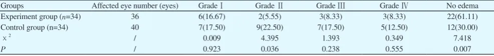
The number of eyes without edema in the experiment group 1d after operation was significantly higher than that in the control group, and the number of eyes with grade Ⅱ edema was significantly less than that in the control group (P<0.05 or P<0.01) (Table 4). After 2 weeks, the corneal endothelial edema was completely vanished.
Table 2.
Comparison of intraocular lens deviation rates after operation [n (%)].
Groups Affected eye number (eyes) 1 d after operation 7 d after operation 1 month after operation 6 months after operation
Experiment group (n=34) 36 9(25.00) 10(27.78) 11(30.56) 12(33.33)
Control group (n=34) 40 12(30.00) 13(32.50) 13(32.50) 15(37.50)
χ2 / 0.237 0.200 0.033 0.144
P / 0.627 0.655 0.856 0.705
Table 1.
Comparison of the number of patients whose uncorrected visual acuity was greater than 0.5 after operation [n (%)].
Groups Affected eye number (eyes) 1 d after operation 7 d after operation 1 month after operation 6 months after operation Experiment group (n=34) 36 18(50.00) 24(66.67) 30(83.33) 31(86.11)
Control group (n=34) 40 19(47.50) 21(52.50) 25(62.50) 26(65.00)
χ2 / 0.047 1.575 4.113 4.504
P / 0.828 0.210 0.043 0.034
Table 3.
Comparison of corneal endothelial cell count before and after operation between the two groups (mean±SD).
Groups Affected eye number
(eyes) Before operation 1 d after operation 7 d after operation
1 month after operation
6 months after operation Experiment group (n=34) 36 2869.4±265.4 2812.4±235.2 2756.4±245.3 2634.1±254.6 2463.4±251.3
Control group (n=34) 40 2863.4±245.8 2801.2±212.4 2735.4±234.6 2659.8±243.7 2415.1±241.3
t / 0.102 0.218 0.381 0.449 0.854
P / 0.919 0.828 0.704 0.654 0.396
Table 4.
Comparison of corneal edema after operation [n (%)].
Groups Affected eye number (eyes) GradeⅠ Grade Ⅱ Grade Ⅲ Grade Ⅳ No edema
Experiment group (n=34) 36 6(16.67) 2(5.55) 3(8.33) 3(8.33) 22(61.11) Control group (n=34) 40 7(17.50) 9(22.50) 7(17.50) 5(12.50) 12(30.00)
χ2 / 0.009 4.395 1.393 0.349 7.418
4. Discussion
Phacoemulsification is the most common method in the treatment of cataract, and can obviously improve the vision in that the lens is smashed by the ultrasound wave and negatively reabsorbed during operation, with small eye surface structure damage, short operation time, and favorable prognosis; therefore, it has achieved an extensive application in the clinic[5,6]. The continuous annular capsulorhexis is commonly adopted in the phacoemulsification. High lens capsular bag elasticity and tension to resist the strong external force, and complete and smooth anterior capsular sac margin are the key requirements for the sequential capsular bag phacoemulsification, but normal lens capsular bag tension and favorable fundus red reflex are the basis for the successful capsulorhexis[7,8]. Due to the complete turbidity of lens cortex, partial cortex decomposition liquidation or absorption, lens nucleus concentration, and hardness increasement, the capsulorhexis sac is easy to be tore in a radial pattern, which can increase the operation difficulty[9-11].
The clinical effect of lens capsules tension reducing capsulorhexis and conventional capsulorhexis in the white cataract phacoemulsification in the study was retrospectively analyzed. Coloring agent was used for dying before capsulorhexis in order to enhance the visuality of anterior capsule[12,13]. Not too much
viscoelastics should be filled the anterior chamber in order to prevent the excessive anterior capsular pressure[14]. By adoption of tension reducing capsulorhexis, the syringe is slowly punctured into the lens capsular bag, slight moving in order to absorb partial liquidation cortex; meanwhile, the lens nucleus is rotated to absorb most liquidation cortex in the capsular bag as much as possible. Moderate speed is adopted during the capsulorhexis process in order to favorably control the anterior capsule direction. The viscoelastics in the anterior chamber should be supplemented at any time in order to maintain the controllability of anterior capsule and enhance the success rate of capsulorhexis[15]. The results in the study showed that phacoemulsification and artificial lens implantation were successfully performed in the two groups; a total of 34 eyes in the experiment group and 35 eyes in the control group successfully completed continuous annular capsulorhexis, and there was no significant difference between the 2 groups (P>0.05); a total of 3 eyes in the experiment group and 5 eyes in the control group had posterior capsule membrane rupture, and the comparison between the two groups was not statistically significant (P>0.05), indicating that the two capsulorhexis methods have a preferable continuous annular capsulorhexis success rate, with no significant difference in the occurrence rate of postoperative capsule rupture.
The mechanical damage and ultrasound factor in the phacoemulsification can cause the reduction of corneal endothelial function, corneal edema, and even the postoperative corneal endothelial decompensation, which can affect the visual effect[16]. Some researches demonstrate that[17] the endothelial loss rate of conventional phacoemulsification is 6%-11%. After successful tension reducing capsulorhexis, the routine two-hand interception separation technology is adopted for emulsification. An auxiliary instrument is used to pick up the nucleus which is slightly leaned. The lens nucleus is emulsified from the corresponding opposite side. The nucleus is divided into several fragments and gradually extracted. The mechanical fragmentation nucleus is increased during operation, and the phacoemulsification energy is reduced. During operation, the fragmentation nucleus far from the corneal endothelial surface can reduce the phacoemulsification energy and the damage of liquid flow and lens fragmentation nucleus on the endothelial cells, effectively decrease the corneal endothelial cell loss and the edema degree of affected eye, and improve the visual function. The results in the study showed that the corneal endothelial cell count 1 d-6 months after operation in the 2 groups was significantly decreased when compared with before operation, but the difference between the 2 groups was not significant (P>0.05), indicating that the two capsulorhexis methods can partially affect the postoperative corneal endothelial function. The results in the study showed that the number of patients whose uncorrected visual acuity was greater than 0.5 increased 1 d-6 months after operation, and that in the experiment group was significantly higher than that in the control group (P<0.05); the difference of intraocular lens deviation rates between the 2 groups at different times after operation was not significant (P>0.05); the number of eyes without edema in the experiment group 1d after operation was significantly higher than that in the control group, and the number of eyes with grade Ⅱ
edema was significantly less than that in the control group (P<0.05 or P<0.01); the corneal endothelial edema 2 weeks after operation in the two groups was completely vanished, indicating that the tension reducing capsulorhexis can significantly improve the vision, alleviate the clinical symptoms, and reduce the corneal edema when compared with the conventional capsulorhexis, but there is no significant difference in the intraocular lens deviation rates by the two methods.
In conclusion, application of cataract phacoemulsification in
References
[1] Shenjie L, Mingxi S, Jian J. Correlation analysis of clinical biochemical
indicators in middle-aged patients with cataract. Chin J Lab Med 2016;
39(6): 448-453.
[2] Yujuan W, Fang C, Jing M. Effect evaluation of meditation therapy
applied in cataract patients. Chin J Nurs2016; 51(3): 321-325.
[3] Linbi X, Yi S, Min W. Application of basic anesthesia in
phacoemulsification in elder cataract patients with poor compatibility.
Rec Adv Ophthal 2015; 35(3): 243-245.
[4] Tao W. Observation on the clinical effect of Beifushu eye drops in
combined with Dianbishu eye drops in the treatment of corneal edema
after cataract phacoemulsification. Zhejiang J Traum Surg 2016; 21(1): 51-53.
[5] Gogate P, Optom JJB, Deshpande S. Meta-analysis to compare the
safety and efficacy of manual small incision cataract surgery and
phacoemulsification. Middle East Afr J Ophthalmol 2015; 22(3): 362-369. [6] Chen M, Swinney C, Chen M. Comparing the intraoperative
complication rate of femtosecond laser-assisted cataract surgery to
traditional phacoemulsification. Int J Ophthalmol 2015; 8(1): 201-203.
[7] Yinping L, Dandan S, Xinyuan P. Novel T-shaped capsulorrhexis in
phacoemulsification surgery for patients with white cataract. J Wannan
Med Coll 2016; 35(2): 143-145.
[8] Hwang YH, Yong YK, Kirti K. Capsule wrinkling during capsulorhexis
in patients with primary angle-closure glaucoma and cataract. Jpn J
Ophthalmol 2010; 54(5): 401-406.
[9] Yune Z, Ming X, Ayong Y. Application of lens capsules tension
reducing capsulorhexis in the white cataract phacoemulsification.Chin J
Ophthamol2004; 40(1): 56-57.
[10] Lei Z, Hong Z, Fang T. Observation of corneal endothelial cell loss after
femtosecond laser-assisted cataract surgery. Chin J Pract Ophthamol
2014; 32(12): 1433-1436.
[11] Li L, Xu Y. Efficacy analysis of corneal endothelial dysfunction treatment
after cataract operation.J Otolaryngol Ophthalmol Shandong Univ 2016;
30(2): 84-86.
[12] Yu J. Application of capsular tension ring in the micro-incision cataract
phacoemulsification and artificial lens implantation. China Med Pharm
2017; 7(9): 223-225.
[13] Wenlin L, Haibo L, Mudan C. Application of double capsulorhexis in
combined with anterior vitrectomy in the congenital cataract surgery in
infants. Chin J Strabis Pediatr Ophthalmol 2017; 25(2): 1-5.
[14] We i Z , Yo n g Y. Tr e a t m e n t o f hy p e r m a t u r e c a t a r a c t w i t h
phacoemulsification. J Clin Ophthalmol 2017; 25(2): 114-116.
[15] Jia L, Xuan L, Changjun L. Changes of intraocular pressure during
phacoemulsification and its influence. Int Rev Ophthalmol 2017; 41(2): 106-109.
[16] Zhiguo L, Chunli W. Early cataract phacoemulsification surgery for the
treatment of a preliminary study of the primary angle-closure suspect.
Chin J Pract Ophthalmol 2016; 34(2): 156-158.
[17] Dongbin C, Wanjiang D, Wenyong L. Comparison of pre-chop and
stop-and-chop techniques in phacoemulsification of hard-nucleus catarac.