R E S E A R C H
Open Access
EGF-activated PI3K/Akt signalling
coordinates leucine uptake by regulating
LAT3 expression in prostate cancer
Blake K. Zhang
1,2, Anne M. Moran
1,2, Charles G. Bailey
2,3, John E. J. Rasko
2,3,4, Jeff Holst
5,6*†and Qian Wang
2,5*†Abstract
Background:Growth factors, such as EGF, activate the PI3K/Akt/mTORC1 signalling pathway, which regulates a distinct program of protein synthesis leading to cell growth. This pathway relies on mTORC1 sensing sufficient levels of intracellular amino acids, such as leucine, which are required for mTORC1 activation. However, it is currently unknown whether there is a direct link between these external growth signals and intracellular amino acid levels. In primary prostate cancer cells, intracellular leucine levels are regulated by L-type amino acid
transporter 3 (LAT3/SLC43A1), and we therefore investigated whether LAT3 is regulated by growth factor signalling. Methods:To investigate how PI3K/Akt signalling regulates leucine transport, prostate cancer cells were treated with different PI3K/Akt inhibitors, or stable knock down of LAT3 by shRNA, followed by analysis of leucine uptake, western blotting, immunofluorescent staining and proximity ligation assay.
Results:Inhibition of PI3K/Akt signalling significantly reduced leucine transport in LNCaP and PC-3 human prostate cancer cell lines, while growth factor addition significantly increased leucine uptake. These effects appeared to be mediated by LAT3 transport, as LAT3 knockdown blocked leucine uptake, and was not rescued by growth factor activation or further inhibited by signalling pathway inhibition. We further demonstrated that EGF significantly increased LAT3 protein levels when Akt was phosphorylated, and that Akt and LAT3 co-localised on the plasma membrane in EGF-activated LNCaP cells. These effects were likely due to stabilisation of LAT3 protein levels on the plasma membrane, with EGF treatment preventing ubiquitin-mediated LAT3 degradation.
Conclusion:Growth factor-activated PI3K/Akt signalling pathway regulates leucine transport through LAT3 in prostate cancer cell lines. These data support a direct link between growth factor and amino acid uptake, providing a mechanism by which the cells rapidly coordinate amino acid uptake for cell growth.
Keywords:EGF, PI3K/Akt signalling pathway, L-type amino acids transporter 3, LAT3, SLC43A1, Prostate cancer
Background
Binding of growth factors to the extracellular ligand binding domain of their membrane-bound receptors leads to a conformational change of the receptors, thereby activating tyrosine or serine/threonine kinase domains. This activation enables the recruitment of di-verse substrates, propagating signals that mediate a
plethora of cellular activities ultimately leading to cell growth [1]. The uptake and metabolism of extracellular nutrients is one of the most critical cellular activities required to provide the building blocks and energy ne-cessary to produce new cells [2]. While numerous stud-ies have suggested that growth factors can regulate uptake of nutrients, whether by transporters, or by macropinocytosis, a direct link to transport has not yet been established [3–5].
Growth factors and their receptors are commonly in-creased in a variety of cancers, with expression of epi-dermal growth factor (EGF) and its receptor (EGFR) significantly increased in prostate cancer [6]. Binding of
© The Author(s). 2019Open AccessThis article is distributed under the terms of the Creative Commons Attribution 4.0 International License (http://creativecommons.org/licenses/by/4.0/), which permits unrestricted use, distribution, and reproduction in any medium, provided you give appropriate credit to the original author(s) and the source, provide a link to the Creative Commons license, and indicate if changes were made. The Creative Commons Public Domain Dedication waiver (http://creativecommons.org/publicdomain/zero/1.0/) applies to the data made available in this article, unless otherwise stated. * Correspondence:[email protected];[email protected]
†Jeff Holst and Qian Wang contributed equally to this work.
5
Translational Cancer Metabolism Laboratory, Lowy Cancer Research Centre, School of Medical Sciences and Prince of Wales Clinical School, University of New South Wales, Sydney, Australia
2Sydney Medical School, University of Sydney, Camperdown, Australia
EGF to EGFR stimulates downstream signalling path-ways including the mitogen-activated protein kinase (MAPK) and phosphoinositide 3 kinase (PI3K)/Akt pathways. In addition, the PI3K/Akt signalling pathway is commonly activated in cancers, either through activat-ing mutations or inactivation of the tumour suppressor
phosphatase and tensin homolog (PTEN) [7,8]. In
pros-tate cancer, up to 70% patients have PTEN mutation or deletion [9], thereby allowing unconstrained growth fac-tor activated PI3K/Akt signalling, cell proliferation and tumour growth.
The PI3K/Akt signalling axis activates mechanistic tar-get of rapamycin complex 1 (mTORC1) through phos-phorylation, thus negatively regulating tuberous sclerosis complex 1/2 (TSC1/2) formation and releasing Rheb, a GTPase activating protein (GAP), to bind to the kinase domain of mTORC1 on the surface of lysosomes, lead-ing to mTORC1 activation [10]. In addition, intracellular levels of free amino acids, in particular leucine, arginine
and glutamine, regulate mTORC1 activation [11, 12].
Amino acids sufficiency can be sensed by mTORC1 through the interaction between Rag GTPase
heterodi-mers and Ragulator on the surface of lysosomes [13–17].
Sestrin2 has been identified as a leucine sensor upstream of mTORC1 by binding with leucine which is required
for mTORC1 activation [18, 19]. Thus, mTORC1
inte-grates upstream signalling pathways as well as amino acid availability to mediate protein synthesis, cell growth and proliferation.
The supply of intracellular amino acids is mediated by amino acid transporters, which are commonly upregulated in cancer cells. One such transporter, LAT3, mediates up-take of large neutral branched chain amino acids (BCAA)
including leucine. LAT3, encoded by the gene SLC43A1,
was originally named prostate cancer overexpressed gene 1 (POV1) [20], and was later identified as a major unipor-ter of leucine, isoleucine, valine, phenylalanine and me-thionine [21]. Human LAT3 is predicted to contain 12 transmembrane (TM) domains. A long intracellular loop between transmembrane domains 6 and 7 contains puta-tive serine phosphorylation sites and a tyrosine phosphor-ylation site [22]. LAT3 protein expression is high in primary and recurrent prostate cancer, driven by direct
androgen receptor (AR) transcription [23, 24]. Increased
LAT3 levels result in increased intracellular leucine levels
and subsequent activation of mTORC1 [23,24]. Moreover,
shRNA knockdown of LAT3 blocks leucine uptake and cell growth in prostate cancer cell lines both in vitro [23] and in vivo [24].
Since both the growth factor-activated PI3K/Akt sig-nalling pathway and amino acid transporters are re-quired for mTORC1 activation, we investigated whether there were any direct links between PI3K/Akt signalling and LAT3 activity. In this study, we show that
EGF-activated PI3K/Akt signalling directly regulates leucine transport in prostate cancer cells by stabilising LAT3 ex-pression on the cell surface. These data suggest that there is a coupling of growth signals to amino acid up-take in prostate cancer, which may provide new avenues to control cancer cell growth, and should be examined across a range of transporters and cancer types.
Materials and methods
Cell lines
Human prostate cancer cell lines LNCaP-FGC and PC-3 were purchased from ATCC (Manassas, VA, USA). We used low passage original stocks, and confirmed LNCaP and PC-3 cell identity by short tandem repeat profiling in 2014 (Cellbank, Australia). Cells are tested monthly by PCR to ensure they are free of mycoplasma contam-ination. Cells were cultured in RPMI 1640 medium (Invitrogen) containing 10% (v/v) fetal bovine serum (FBS), penicillin-streptomycin solution (Sigma-Aldrich) and 1 mM sodium pyruvate (Invitrogen). Cells were maintained at 37 °C in a fully humidified atmosphere
containing 5% CO2.
Leucine uptake assay
The [3H]-L-leucine uptake was performed as detailed
previously [23]. Briefly, cells were cultured in 6-well plates in RPMI media. After serum starvation for 2 h, cells (3 × 10⁴/well) were incubated with 0.3μCi [3 H]-L-leucine (200 nM; PerkinElmer) in H]-L-leucine-free RPMI media (Invitrogen) in the absence or presence of EGF (10 ng/mL, Sapphire Bioscience) or dialyzed FBS, or
in-hibitors, including MK2206 (10μM, Selleck Chemicals),
LY294002 (20μM, Merck), 2-amino-bicyclo [2,2,1]
hepta-2-carboxylic acid (BCH, 10 mM, Sigma),
rapamy-cin (50 nM, Merck), JPH203 (10μM, Selleck Chemicals),
for the required time at 37 °C. Cells were collected, transferred to filter paper using a MicroBeta FilterMat-96 Cell Harvester (PerkinElmer), dried, exposed to scin-tillation fluid and counts were measured using a
MicroBeta2liquid scintillation counter (PerkinElmer).
Western blot
Cells were seeded at a density of 5 × 10⁵in 6-well plates, allowed to adhere overnight, before incubation with EGF and/or inhibitors, MK2206, LY294002, BCH, rapamycin,
MG132 (50μM, Sigma) for required time. Cells were
lysed by addition of lysis buffer (200μL) with
EDTA-free Protease Inhibitor Cocktail III (cOmplete™; Roche
albumin (BSA, Sigma-Aldrich) in PBS containing 0.1% (v/ v) Tween 20 (0.1% PBS-T), and incubated with the pri-mary antibodies overnight. The next day, membrane was washed with 0.1% PBS-T three times and incubated with secondary antibodies. After washing, the membrane was visualised using enhanced chemiluminescence reagents
(ECL, Pierce™, Thermo Scientific) on a ChemiDoc™
TOUCH imager (Bio-Rad). Antibodies used in this study
include phospho-P70S6K (Thr398, #9205, 1:1000),
P70S6K (#9202, 1:1000), phospho-Akt (Ser473, #4051, 1: 1000), Akt (#9272, 1:1000), Na, K-ATPase (#3010, 1:1000) from Cell Signalling Technology (CST); ubiquitin (ab7780,
1:1000), glyceraldehyde-3-phosphate dehydrogenase
(GAPDH, ab8245, 1:2000) from Abcam. LAT3 rabbit polyclonal antibody was generated against a peptide (TGGKERETNEQRQ) from mouse LAT3 (The Institute of Medical and Veterinary Science, Adelaide, Australia) and affinity purified from rabbit serum [22]. Horseradish peroxidase (HRP)-conjugated donkey anti-mouse IgG (AP192P, 1:5000), and donkey anti-rabbit IgG (AP182P, 1: 5000) from Millipore were used as secondary antibodies. Densitometry of western blots was analysed by ImageJ (NIH).
Immunofluorescent staining
Cells were seeded on BD Falcon™ culture slides (Becton
Dickinson) at a density of 1 × 10⁴cells/well and allowed
to adhere overnight. Fresh media was added, and the cells were incubated for 24 h before fixation using 4% (w/v) paraformaldehyde (Sigma-Aldrich) for 20 min, and permeabilisation using 0.1% PBS-T for 15 min. Cells were washed and incubated with 5% (v/v) normal goat serum in 2% (w/v) BSA/PBS for 30 min before addition of the LAT3 antibody at 4 °C for overnight incubation. The cells were washed in PBS before addition of a goat anti-rabbit IgG conjugated with AlexaFluor 594 (Invitro-gen, A12381) for LAT3 antibody or a goat anti-mouse
IgG conjugated with AlexaFluor 488 (Invitrogen,
A12379) for pAkt antibody for 1 h at room temperature,
and nuclear staining with DAPI (5μg/mL, Thermo
Fisher) for 5 min. Cells were washed in PBS, and cover-slips were immersed in glycerol and placed on a slide, and visualized using a DeltaVision confocal microscope (GE Healthcare Life Science).
Proximity ligation assay (PLA)
Cells were seeded on coverslip which has been immersed in poly-L-lysine (Sigma-Aldrich) at a density
of 5 × 10⁵ cells/well and allowed to adhere overnight.
Cells were incubated in serum free media for 30 min,
followed by EGF (10 ng/μL) treatment for 30 min. The
cells were fixed using 4% (w/v) paraformaldehyde for 20 min, and permeabilisation using 0.1% PBS-T for 15 min. Cells were washed and incubated with 5% (v/v) normal
goat serum in 5% (w/v) BSA/PBS for 30 min before addition of the rabbit LAT3 antibody (1:50) and mouse pAkt antibody (1:50) at 4 °C overnight. For negative con-trol, mouse pAkt antibody and rabbit IgG (Santa Cruz, sc-2027) or rabbit LAT3 antibody and mouse IgG (Santa Cruz, sc-2025) were added (1:50). As a positive control, pAkt and Akt antibody (1:50) were added. PLA was
performed using the DUOlink™ kit (OLINK, Uppsala,
Sweden) following the manufacturer’s instruction. The
cells were visualized using a DM6000B microscope (Leica, Germany). Quantification of PLA signals was
measured using Volocity software (Version 6.3,
PerkinElmer).
Cell surface protein isolation
LNCaP cells were cultured in T75 flasks with RPMI 1640 medium containing 10% FBS, penicillin-streptomycin
solution and 1 mM sodium pyruvate to reach 90–95%
confluence. LNCaP cells were incubated with serum free
media for 2 h, followed by EGF (10 ng/μL) treatment for
30 min. Surface protein isolation was performed using the
Pierce™Cell Surface Protein Isolation Kit (Thermo
Scien-tific) following the manufacturer’s instructions. Briefly, cells were biotin-labelled, harvested, and lysed, and NeutrAvidin resin was used to isolate the protein. Protein
was eluted with SDS-PAGE sample buffer (NuPAGE™Life
Technologies) containing 50 mM 1,4-dithiothreitol (DTT, Sigma-Aldrich). The eluate was analysed by SDS-PAGE and western blot.
Immunoprecipitation
LNCaP cells were seeded at a density of 5 × 10⁵in 6-well
plates and allowed to adhere overnight. The next day,
cells were pre-treated with MK2206 or MG132 (50μM,
Merck) in serum free RPMI 1640 media for 30 min,
before addition of EGF (10 ng/μL) to each well for 30
min. Cells were lysed by addition of lysis buffer with Protease Inhibitor Cocktail and Phosphatase Inhibitor Cocktail. Protein concentration of cell lysates was deter-mined by the micro-BCA method. Cell lysates were
incubated with magnetic protein A/G beads (Surebeads™
Bio-Rad) coupled with LAT3 antibody or rabbit IgG at 4 °C overnight. Protein-bound beads were washed with E1A buffer (50 mM HEPES, 150 mM NaCl, 0.1% NP-40,
1 mM Na3VO4) three times. Protein was eluted with
sample buffer (NuPAGE™, Life Technologies) containing
report for other transporters [25], which may be due to altered conformation of the antibody binding sites on the antigen due to detergents in the immunoprecipita-tion buffer [26].
Knockdown of LAT3
LAT3 shRNA knockdown was performed as previously described [23]. Briefly, empty vector pLKO.1 or contain-ing short hairpin (shRNA) targetcontain-ing LAT3 (CCGGCA AATCCATCAGACCACGCTACTCGAGTAGCGTGGT CTGATGGATTTGTTTTTG) was mixed with pMDLg/ prre, pRSVRev and pMD2.VSV-G packaging plasmids and the calcium phosphate precipitation method used to transfect 70% confluent HEK293T cells [23]. After 8 h, the media was changed, and viral supernatant was col-lected and filtered 24 h later. The viral supernatant was
used to transduce 70% confluent LNCaP with 8μg/mL
polybrene. After 2 days, transduced cells were selected
by 10μg/mL puromycin for 7 days. Expression of the
LAT3 protein was determined using SDS-PAGE and western blot as described above.
Statistical analysis
Data are shown as mean ± SEM. All experiments were performed with at least 3 biological replicates. All
exper-iments were analysed by two-tailed student’s t-test or
two-way ANOVA or one-way ANOVA in GraphPad Prism 6.
Results
PI3K/Akt signalling pathway regulates leucine transport To model the effects of growth factors on prostate can-cer, we utilized the androgen-sensitive prostate adeno-carcinoma cell line LNCaP, which contains one deleted allele and one mutated allele of PTEN [27], and the an-drogen-insensitive cell line PC-3, which has a
homozy-gous deletion of PTEN [27, 28], leading to the
hyperactivation of the PI3K/Akt pathway. Leucine up-take is predominantly mediated by LAT3 in LNCaP cells, while PC-3 cells rely on both LAT3 and LAT1 (SLC7A5) for leucine uptake [23]. To investigate how PI3K/Akt signalling affects LAT3-mediated leucine uptake in the presence of serum, we utilized a PI3K inhibitor LY294002, an Akt inhibitor MK2206, an L-type amino acid transporter inhibitor BCH and a LAT1 selective
inhibitor JPH203 [29–32]. In LNCaP cells, leucine
trans-port was inhibited to 59% of control in the presence of LY294002, 67% of control with MK2206 and 54% of con-trol with JPH203, whereas BCH reduced leucine uptake
to 24% of control (Fig. 1a). In PC-3 cells, the leucine
transport was reduced to 81% of control by LY294002, and 94% of control by MK2206, which exhibited less in-hibitory effect compared to BCH (12% of control) and
JPH203 (6% of control; Fig. 1b), consistent with the
dominant role of LAT1 in mediating leucine uptake in PC-3 cells [23]. Western blots showed that both LY294002 and MK2206 reduced Akt phosphorylation (pS473-Akt) in LNCaP and PC-3 cells after 30 min
A
B
C
D
treatment. The LAT inhibitors, BCH and JPH203, had
no effect on Akt phosphorylation (Fig. 1c, d). These
re-sults suggest that PI3K/Akt signalling pathway regulates leucine uptake, which is more likely through LAT3 ra-ther than LAT1.
LAT3 is required for PI3K/Akt regulated leucine uptake To determine if LAT3 is the target of PI3K/Akt signal-ling, we used shRNA to knock down LAT3 expression in LNCaP cells, which have higher LAT3 expression compared to PC-3 cells [23]. Western blots showed that LAT3 expression levels were decreased in LNCaP
shLAT3 comparing to shControl (Fig. 2a). Leucine
up-take assays were performed in both LNCaP shControl and shLAT3 cells. RPMI media supplemented with dia-lyzed FBS was used as control, and was compared to serum-free media, or addition of MK2206 or BCH. Dia-lyzed FBS contains normal level of growth factors but low levels of amino acids. In shControl cells, leucine up-take was reduced in serum-free media, MK2206 and
BCH treatment groups (Fig.2b). Under the same
condi-tions, knockdown of LAT3 in LNCaP cells showed a sig-nificant reduction in leucine uptake equivalent to 16% of
control in shControl cells (Fig. 2b), suggesting that
LAT3 has been efficiently knocked down in LNCaP cells. Serum depletion and inhibition by MK2206 for shLAT3 cells showed no significant difference in leucine uptake
compared to control (Fig. 2b), indicating that these
sig-nalling pathways activate leucine uptake in a
LAT3-dependent manner (Fig.2b).
EGF stimulation regulates leucine uptake
To specifically test whether EGF mediates activation of PI3K/Akt signalling pathway and leucine uptake, EGF treatment was carried out on LNCaP and PC-3 cells. EGF stimulation for 5, 15 or 30 min significantly in-creased leucine uptake in LNCaP cells by 30%, 33% and
79%, respectively (Fig. 3a). In PC-3 cells, there was no
change in leucine uptake at either 5 or 15 min post EGF, however, there was a significant increase after 30 min
(Fig. 3b). To determine whether this corresponded with
downstream activation of signalling from the EGF recep-tor, we next examined the level of phosphorylated Akt (pS473-Akt) over the same time course. In LNCaP cells, Akt phosphorylation increased by 5 min, and remained
at similar levels between 15 min and 30 min (Fig. 3c).
PC-3 cells also rapidly increased Akt phosphorylation at 5 min, however, this phosphorylation was downregulated
by 15 and 30 min post stimulation (Fig.3d).
Leucine transport is dependent on EGF-activated PI3K/Akt signalling
EGF stimulates the PI3K/Akt signalling pathway, subse-quently activating mTORC1 signalling and driving pro-tein synthesis. To determine which kinase in the PI3K/ Akt/mTORC1 pathway axis was involved, we tested a PI3K inhibitor, LY294002, and an mTORC1 inhibitor, rapamycin, in combination with EGF treatment. As ex-pected, 30 min pre-incubation with LY294002 blocked the activation of both Akt and downstream P70S6K
(Fig. 4a, b) signalling, while rapamycin treatment
de-creased phosphorylation of P70S6K (Fig. 4a, b), but had
no effect on upstream Akt phosphorylation. In addition, while LY294002 treatment significantly reduced leucine
transport in both LNCaP and PC-3 cells (Fig. 4c, d),
rapamycin showed no effect on leucine uptake in either
cell line (Fig. 4c, d). These data suggest that the PI3K/
Akt signalling pathway can regulate leucine transport, and this occurs upstream of mTORC1 signalling.
EGF stimulation promotes co-localisation of LAT3 with pAkt
To determine the relationship between PI3K/Akt signal-ling and LAT3, we examined the co-localisation of
A
B
Fig. 2LAT3 is required for PI3K/Akt stimulated leucine uptake.aLAT3 expression levels were examined by western blot in shControl and shLAT3 of LNCaP cells. All western blot images are representative of three independent experiments.bleucine uptake was examined in RPMI
phosphorylated Akt and LAT3 using confocal micros-copy in LNCaP, as LAT3 is highly expressed in LNCaP cells but not in PC-3 cells. LAT3 was strongly expressed at the plasma membrane of cells that were in contact with the extracellular environment, but showed lower expression in areas of cell-cell contact (Fig.5a). By con-trast, pAkt was detected in the majority of the plasma
membrane of LNCaP cells (Fig. 5a). The merged image
showed a number of areas of co-localisation of LAT3
and pAkt (Fig. 5a, arrows), as well as distinct areas of
single staining of each protein/phosphoprotein.
To confirm these confocal microscopy data, we next performed a proximity ligation assay (PLA) to determine whether pAkt and LAT3 were closely associated in LNCaP cells. To demonstrate the value of this tech-nique, mouse pAkt and rabbit Akt antibodies bound to different antigens on the same target, resulted in a high
co-localisation signal (Fig. 5b). Next, we used rabbit IgG
with mouse pAkt antibody or rabbit LAT3 antibody with mouse IgG as negative controls to establish background
PLA signals (Fig. 5b). LNCaP cells demonstrate a low
level basal amount of PLA signal with LAT3 and pAkt, however after EGF treatment the PLA signals of LAT3
and pAkt were dramatically enhanced (Fig. 5b, c),
indi-cating that EGF enhanced the co-localisation of phos-phorylated Akt and LAT3. Given the fact that PLA
resolution is within 40 nm [33,34] pAkt and LAT3 were
in close proximity and they may form a multi-protein complex in response to the extracellular signals as well as to increase amino acid uptake required for cell division.
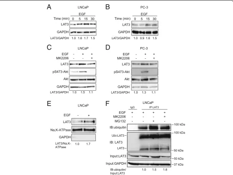
EGF stimulation enhanced surface expression of LAT3 To determine the mechanism of PI3K/Akt regulated leu-cine transport via LAT3, we examined LAT3 expression by western blots in the presence of EGF. LAT3 protein levels were elevated within 5 min of EGF treatment and
maintained for 30 min in LNCaP cells (Fig. 6a). In PC-3
cells, LAT3 protein levels increased at 15 min and
main-tained this increase for 30 min (Fig. 6b), indicating that
EGF-induced leucine uptake may result from upregulated LAT3 surface expression. This late elevation of LAT3 in PC-3 cells might explain why changes in leucine uptake are delayed, although this may also be due to physical dif-ferences in the analysis of adherent (western blot) and
sus-pension (uptake assay) cells (Fig. 3b). As LAT1 is the
major transporter in PC-3 cells [23], we examined LAT1 expression after EGF treatment. LAT1 protein levels did not change after 30 min EGF treatment, suggesting that LAT1 is not involved in the EGF-stimulated upregulation
of leucine transport (Additional file 1: Figure S1A). To
confirm this, we next examined EGF stimulation of leu-cine transport in the presence of the LAT1 inhibitor
JPH203 (Additional file 1: Figure S1B). Despite blocking
A
B
C
D
A
B
C
D
Fig. 4Leucine transport is dependent on EGF-stimulated PI3K/Akt signalling.aandb, Akt and P70S6K activation was examined in the presence of rapamycin and LY294002 in LNCaP (a) or PC-3 cells (b). GAPDH was used as loading control. All western blot images are representative of three independent experiments.candd, leucine transport was examined in the presence of rapamycin or LY294002 in LNCaP (c) and PC-3 cells (d). Two-tailed student’s t-test was performed. Data are the mean ± SEM,n= 3
A
B
C
LAT1-mediated leucine transport, JPH203 had no effect on the EGF-stimulated leucine uptake, confirming that LAT1 does not play a role in EGF stimulation of PC-3 cells. Importantly, the Akt inhibitor MK2206 suppressed Akt phosphorylation as well as LAT3 expression even in
the presence of EGF in LNCaP and PC-3 cells (Fig.6c, d),
suggesting EGF-activated Akt is required for increased LAT3 expression.
Since LAT3 needs to be stabilized at the plasma mem-brane to fulfil its transport function, the expression level at cell surface is critical in response to EGF stimulation. Cell surface proteins were isolated and LAT3 was exam-ined by western blot in the presence or absence of EGF. After 30 min treatment with EGF, LAT3 expression was increased 1.7-fold at the plasma membrane compared to
control (Fig.6e and Additional file1: Figure S1C), suggest-ing that LAT3 levels were increased at the cell surface. To determine whether EGF treatment affects either LAT3 synthesis or LAT3 degradation, we next treated EGF-stim-ulated cells with MK2206 or a proteasome inhibitor MG132, which reduces the ubiquitin-mediated degrad-ation of protein in the proteasome. After 30 min MG132 and MK2206 treatment, ubiquitinated LAT3 (~ 90 kDa) was increased 1.5-fold and 1.8-fold in the anti-LAT3
immunoprecipitates respectively (Fig. 6f). In addition, we
showed that ubiquitinated LAT3 (~ 90 kDa) is decreased after EGF treatment in both LNCaP and PC-3 cells
(Additional file1: Figure S1D, E). After MG132 treatment,
unmodified LAT3 protein levels (~ 55 kDa) are increased
compared to control (Additional file1: Figure S1F). These
A
B
C
D
E
F
results suggest that inhibition of EGF-activated PI3K/Akt signalling may induce ubiquitin-mediated degradation of LAT3.
Discussion
Rapidly proliferating cancer cells require sustained growth factor stimulation and more nutrient supply for protein synthesis and cell mass accumulation [2]. Growth factors, such as EGF, activate multiple downstream signalling pathways and have an important role in cancer progres-sion, including proliferation, invasion and migration [35]. Its downstream PI3K/Akt signalling, and amino acids are required for mTORC1 to regulate protein synthesis. In this study, we firstly reported that EGF-activated PI3K/ Akt signalling pathway regulates leucine uptake through the amino acid transporter LAT3 in prostate cancer.
Cells have many ways of uptaking nutrients that are available in the extracellular matrix, such as transporter-based nutrient uptake, receptor-mediated endocytosis, macropinocytosis, as well as entosis [36]. The transport of majority extracellular amino acids relies on their mem-brane-bound transporters due to their hydrophilicity, and the expression of transporters is highly upregulated in many cancer types, such as Alanine-Serine-Cysteine Transporter 2 (ASCT2/SLC1A5) in triple negative breast
cancer, prostate cancer and melanoma [37–39], LAT1
(SLC7A5) in endometrial cancer [40], ATB0,+(SLC6A14)
in colorectal cancer [41], xCT (SLC7A11) in glioma [42]. Growth factor signalling pathways play an instructive role in regulating nutrient transporters via PI3K/Akt, which in-duces the phosphorylation and translocation of glucose transporter 1 (GLUT1) to cell surface to increase glucose
uptake upon growth factor stimulation [43,44]. PI3K/Akt
signalling also affects transcription factor MYC, which in-duces the expression of glutamine transporters ASCT2 and SNAT5 and promotes glutaminolysis to provide glu-tamine carbon for the TCA cycle in response to growth
factor [45, 46]. A recent study has showed that active
MYC could preferentially upregulate LAT3 among many other SLC transporters in neuroblastoma cells, and that inhibition of LAT3 would in turn downregulate MYC mRNA levels [47]. In this study, we have observed that total and surface LAT3 protein levels are increased within
5–30 min after EGF stimulation. When LAT3 is knocked
down in LNCaP cells, leucine transport becomes less sen-sitive to the stimulation or inhibition of the signalling pathway. Importantly, ubiquitinated LAT3 levels (~ 90 kDa) are increased after inhibition PI3K/Akt signalling, suggesting that EGF-activated PI3K/Akt signalling may affect the protein stabilisation or localisation of LAT3 to maintain its activity, therefore regulating leucine uptake. Activated Akt has been shown to be able to maintain the surface expression of other transporters [48], which is consistent with what we observed in this study.
Akt, as a major effector in PI3K/Akt signalling axis, has an important role in activating mTORC1 signalling. Recent studies have shown that inhibition of mTORC1 signalling increases ubiquitin ligase Nedd4-2 expression, therefore upregulating ubiquitination of SNAT2 and LAT1 transporters in primary human trophoblast cells [25]. However, our data clearly show that LAT3-mediated leucine transport does not rely on downstream mTORC1 signalling, but instead is regulated by Akt or other up-stream molecules. Akt contains a Pleckstrin homology (PH) domain which binds to phosphoinositides on the plasma membrane [49], thereby allowing it to phosphoryl-ate several multi-transmembrane proteins, such as G
pro-tein coupled receptor EDG-1 [50], and Na+/H+exchanger
SLC9A1 (NHE1) [51]. Our data showed that phosphory-lated Akt co-located with LAT3 at the plasma membrane of LNCaP cells. Although mass spectrometry data have shown that LAT3 possesses multiple phosphorylation sites at Y251, S262 and S267, and one ubiquitination site at
K264 within its transmembrane domains [52–54], we have
no direct evidence to show that Akt binds and phosphory-lates LAT3 directly. It is possible that LAT3 and Akt are part of a larger membrane-localised protein complex formed after EGF stimulation. Subsequent Akt activation in that complex may induce another kinase to phosphoryl-ate LAT3 at residues S262 or S267. This may then inhibit K264 ubiquitination, thus permitting LAT3 stabilisation at the plasma membrane.
Our study emphasizes a central role of growth factor activated PI3K/Akt signalling in response to environ-ment changes. With growth factor stimulation, PI3K/ Akt signalling increased stabilisation of the LAT3 trans-porter and its localisation on the plasma membrane, thereby enhancing leucine transport. Elevated intracellu-lar levels of leucine are then available for mTORC1 sig-nalling activated by PI3K/Akt sigsig-nalling. At low level growth factor-stimulation, our data suggest that more LAT3 will be degraded and less proteins recycled to the plasma membrane, resulting in decreased leucine trans-port and protein synthesis. This would be an economic strategy for cells, while still allowing rapid upregulation of leucine transport upon growth factor stimulation.
Conclusion
This is the first study to show that growth factor-acti-vated PI3K/Akt signalling pathway regulates leucine transport through LAT3 in prostate cancer cell lines. These data support a direct link between growth factor and amino acid uptake, providing a mechanism by which the cells rapidly coordinate amino acid uptake for cell growth. Previous studies have shown that LAT3 protein expression is increased in the primary and recurrent prostate cancer patient samples and regulates cancer cell
pancreas and muscle cells [22]. Leucine is critical for
in-sulin secretion in pancreatic β cells [55]. LAT3 is also
important for podocyte development and function in kidney [56]. Therefore, our study on the regulatory mechanisms of LAT3 is important for understanding the metabolism of leucine across many systems, and poten-tially for developing novel cancer therapies targeting the LAT3 transporter.
Additional file
Additional file 1:Figure S1.A, LAT1 expression after EGF treatment in PC-3 cells. GAPDH was used as loading control. B, leucine uptake in PC-3 cells after JPH203 treatment in the absence or presence of EGF for 30 min. Two tailed Student’s t-test was performed. Data are the mean ± SEM,
n= 4. C, GAPDH is examined in cell surface fraction in LNCaP cells. Cell lysates of LNCaP cells are used as positive control. D and E, Ubiquitin and LAT3 were examined after immunoprecipitation with anti-LAT3 in the presence or absence of EGF in LNCaP (D) and PC-3 (E) cells. Ratio of IB: ubiquitin expression level is shown relative to Input: LAT3. F, LAT3 expression levels were examined in the absence or presence of MG132 in EGF treated LNCaP or PC-3 cells. GAPDH was used as loading control. Protein expression levels were normalised to loading control. (PDF 1131 kb)
Abbreviations
BCAA:Branched chain amino acid; EGF: Epidermal growth factor; EGFR: Epidermal growth factor receptor; GAP: GTPase activating protein; LAT3: L-type amino acid transporter 3; MAPK: Mitogen-activated protein kinase; mTORC1: Mechanistic target of rapamycin complex 1; PBS: Phosphate buffered saline; PI3K: Phosphoinositide 3 kinase; PIP2: Phosphatidylinositol (3,4)-bisphosphate; PIP3: Phosphatidylinositol (3,4,5)-trisphosphate; PLA: Proximity ligation assay; PTEN: Phosphatase and tensin homolog; SDS-PAGE: Sodium dodecyl sulfate polyacrylamide gel electrophoresis; SLC43A1: BCH: 2-amino-bicyclo [2,2,1] hepta-2-carboxylic acid; TSC1/ 2: Tuberous sclerosis complex 1/2
Acknowledgements
We thank Dr. Amy Marshall (PLA) and Dr. Kristina Jahn (confocal imaging) at Centenary Institute for technical assistance.
Authors’contributions
JH, QW participated in the design of the study and data analysis. BKZ, JH, QW wrote the manuscript. BKZ, AMM, QW carried out the assays and analysis. CGB, JEJR participated in data analysis. All authors read and approved the final manuscript.
Funding
The authors acknowledge the Movember Foundation through the Prostate Cancer Foundation of Australia (YI0813 to Q.W.; PG2910 to J.H.; YI0707 to J.H.) and the Movember Revolutionary Team Award Targeting Advanced Prostate Cancer (Q.W. and J.H.), Cancer Council NSW (RG15-04 to J.H.), the University of Sydney (Sydney Medical School Ph.D. ECR supervisor grant, Q.W.) and Tour de Cure project grants (C.B. and J.R.).
Availability of data and materials
All data generated or analyzed during this study are included in this published article.
Ethics approval and consent to participate
Not applicable.
Consent for publication
All authors have read this manuscript and approved for the submission.
Competing interests
The authors declare that they have no competing interests.
Author details
1Centenary Institute, University of Sydney, Camperdown, Australia.2Sydney
Medical School, University of Sydney, Camperdown, Australia.3Gene & Stem
Cell Therapy Program Centenary Institute, University of Sydney, Camperdown, Australia.4Cell and Molecular Therapies, Royal Prince Alfred
Hospital, Camperdown, Australia.5Translational Cancer Metabolism
Laboratory, Lowy Cancer Research Centre, School of Medical Sciences and Prince of Wales Clinical School, University of New South Wales, Sydney, Australia.6Origins of Cancer Program Centenary Institute, University of
Sydney, Camperdown, Australia.
Received: 20 February 2019 Accepted: 16 July 2019
References
1. Witsch E, Sela M, Yarden Y. Roles for growth factors in cancer progression. Physiology (Bethesda). 2010;25(2):85–101.
2. Hosios AM, Hecht VC, Danai LV, Johnson MO, Rathmell JC, Steinhauser ML, et al. Amino acids rather than glucose account for the majority of cell mass in proliferating mammalian cells. Dev Cell. 2016;36(5):540–9.
3. Yoshida S, Pacitto R, Yao Y, Inoki K, Swanson JA. Growth factor signaling to mTORC1 by amino acid-laden macropinosomes. J Cell Biol. 2015;211(1):159–72. 4. Palm W, Park Y, Wright K, Pavlova NN, Tuveson DA, Thompson CB. The
utilization of extracellular proteins as nutrients is suppressed by mTORC1. Cell. 2015;162(2):259–70.
5. v Torres-Zamorano, Leibach FH, Ganapathy V. Tyrosine phosphorylation-and epidermal growth factor-dependent regulation of the sodium-coupled amino acid transporter B0 in the human placental choriocarcinoma cell line JAR. Biochim Biophys Acta 1997;1356(3):258–270.
6. De Miguel P. Royuela, Bethencourt R, Ruiz a, Fraile B, Paniagua R. Immunohistochemical comparative analysis of transforming growth factor alpha, epidermal growth factor, and epidermal growth factor receptor in normal, hyperplastic and neoplastic human prostates. Cytokine. 1999;11(9):722–7.
7. Dibble CC, Cantley LC. Regulation of mTORC1 by PI3K signaling. Trends Cell Biol. 2015;25(9):545–55.
8. Manning BD, Cantley LC. AKT/PKB signaling: navigating downstream. Cell. 2007;129(7):1261–74.
9. Chen Z, Trotman LC, Shaffer D, Lin HK, Dotan ZA, Niki M, et al. Crucial role of p53-dependent cellular senescence in suppression of Pten-deficient tumorigenesis. Nature. 2005;436(7051):725–30.
10. Long X, Lin Y, Ortiz-Vega S, Yonezawa K, Avruch J. Rheb binds and regulates the mTOR kinase. Curr Biol. 2005;15(8):702–13.
11. Wise DR, Thompson CB. Glutamine addiction: a new therapeutic target in cancer. Trends Biochem Sci. 2010;35(8):427–33.
12. Hara K, Yonezawa K, Weng QP, Kozlowski MT, Belham C, Avruch J. Amino acid sufficiency and mTOR regulate p70 S6 kinase and eIF-4E BP1 through a common effector mechanism. J Biol Chem. 1998;273(23):14484–94. 13. Han JM, Jeong SJ, Park MC, Kim G, Kwon NH, Kim HK, et al. Leucyl-tRNA
synthetase is an intracellular leucine sensor for the mTORC1-signaling pathway. Cell. 2012;149(2):410–24.
14. Bonfils G, Jaquenoud M, Bontron S, Ostrowicz C, Ungermann C, De Virgilio C. Leucyl-tRNA synthetase controls TORC1 via the EGO complex. Mol Cell. 2012;46(1):105–10.
15. Sheen JH, Zoncu R, Kim D, Sabatini DM. Defective regulation of autophagy upon leucine deprivation reveals a targetable liability of human melanoma cells in vitro and in vivo. Cancer Cell. 2011;19(5):613–28.
16. Sancak Y, Bar-Peled L, Zoncu R, Markhard AL, Nada S, Sabatini DM. Ragulator-rag complex targets mTORC1 to the lysosomal surface and is necessary for its activation by amino acids. Cell. 2010;141(2):290–303. 17. Kim E, Goraksha-Hicks P, Li L, Neufeld TP, Guan KL. Regulation of TORC1 by
rag GTPases in nutrient response. Nat Cell Biol. 2008;10(8):935–45. 18. Saxton RA, Knockenhauer KE, Wolfson RL, Chantranupong L, Pacold ME,
Wang T, et al. Structural basis for leucine sensing by the Sestrin2-mTORC1 pathway. Science. 2016;351(6268):53–8.
19. Wolfson RL, Chantranupong L, Saxton RA, Shen K, Scaria SM, Cantor JR, et al. Sestrin2 is a leucine sensor for the mTORC1 pathway. Science. 2016; 351(6268):43–8.
21. Babu E, Kanai Y, Chairoungdua A, Kim DK, Iribe Y, Tangtrongsup S, et al. Identification of a novel system L amino acid transporter structurally distinct from heterodimeric amino acid transporters. J Biol Chem. 2003;278(44):43838–45. 22. Fukuhara D, Kanai Y, Chairoungdua A, Babu E, Bessho F, Kawano T, et al.
Protein characterization of NA+-independent system L amino acid transporter 3 in mice: a potential role in supply of branched-chain amino acids under nutrient starvation. Am J Pathol. 2007;170(3):888–98. 23. Wang Q, Bailey CG, Ng C, Tiffen J, Thoeng A, Minhas V, et al. Androgen
receptor and nutrient signaling pathways coordinate the demand for increased amino acid transport during prostate cancer progression. Cancer Res. 2011;71(24):7525–36.
24. Wang Q, Tiffen J, Bailey CG, Lehman ML, Ritchie W, Fazli L, et al. Targeting amino acid transport in metastatic castration-resistant prostate cancer: effects on cell cycle, cell growth, and tumor development. J Natl Cancer Inst. 2013;105(19):1463–73.
25. Rosario FJ, Dimasuay KG, Kanai Y, Powell TL, Jansson T. Regulation of amino acid transporter trafficking by mTORC1 in primary human trophoblast cells is mediated by the ubiquitin ligase Nedd4-2. Clin Sci (Lond). 2016;130(7):499–512. 26. Lee YC, Baath JA, Bastle RM, Bhattacharjee S, Cantoria MJ, Dornan M, et al. Impact of detergents on membrane protein complex isolation. J Proteome Res. 2018;17(1):348–58.
27. Vlietstra RJ, van Alewijk DC, Hermans KG, van Steenbrugge GJ, Trapman J. Frequent inactivation of PTEN in prostate cancer cell lines and xenografts. Cancer Res. 1998;58(13):2720–3.
28. McMenamin ME, Soung P, Perera S, Kaplan I, Loda M, Sellers WR. Loss of PTEN expression in paraffin-embedded primary prostate cancer correlates with high Gleason score and advanced stage. Cancer Res. 1999;59(17):4291–6. 29. Kanai Y, Segawa H, Miyamoto K, Uchino H, Takeda E, Endou H. Expression
cloning and characterization of a transporter for large neutral amino acids activated by the heavy chain of 4F2 antigen (CD98). J Biol Chem. 1998; 273(37):23629–32.
30. Wempe MF, Rice PJ, Lightner JW, Jutabha P, Hayashi M, Anzai N, et al. Metabolism and pharmacokinetic studies of JPH203, an L-amino acid transporter 1 (LAT1) selective compound. Drug Metab Pharmacokinet. 2012;27(1):155–61. 31. Toyoshima J, Kusuhara H, Wempe MF, Endou H, Sugiyama Y. Investigation
of the role of transporters on the hepatic elimination of an LAT1 selective inhibitor JPH203. J Pharm Sci. 2013;102(9):3228–38.
32. Yun D-W, Lee SA, Park M-G, Kim J-S, Yu S-K, Park M-R, et al. JPH203, an L-type amino acid transporter 1–selective compound, induces apoptosis of YD-38 human Oral Cancer cells. J Pharmacol Sci. 2014;124(2):208–17. 33. Zatloukal B, Kufferath I, Thueringer A, Landegren U, Zatloukal K, Haybaeck J.
Sensitivity and specificity of in situ proximity ligation for protein interaction analysis in a model of steatohepatitis with Mallory-Denk bodies. PLoS One. 2014;9(5):e96690.
34. Lonn P, Landegren U. Close encounters - probing proximal proteins in live or fixed cells. Trends Biochem Sci. 2017;42(7):504–15.
35. Russell PJ, Bennett S, Stricker P. Growth factor involvement in progression of prostate cancer. Clin Chem. 1998;44(4):705–23.
36. Palm W, Thompson CB. Nutrient acquisition strategies of mammalian cells. Nature. 2017;546(7657):234–42.
37. van Geldermalsen M, Wang Q, Nagarajah R, Marshall AD, Thoeng A, Gao D, et al. ASCT2/SLC1A5 controls glutamine uptake and tumour growth in triple-negative basal-like breast cancer. Oncogene. 2016;35(24):3201–8. 38. Wang Q, Beaumont KA, Otte NJ, Font J, Bailey CG, van Geldermalsen M, et
al. Targeting glutamine transport to suppress melanoma cell growth. Int J Cancer. 2014;135:1060–71.
39. Wang Q, Hardie RA, Hoy AJ, van Geldermalsen M, Gao D, Fazli L, et al. Targeting ASCT2-mediated glutamine uptake blocks prostate cancer growth and tumour development. J Pathol. 2015;236(3):278–89.
40. Marshall AD, van Geldermalsen M, Otte NJ, Anderson LA, Lum T, Vellozzi MA, et al. LAT1 is a putative therapeutic target in endometrioid endometrial carcinoma. Int J Cancer. 2016;139(11):2529–39.
41. Gupta N, Miyauchi S, Martindale RG, Herdman AV, Podolsky R, Miyake K, et al. Upregulation of the amino acid transporter ATB0,+ (SLC6A14) in colorectal cancer and metastasis in humans. Biochim Biophys Acta. 2005; 1741(1–2):215–23.
42. Lyons SA, Chung WJ, Weaver AK, Ogunrinu T, Sontheimer H. Autocrine glutamate signaling promotes glioma cell invasion. Cancer Res. 2007;67(19):9463–71. 43. Wieman HL, Wofford JA, Rathmell JC. Cytokine stimulation promotes
glucose uptake via phosphatidylinositol-3 kinase/Akt regulation of Glut1 activity and trafficking. Mol Biol Cell. 2007;18(4):1437–46.
44. Lee EE, Ma J, Sacharidou A, Mi W, Salato VK, Nguyen N, et al. A protein kinase C phosphorylation motif in GLUT1 affects glucose transport and is mutated in GLUT1 deficiency syndrome. Mol Cell. 2015;58(5):845–53. 45. Wise DR, DeBerardinis RJ, Mancuso A, Sayed N, Zhang XY, Pfeiffer HK, et al.
Myc regulates a transcriptional program that stimulates mitochondrial glutaminolysis and leads to glutamine addiction. Proc Natl Acad Sci U S A. 2008;105(48):18782–7.
46. Gao P, Tchernyshyov I, Chang TC, Lee YS, Kita K, Ochi T, et al. C-Myc suppression of miR-23a/b enhances mitochondrial glutaminase expression and glutamine metabolism. Nature. 2009;458(7239):762–5.
47. Yue M, Jiang J, Gao P, Liu H, Qing G. Oncogenic MYC activates a feedforward regulatory loop promoting essential amino acid metabolism and tumorigenesis. Cell Rep. 2017;21(13):3819–32.
48. Edinger AL, Thompson CB. Akt maintains cell size and survival by increasing mTOR-dependent nutrient uptake. Mol Biol Cell. 2002;13(7):2276–88. 49. Datta K, Franke TF, Chan TO, Makris A, Yang SI, Kaplan DR, et al. AH/PH
domain-mediated interaction between Akt molecules and its potential role in Akt regulation. Mol Cell Biol. 1995;15(4):2304–10.
50. Lee MJ, Thangada S, Paik JH, Sapkota GP, Ancellin N, Chae SS, et al. Akt-mediated phosphorylation of the G protein-coupled receptor EDG-1 is required for endothelial cell chemotaxis. Mol Cell. 2001;8(3):693–704. 51. Snabaitis AK, Cuello F, Avkiran M. Protein kinase B/Akt phosphorylates and
inhibits the cardiac Na+/H+ exchanger NHE1. Circ Res. 2008;103(8):881–90. 52. Zhou H, Di Palma S, Preisinger C, Peng M, Polat AN, Heck AJ, et al. Toward a
comprehensive characterization of a human cancer cell phosphoproteome. J Proteome Res. 2013;12(1):260–71.
53. Phanstiel DH, Brumbaugh J, Wenger CD, Tian S, Probasco MD, Bailey DJ, et al. Proteomic and phosphoproteomic comparison of human ES and iPS cells. Nat Methods. 2011;8(10):821–7.
54. Wilson-Grady JT, Haas W, Gygi SP. Quantitative comparison of the fasted and re-fed mouse liver phosphoproteomes using lower pH reductive dimethylation. Methods. 2013;61(3):277–86.
55. Yang J, Wong RK, Park M, Wu J, Cook JR, York DA, et al. Leucine regulation of glucokinase and ATP synthase sensitizes glucose-induced insulin secretion in pancreatic beta-cells. Diabetes. 2006;55(1):193–201. 56. Sekine Y, Nishibori Y, Akimoto Y, Kudo A, Ito N, Fukuhara D, et al. Amino
acid transporter LAT3 is required for podocyte development and function. J Am Soc Nephrol. 2009;20(7):1586–96.
Publisher’s Note