R E S E A R C H
Open Access
High expression of
α
-synuclein in damaged
mitochondria with
PLA2G6
dysfunction
Hisae Sumi-Akamaru
1*, Goichi Beck
1, Koei Shinzawa
2, Shinsuke Kato
3, Yuichi Riku
4,5, Mari Yoshida
5,
Harutoshi Fujimura
6, Yoshihide Tsujimoto
2,7, Saburo Sakoda
6and Hideki Mochizuki
1*Abstract
To clarify the role ofα-synuclein (αSyn) in neuronal membrane remodeling, we analyzed the expression ofαSyn in neurons with a dysfunction of PLA2G6, which is indispensable for membrane remodeling.αSyn/phosphorylated-αSyn (PαSyn) distribution and neurodegeneration were quantitatively estimated inPLA2G6-knockout (KO) mice, which demonstrate marked mitochondrial membrane degeneration. We also assessed the relationship betweenαSyn deposits and mitochondria in brain tissue from patients with PLA2G6-associated neurodegeneration (PLAN) and Parkinson’s disease (PD), and quantitatively examined Lewy bodies (LBs) and neurons. The expression level of αSyn was elevated inPLA2G6-knockdown cells and KO mouse neurons. Strong PαSyn expression was observed in neuronal granules in KO mice before onset of motor symptoms. The granules were mitochondrial outer membrane protein (TOM20)-positive. Ultramicroscopy revealed that PαSyn-positive granules were localized to mitochondria with degenerated inner membranes. After symptom onset, TOM20-positive granules were frequently found in ubiquitinated spheroids, where PαSyn expression was low. Axons were atrophic, but the neuronal loss was not evident in KO mice. In PLAN neurons, small PαSyn-positive inclusions with a TOM20-positive edge were frequently observed and clustered into LBs. The surfaces of most LBs were TOM20-positive in PLAN and TOM20-negative in PD brains. The high proportion of LB-bearing neurons and the preserved neuronal number in PLAN suggested long-term survival of LB-bearing neurons. Elevated expression ofαSyn/PαSyn in mitochondria appears to be the early response to PLA2G6-deficiency in neurons. The strong affinity ofαSyn for damaged mitochondrial membranes may promote membrane stabilization of mitochondria and neuronal survival in neurons.
Keywords:PLA2G6,α-synuclein, Mitochondrial membrane, Lewy body
Introduction
It is well known thatα-synuclein (αSyn) is a pathological marker of Parkinson disease (PD), because α Syn/phos-phorylated αSyn (PαSyn) is a main component of Lewy bodies (LBs). The physiological function of αSyn has recently become known. As a pre-synaptic chaperone, αSyn promotes soluble NSF-attachment protein receptor (SNARE)-complex assembly [9, 22].αSyn also localizes to nuclei and subcellular organelles, including mitochondria and mitochondrion-associated endoplasmic reticulum (ER) membranes [5, 16, 37]. αSyn binds to lipid mem-branes [42, 43], in particular, to memmem-branes with high
curvature, such as synaptic vesicles and mitochondrial inner membranes [5, 25, 52]. The N-terminus ofαSyn lies along the surface of the membrane [5, 43], where it senses lipid-packing defects and leads to membrane remodeling and stabilization [7, 29].
Mitochondria comprise an inner and an outer mem-brane that separate the intermemmem-brane space and the matrix. Mitochondria have various functions, including oxidative phosphorylation, lipid metabolism, endocytosis [24], apoptosis, and calcium and iron homeostasis [32]. A mitochondrial inner membrane-specific phospholipid, cardiolipin, is crucial for the integrity and function of mitochondria [25, 32]. In the brains of mice lacking αSyn, the mitochondrial lipid composition changes, and complex I/III activity is reduced [12]. Moreover, the N-terminus of αSyn regulates mitochondrial membrane
* Correspondence:[email protected];hmochizuki@neurol. med.osaka-u.ac.jp
1Department of Neurology, Graduate School of Medicine, Osaka University,
2-2 Yamadaoka, Suita 565-0871, Japan
Full list of author information is available at the end of the article
permeability [40]. Together, these findings suggest that αSyn is integral to maintaining mitochondrial function.
Calcium-independent phospholipase A2β, encoded by PLA2G6, has diverse functions, such as releasing lipid me-diators, inflammation, vascular relaxation, and secretion, by hydrolyzing thesn-2ester bond in phospholipids [8, 10, 27, 28, 51]. InPLA2G6-associated neurodegeneration (PLAN) [19, 26], formerly called Seitelberger disease, a variety of neurological deficits are present from infancy, suggesting the importance of PLA2G6 in the brain. PLAN encom-passes a number of phenotypes, such as infantile neuroaxo-nal dystrophy and adult-onset dystonia-parkinsonism (Park14). Although these phenotypes differ in the degree and distribution of neurodegeneration, αSyn/Lewy-related pathology is commonly observed [15, 31, 36].
The pathogenesis of PLA2G6 deficiency is thought to involve dysfunction of mitochondria and membrane re-modeling [13, 20].Pla2g6-knockout (KO) mice show slow progression of motor deficits, and there is a progressive formation of spheroids and tubulovesicular structures [23, 41], similar to that seen in PLAN [11, 17]. Recently, we reported that the spinal cord neurons in Pla2g6-KO mice have ultra-microscopically abnormal mitochondria, with degenerated inner membranes, which are periodic acid Schiff (PAS)-positive, negative for an inner membrane pro-tein (cytochrome c oxidase, CCO), and positive for an outer membrane protein (translocase of the outer mitochondrial membrane, TOM20) on immunohistochemistry [4, 45]. In mass spectrometry, the content of mitochondria-specific phospholipid, cardiolipin, is high inPla2g6-KO mice. These findings suggest dysregulation of phospholipid metabolism in the mitochondrial inner membrane. We also previously demonstrated low expression of CCO and low production of ATP inPLA2G6-knockdown cells, suggesting mitochon-drial dysfunction [3].
Genetic mutation of SNCA [34] and multiple copies of
SNCA[38] cause PD, suggesting the toxic function due to a genetic abnormality ofαSyn [5, 48]. On the other hand, in the brains of elderly people without neuropsychiatric symp-toms, where widespread αSyn/Lewy-related pathology is seen [39], the severity of αSyn/Lewy-related pathology is not associated with the clinical course in PD [14, 18]. In spite of many intensive researches [1, 5, 14, 18, 35, 48], the biological significance of LBs in sporadic PD and other fa-milial PD is not yet fully understood. In this study, we aimed to clarify the reason forαSyn accumulation in neu-rons, and pathologically analyzed the relationship between αSyn and mitochondrial membranes in PLAN and in
Pla2g6-KO mice.
Materials and methods
Generation of Pla2g6-Kd cells
We generated PLA2G6gene knockdown (Kd) SH-SY5Y human neuroblastoma cells, as described before [3].
Briefly, SH-SY5Y neuroblastoma cell line was obtained from American Tissue Culture Collection (ATCC, Ma-nassas, VA). Cells were grown in Dulbecco’s modified Eagle’s medium high glucose (high-glucose formulation, Nacalai Tesque, Kyoto, Japan) supplemented with 10 % fetal bovine serum, 100 units/ml penicillin, and 100μg/ ml streptomycin. Cell cultures were all kept at 37 °C. The small interfering RNA (siRNA) targeting human
PLA2G6 gene (Life technologies, Carlsbad, CA) and negative control siRNA (Qiagen, Hilden, Germany), were obtained. Subconfluent SH-SY5Y cells were transfected with siRNAs using Lipofectamine RNAiMax (Invitrogen, Carlsbad, CA). The targeting sense sequence for human
PLA2G6 in SH-SY5Y cells is 5′-GACCAAAGAGCAAG UGACAAAUGUU-3′.
RNA expression analysis
The absence of thePLA2G6expression was confirmed in
Pla2g6-Kd cells, as described before [3]. Briefly, total RNA was extracted from siRNA-transfected SH-SY5Y cells using the RNeasy Kit (Qiagen, Hilden, Germany), and the RNA concentrations were determined spectrophotometrically. cDNA was generated using the SuperScript VILO cDNA Synthesis Kit (Invitrogen, Carlsbad, CA) from 100 ng of each RNA sample. RT-PCR was used to confirm reduced expression levels ofPLA2G6gene (data not shown).
Western blotting
Cells were collected after transfection for 48 h. Samples (n= 6 per group) were prepared as described before [3]. Cells, which were transfected with negative control siRNA, were used as control. The protein (10 μg) was separated on 15 % SDS-PAGE, electrotransferred to a polyvinylidene difluoride (PVDF) membrane (Bio-Rad, CA, USA), blocked with 5 % nonfat milk and incubated overnight at 4 °C with the primary antibody against GAPDH (1:1000, Millipore) and α synuclein (αSyn, 1:200, IBL, Fujioka, Japan). The bands were visualized with enhanced chemiluminescence’s reagents and ex-posed to X-ray film.
Immunocytochemistry
(Life Technologies) were used as the secondary antibodies. Confocal laser-scanned images were obtained using an LSM 510 META (Carl Zeiss, Oberkochen, Germany).
Animals
All animals were handled in accordance with the Guide-lines for Animal Experimentation of Osaka University (No.26-044-000).
Mice carried a homozygous disruption of Pla2g6on a C57BL/6 background [41], aged 15 weeks (n= 3, pre-clinical stage, 1 male and 2 females); 1 year (n= 4, early symptomatic stage, 1 male and 3 females); and 2 years (n= 5, end stage, 2 males and 3 females); and wild-type mice, aged 15 weeks (n= 3, 1 male and 2 females); and 2 years (n= 4, 1 male and 3 females) were obtained. After perfusion of 4 % paraformaldehyde, spinal cords were removed and immersed in the same fixative overnight at 4 °C, after which 4-μm–thick paraffin sections were pre-pared. Sciatic nerves were also obtained. The sciatic nerves and small pieces of the spinal cord were fixed with 2.5 % glutaraldehyde and processed to epon blocks, as described previously [44]. Epon sections (1-μm–thick) were stained with thionine and PAS.
Autopsy
The research presented in this study has been approved by the University Ethics Committee Osaka University Graduate School of Medicine (No. 10038). Consent for autopsies were obtained from legal representatives for all subjects in accordance with local institutional review board requirements, which was approved by the University Ethics Committee (Osaka University Graduate School of Medicine, Osaka, Japan).
Autopsy samples of one PLAN case (age at death: 20 years; disease duration: 17 years), seven Parkinson’s disease cases (age at death: 73 ± 9 years; disease dur-ation: 13 ± 8 years), and five non-neurodegenerative control cases (age at death: 61 ± 16 years) were ob-tained. Paraffin-embedded 6-μm–thick sections of the midbrain, at the level of the red nucleus, upper pons, and upper medulla, were prepared and examined, be-cause of the high frequency of LBs in those area in both PLAN [15, 31, 36] and PD.
Immunohistochemistry
Deparaffinized sections were incubated for 30 min with 0.3 % H2O2to quench endogenous peroxidase activity,
and were then washed with phosphate-buffered saline (PBS). The primary antibodies used were rabbit poly-clonal antibodies against α-Syn (1:2000 dilution for mouse, 1:5000 dilution for human; Sigma−Aldrich, St. Louis, MO), TOM20 (1:40 dilution for mouse, 1:100 di-lution for human, Santa Cruz), cathepsin D (lysosomal enzyme, 1:400 dilution, Dako, Glostrup, Denmark),
ubiquitin (1:2000 dilution, DAKO), and mouse mono-clonal antibodies against PαSyn (specific for Ser129-phosphortlated αSyn, 1:800 dilution for mouse, 1:2000 dilution for human, pSyn#64; Wako, Osaka, Japan) and cytochrome c oxidase subunit IV (CCO, respiratory complex IV, expressed on the mitochondrial inner membrane; 1:300 dilution for mouse, 1: 1000 dilution for human; Invitrogen) and KDEL (Lys-Asp-Glu-Leu, 1:500 dilution, Enzo Life Sciences, Farmingdale, NY). Goat anti-rabbit and anti-mouse immunoglobulins con-jugated to peroxidase-labeled dextran polymer (Dako Envision+, Dako) were used as secondary antibodies. Reaction products were visualized with 3,3′-diamino-benzidine tetrahydrochloride (Vector Laboratories, Burlingame, CA), and hematoxylin was used to coun-terstain the cell nuclei. The immunostaining patterns were compared in serial sections. Some sections were additionally stained with Luxol Fast Blue (LFB) or PAS.
Double immunohistochemistry
For double immunohistochemistry, two primary anti-bodies were combined, including antianti-bodies for αSyn (αSyn or PαSyn), mitochondrial membrane markers (CCO or TOM20), ubiquitin, and tyrosine hydroxylase (TH). The VECTASTAIN ABC-AP kit (Vector Laboratories) and ALKALINE PHOSPHATASE SUBSTRATE KIT IV BCIP/NBT (Vector Laboratories) were used for the secondary antibody and visualization of reaction products, respectively.
Quantitative pathological analysis of anterior horn cells and sciatic nerves of mice
calculated for each mouse. The differences in the num-ber of motoneurons and the finum-ber density between wild-type mice and Pla2g6-KO mice were statistically analyzed using the Wilcoxon rank sum test.
Ultrastructural analysis
Ultrathin sections of the spinal cord from Pla2G6-KO mice aged 15 weeks, 56 weeks, or 100 weeks were pre-pared and stained with uranyl acetate and lead citrate, and examined using a transmission electron microscope (H-7650; Hitachi High-Technologies Co., Tokyo, Japan).
Quantitative pathological analysis of TOM20-expression on the surface of human LBs
Double immunohistochemistry was performed for PαSyn (in brown) and for TOM20 (in blue) in the substantia nigra, in the locus coeruleus, or in the dorsal motor nuclei of the vagus nerve. Neuronal round or spherical PαSyn-positive inclusions with a clear contour were de-fined as LBs. In double immunohistochemistry, Pα Syn-positive LBs (brown) were separated into three types, according to the expression pattern of TOM20 (blue) on the surface: TOM20-negative, partially TOM20-positive, and completely TOM20-positive (Fig. 7). Where less than half of the LB surface was positive for TOM20, the LB was classified as “partially TOM20-positive” and where more than half of the LB surface was positive for TOM20, the LB was classified as “completely TOM20-positive”. We counted the number of each type of LB in the substantia nigra, the locus coeruleus, and dorsal motor nuclei of the vagus in each patient. Slides were examined with a × 40 objective, and the microscope im-ages were viewed on video-screen. The contours of the substantia nigra and the locus coeruleus, and dorsal motor nuclei of the vagus were drawn on the slide. The number of LBs with various surface expression levels of TOM20 was compared between PLAN and PD sections.
Estimation of dopaminergic neuron density and the proportion of LB-bearing neurons in controls and PD and PLAN patients
Double immunohistochemistry was performed against PαSyn (brown) and TH (blue) in human brainstem sec-tions. In double immunohistochemistry, we counted the number of TH-positive (dopaminergic) neurons bearing LBs and total TH-positive neurons in the substantia nigra and the locus coeruleus [47]. Slides were examined with a × 40 objective, and the microscope image was viewed on a video-screen. The contours of the substantia nigra and the locus coeruleus were drawn on the slide. The neuron density over three fields was calculated for each patient. The density (per mm2) of TH-positive neurons bearing LBs and total TH-positive neurons
were compared between PLAN, PD (n= 7), and control (n= 5) individuals.
Estimation of the influence of LBs on TH expression levels in dopaminergic neurons
Double immunohistochemistry was performed for PαSyn (brown) and TH (blue). The immunostaining of TH was compared between dopaminergic neurons with LBs and those without LBs, in the substantia nigra and locus coeruleus. The brainstem was compared between PLAN and PD (n= 7) individuals.
Statistical analysis
All statistical analyses of histopathological data in mice were performed using SPSS Statistics software for Win-dows version 20.0 (SPSS, Chicago, IL). Data are expressed as the mean (SD). Mann–WhitneyUtest was
used to analyze the ratio of αSyn/GADPH in cultured cells, the number of neurons and the density of large myelinated fibers and total myelinated fibers in mice. P values of less than 0.05 were considered as statistically significant.
a
b
c
d
e
f
g
h
i
j
k
l
m
n
Results
Cultured cells
High expression ofαSyn in Pla2g6-knockdown cells
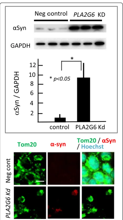
To clarify the relationship between αSyn and PLA2G6 dysfunction in cultured neurons, we analyzed the ex-pression level of αSyn in Pla2g6-Kd SH-SY5Y human neuroblastoma cells. In the western blotting analysis of
Pla2g6-Kd cells, αSyn expression was strikingly high (Fig. 1). The difference between Kd cells and negative control cells was significant (n= 6 per group, p< 0.05). Colocalization of αSyn and translocator of outer mito-chondrial membrane 20 (TOM20) was demonstrated by immunocytochemistry.
Mice
Strong expression ofαSyn in the spinal cord of Pla2g6-KO mice at the preclinical-stage
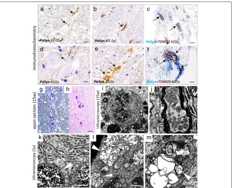
To clarify the relationship between αSyn and PLA2G6 dysfunction in mice in vivo, we analyzed the distribution of αSyn in Pla2g6-KO mice using immunohistochemis-try. In wild-type mice of both 15 weeks and 2 years of age, the neuropil was moderately stained for αSyn, with punctate pattern (Fig. 2a). In Pla2g6-KO mice at the pre-clinical stage (15 weeks), the immunoreactivity of αSyn was highly diffuse in the gray matter (Fig. 2b). αSyn staining in the neuropil had a punctate pattern, similar to that in wild-type mice, but the cytoplasm of some swollen neurons was also mildly positive forαSyn (Fig. 2c). PAS staining showed thatαSyn-positive swollen neurons were filled with PAS-positive granules (Fig. 2d). With age, large vacuoles and spheroids in the neuropil were increased in number, where the expression level of αSyn was almost none or low (Fig. 2e).
Strong expression of PαSyn in CCO-negative, TOM20-positive granules in young Pla2g6-KO mice
To determine if there was a relationship between sub-cellular micro-organelle localization and accumulation of PαSyn, the most important modified-form of αSyn after transcription, we compared the distribution of
PαSyn with markers of subcellular micro-organelles in neurons of Pla2g6-KO mice. Immunohistochemistry with an antibody specifically directed against Ser129-phosphorylatedαSyn (pSyn#64) revealed the absence of PαSyn in the wild-type mice (Fig. 2f ). In Pla2g6-KO mice at 15 weeks, very small PαSyn-positive granules were prominent in the cytoplasm and proximal axons of anterior horn cells (Fig. 2g, h, j) and dorsal root gan-glion cells (data not shown). In some of the swollen neurons filled with small PαSyn-positive granules, the nuclei were atrophic (Fig. 2h, j). By comparing serial sections, we found that the neurons filled with Pα Syn-positive granules were CCO-negative (Fig. 2i) and TOM20-positive (Fig. 2k). The granules were PAS-positive (Fig. 2l), KDEL-negative (Fig. 2m), and cathep-sin D-negative (Fig. 2n). The number of neurons filled with PαSyn-positive granules is shown in the graph (Fig. 2) and Table 1.
Morphological changes of PαSyn-positive granules in Pla2g6-KO mice with age
To assess the relationships between high PαSyn expres-sion and neurodegeneration in Pla2g6-KO mice, we ana-lyzed the age-dependent distribution of PαSyn. In
Pla2g6-KO mice at 15 weeks, very small PαSyn-positive granules were prominently observed in the proximal axons (Fig. 3a) and cytoplasm of anterior horn cells. At the early symptomatic stage (1 year), there were only a few PαSyn-positive granules in neuronal cytoplasm. Swollen neurons, filled with PαSyn-positive granules, were completely absent (Table 1). In the proximal axons, there were many PαSyn-positive granules, some of which were large and deformed (Fig. 3b). Double immunohis-tochemistry revealed PαSyn co-localization with TOM20 in the granules (Fig. 3c).
At the end stage (2 years), some of the PαSyn-positive granules showed degenerated membranes (Fig. 3d, e). Using double immunohistochemistry, PαSyn was shown to co-localize with TOM20 in the granules and the degenerated membrane of the granules (Fig. 3f ).
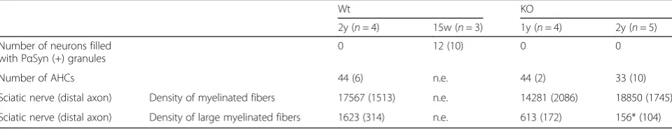
Table 1The time course of Pα-syn expression in neurons and the number of motor neurons and myelinated fibers ofPla2g6-knockout (KO) mice
Wt KO
2y (n= 4) 15w (n= 3) 1y (n= 4) 2y (n= 5)
Number of neurons filled with PαSyn (+) granules
0 12 (10) 0 0
Number of AHCs 44 (6) n.e. 44 (2) 33 (10)
Sciatic nerve (distal axon) Density of myelinated fibers 17567 (1513) n.e. 14281 (2086) 18850 (1745)
Sciatic nerve (distal axon) Density of large myelinated fibers 1623 (314) n.e. 613 (172) 156* (104)
PαSynphosphorylatedα-synuclein,Wtwild-type,KO Pla2g6-knockout,15 w15 weeks,1 y1 year,2y2 years,AHCanterior horn cell,number of anterior horn cells
mean (SD),n.e. not examined,Density of myelinated fibersnumber of myelinated fibers per mm2
Semi-thin and ultra-microscopic image of small Pα Syn-positive granules in Pla2g6-KO mice
To define the structure of small PαSyn-positive granules in Pla2g6-KO mouse neurons, we compared the the structures observed on semi-thin or ultra-microscopic pictures with the small PαSyn-positive granules seen on paraffin sections. In thionine-stained semi-thin sections,
we observed dark colored granules in the cytoplasm and proximal axons of neurons in young KO mice (Fig. 3g). The granules were clearly PAS-positive (Fig. 3h), and showed a similar size and distribution as PαSyn-positive granules. Ultra-microscopically, round mitochondria could be seen in which the cristae were partially replaced by small dense granules both in the perinuclear space of
a
b
c
d
e
f
g
h
i
j
k
l
m
neurons (Fig. 3i) and in axons (Fig. 3j). These abnormal mitochondria and round bodies were of a similar size and had a similar distribution as the PAS-positive granules in semi-thin sections, suggesting that PAS-positive granules were indicative of PαSyn-loading abnormal mitochondria.
In the aged KO mice, fewer round mitochondria could be seen in which the cristae were partially replaced by small dense granules in the perinuclear space of neurons (Fig. 3k). In the axons, mitochondria with branching and tubular cristae were frequently seen, and were clustered together (Fig. 3l). In the spheroid, highly degenerated mitochondria, tubulovesicular structures and degener-ated membranes were combined to form an irregular structure (Fig. 3m), suggesting severe degeneration of mitochondrial membranes.
Different distribution of ubiquitin and PαSyn in Pla2g6-KO mice
To determine whether the ubiquitin-proteasome system is activated in PαSyn-loading mitochondria, we estimated the relationship between ubiquitin and mitochondrial membrane protein by double immunohistochemistry. At the pre-clinical stage, there were rare ubiquitin deposits in the spinal cord ofPla2g6-KO mice (Fig. 4a-c). At the early symptomatic stage (1-year-old), ubiquitin-positive struc-tures were frequently observed in the neuropil and the axons (Fig. 4d), but not in the neuronal cytoplasm. Double immunohistochemistry revealed that the distribution of PαSyn and ubiquitin in the axons was markedly different (Fig. 4e). At the end stage (2-year-old), ubiquitination be-came severe in axons, but no ubiquitination was observed
a
b
c
d
e
f
g
h
i
in the cytoplasm (data not shown). Double immunohisto-chemistry showed almost no colocalization of PαSyn and ubiquitin (Fig. 4g) but showed frequent colocalization of TOM20 and ubiquitin in the spheroids (Fig. 4h, i).
Significant decrease in the density of large myelinated fibers, but not neuron number, in the spinal cord
To clarify the degree of neurodegeneration in axons and neurons that contained PαSyn-positive granules, from young mice, we estimated the number of axons and neu-rons in Pla2g6-KO mice after symptom onset. Axonal degeneration progressed as reported previously [41] and myelinated fibers became atrophic with age. Quantitative analysis (Table 1) revealed a significant decrease in large myelinated fibers (>10 μm in diameter) with age (KO mice 1y, n= 4, p =0.057; KO mice 2 y, n= 5, p <0.05). The density of the total myelinated fibers in Pla2g6-KO mice after onset was similar to that in the wild-type mice. The number of anterior horn cells in Pla2g6-KO mice at the early symptomatic stage was similar to that
of wild-type mice. Even at the end stage, the number of neurons was not significantly different between Pla2g6 -KO and wild-type mice (p= 0.067).
Human
Prominent formation of Lewy Bodies (LBs) in a PLAN patient with a mutation in PLA2G6
In the midbrain, upper pons, and upper medulla of both PD and PLAN patients, numerous neuronal in-clusions that were positive for αSyn and PαSyn (pSyn#64) could be seen. The affected areas included the substantia nigra, dorsal raphe nucleus, locus coeruleus, dorsal motor nuclei of the vagus nerve, and the reticular formation. Morphologically, the in-clusions were round or spherical, similar to Pale bodies or LBs seen in PD (Fig. 5a). In PD, most of the PαSyn-positive LBs were present in the neuropil, particularly in the region showing severe neuronal loss. In the brain of the PLAN patient, LBs were ob-served both in the neurons and in the neuropil
a
b
c
d
e
f
a b c
d e f
a b c
d e f
g h i
(Fig. 5b-f ). The size of LBs in PLAN varied, as it does in PD.
PαSyn -positive small inclusions colocalized with the mitochondrial membrane protein in PLAN
To assess the relationship between αSyn/PαSyn de-posits and mitochondria, we analyzed the neurons affected in PD and in PLAN using a double immuno-histochemical assay. Double immunohistochemistry revealed amorphous deposits of αSyn in the CCO-and TOM20-positive cytoplasm of neurons both in PD and in PLAN patients (PD; Fig. 6a, PLAN; Fig. 6d). As more αSyn accumulated in the neuronal cytoplasm, the CCO-immunoreactivity diminished (PD: Fig. 6b, c, PLAN: Fig. 6a-c). In PLAN neurons, small PαSyn-positive inclusions with a TOM20-positive edge were frequently found, but similar inclusions were rare in PD (PD: Fig. 6d-f, PLAN: Fig. 6e).
Clustering of PαSyn-positive small inclusions to form a larger neuronal inclusion in PLAN
To clarify the relationship between PαSyn-positive small inclusions and LBs, we analyzed the distribution of PαSyn-positive small inclusions in neurons affected in PLAN. In PLAN neurons, small PαSyn-positive in-clusions with a TOM20-positive edge appeared to cluster, and formed a larger narrow (PLAN: Fig. 6f ) or round (PLAN: Fig. 6g) inclusion. In some LBs, small PαSyn-positive inclusions with a TOM20-positive edge appeared to cover the surface (PLAN: Fig. 6h), whereas the immunoreactivity of PαSyn was reduced in the center. PαSyn and TOM20-positive
membranous structures also appeared to cover the surface of LBs (Fig. 6i).
Quantitative analysis of LBs with TOM20 present on their surface
To evaluate the relationship between LBs and mitochon-dria quantitatively, we counted the number of LBs with mitochondrial membrane protein (TOM20) present on their surface in PD and in PLAN. The number of LBs in each area is shown in Fig. 7. In all three investigated areas of the PD brains, most LBs were TOM20-negative on the surface. In the PLAN brain, most LBs were TOM20-positive on the surface. In all three investigated areas of the PLAN brain, more than half of the LBs were completely TOM20-positive. The surfaces of most LBs were TOM20-positive in PLAN and TOM20-negative in PD brains.
Proportion of LB-bearing dopaminergic neurons and neuronal density
To assess the survival rates of LB-bearing dopaminergic neurons, we estimated the neuronal density and the pro-portion of LB-harboring neurons in both PD and PLAN. The density of LB-bearing TH-positive neurons and total number of TH-positive neurons in substantia nigra and locus coeruleus are shown in Table 2. In the PD brains, the density of LB-bearing TH-positive neurons was low, because most LBs were found in the neuropil; this density was 1 ± 1 (mean ± S.D.) in the substantia nigra and 2 ± 2 in the locus coeruleus (number per 1 mm2). In the PLAN brain, the density of LB-bearing neurons was 6 in the sub-stantia nigra and 17 in the locus coeruleus, per 1 mm2. The density of TH-positive neurons in the substantia nigra
Fig. 7Strong expression of a mitochondrial outer membrane protein (TOM20) on the surface of Lewy bodies (LBs) inPLA2G6-associated neurodegeneration (PLAN). The left and the right images show the dorsal nuclei of the vagus in a patient with PLAN. The center image shows the substantia nigra in a patient with Parkinson disease (PD).TOM2020-kDa translocase of the outer mitochondrial membrane,SN
was low, but that in the locus coeruleus was similar to that in the control brains, despite the prominent presence of LBs (Fig. 5e, f ). The proportion of LB-bearing dopamin-ergic neurons was 6/17 in the substantia nigra and 17/81 in the locus coeruleus, respectively. Thus, a high propor-tion of LB-bearing neurons and preserved neuronal num-ber were observed in PLAN.
Preserved expression of TH in dopaminergic neurons, despite the presence of LBs
To determine if LB influences the micro-environment in dopaminergic neurons, we estimated cytoplasmic TH ex-pression of LB-bearing neurons. Both in PD and PLAN brains, most of the cytoplasmic TH expression in LB bearing neurons was preserved, both in substantia nigra and locus coeruleus (Table 3). Some LB-bearing neurons were TH-negative. No relationship between cytoplasmic TH expression and LB presence was found.
Discussion and conclusions
In this study, we demonstrated elevated expression of αSyn both in cultured cells and in mice with PLA2G6 de-ficiency. Prominent accumulation of PαSyn in damaged mitochondria was shown inPla2g6-KO mice, which con-stitute a model of PLA2G6-associated neurodegeneration (PLAN). In PLAN neurons, small PαSyn-positive granules and Lewy Bodies (LBs) were covered with mitochondrial membrane component on the surface. These findings sug-gest thatαSyn/PαSyn associates with damaged mitochon-dria in PLAN.
Strong expression of αSyn was evident in Pla2g6-Kd neuroblastoma cells and KO mouse neurons, suggesting that endogenous αSyn was induced in neurons due to PLA2G6-deficiency. In Pla2g6-KO mice, surprisingly high levels of PαSyn expression were observed in neu-rons; this was never seen in the wild-type mice. PαSyn was detected on the surface of tiny granules, which had a staining pattern that was apparently different from that of the toxic strains ofαSyn, including fibrils and ribbons [6, 33]. Immunohistochemical and ultra-microscopic data suggest that PαSyn-positive and PAS-positive gran-ules were abnormal mitochondria with degenerated inner membranes in KO mice, but not lysosomes or endoplas-mic reticulum, as has been shown previously [4, 45].
Injury of the inner mitochondrial membrane leads to permeabilization of mitochondria [2, 32, 50]. In KO mice from young, many neuronal mitochondria with degener-ated inner membrane were present, but neither autopha-gic degradation (mitophagy) nor ubiquitination of mitochondria were observed. Mitochondrial membrane permeabilization also induces apoptosis [32, 49]. More than 8 months after the appearance of mitochondria with degenerated inner membranes, due to PLA2G6 deficiency, neuronal loss was not significant in mice, suggesting that apoptosis did not occur to neurons. TOM20-positive granules were ubiquitin-negative in neurons and ubiquitin-positive in spheroids, suggesting ubiquitination of mitochondria occurs in degenerated axons, but not in neurons.
In aged KO mice, PαSyn-positive granules became more degenerated morphologically, but the PαSyn expression
Table 2The dopaminergic neuron density and the proportion of Lewy body-bearing neurons
Area Density of dopaminergic neurons
Control PD PLAN
(n= 5) (n= 7) (n= 1)
Substantia nigra LB harbouring neuron 0 1 (1) 6
total neuron 84 (14) 13 (6) 17
Locus coeruleus LB harbouring neuron 0 1 (3) 17
total neuron 77 (13) 9 (10) 81
THtyrosine hydroxylase,Density of TH(+)neuronsmean (SD) per mm2
,controlnon-neurodegenerative disease,PDParkinson disease,PLANPLA2G6-associated neurodegeneration,LBLewy body
Table 3The expression of tyrosine hydroxylase in dopaminergic neurons bearing Lewy bodies (LBs)
SN LC
PD PLAN PD PLAN
(n= 7) (n= 1) (n= 7) (n= 1)
Number of LB bearing neurons with the variety of tyrosine hydroxylase expression
TH (+) 3 (2)* 34 2 (4) 19
TH (−) 1 (1) 3 1 (1) 0
total number 3 (2) 37 3 (4) 19
LBLewy body,SNsubstantia nigra,LC, locus coeruleus,THtyrosine hydroxylase,TH(+) TH-positve neuron,TH(−) TH-negative neuron,Number of neurons
level was maintained.αSyn/PαSyn seems to have a strong affinity for the mitochondrial membrane [37, 52]. How-ever, considering the different localization of PαSyn and ubiquitin in axons, PαSyn seems to disappear from mito-chondria before mitomito-chondrial ubiquitination, following membrane permeabilization. The elevated expression of αSyn/PαSyn in mitochondria may be related to membrane stabilization [40].
In the substantia nigra of 26S proteasome depleted mice, neurodegeneration is accompanied by neuronal in-clusions similar to Pale bodies [30], the precursors of LB [46]. The inclusions contain mainly abnormal mitochon-dria [30], suggesting that Pale body or LB might be associated with abnormal mitochondria with bad quality control owing to dysfunction of ubiquitin proteasome sys-tem. As previously reported, LB formation is prominent in
h
cell death
proapoptotic materials
Lipid peroxides
ROS
Lewy body with broken barrier
g
cell alive Lipid
peroxides ROS
f
Lipid peroxides
ROS
e
Lewy body formation
d
c
Mitochondrion(Mit) Clustering of αSyn/PαSyn
loading Mit
b
αSyn/PαSyn
cyt C ROS
a
Mit with degenerated inner membranePLAN [15, 31, 36]. Considering the severity of mitochon-drial degeneration inPla2g6-KO mice, the association of Pale bodies or LBs with mitochondria would be strongly expected in PLAN.
Our double immunohistochemical data in PLAN re-vealed that the surface of PαSyn-positive small inclu-sions were TOM20-positive and CCO-negative, which is the same expression pattern of granules seen inPla2g6 -KO mouse neurons. αSyn/PαSyn appears to have a strong affinity for mitochondrial membrane components both inPla2g6-KO mice and in the PLAN case investi-gated here. Moreover, the small inclusions appeared to cluster into a large inclusion, possibly due to the strong affinity ofαSyn for the mitochondrial membrane.
LB extracts from PD brains have been reported to trigger αSyn pathology and motor deficits in mice and monkeys, which suggests that LBs contain neurotoxic materials [35]. However, the presence of LBs in dopa-minergic neurons was not associated with the low cyto-plasmic expression of TH, either in the autopsied PLAN or PD brains studied here, although this occurs early in PD [21]. This suggests that the presence of LB would not necessarily convey negative effects on the micro-environment of neurons, in spite of the neurotoxic ma-terials inside the LB.
A certain proportion of LB-bearing neurons is re-ported to occur in the substantia nigra (3.6 % on average), and this does not correlate with the symp-toms or disease duration [14, 18]. In our case of PLAN, a high proportion of dopaminergic neurons harbor LBs, suggesting the long-term survival of these neurons. In particular, the neurons were preserved well in the locus coeruleus, despite the presence of multiple LBs. This suggests that LBs in PLAN have a low toxicity. As αSyn/PαSyn-loading mitochondria with degenerated inner membrane did not lead to neuronal death in mice, high concentrations of mito-chondrial membrane components on the surface of LBs may be associated with the low toxicity of LBs in PLAN through the stabilization of the mitochondrial lipid membrane [7, 29].
We illustrate this hypothesis in Fig. 8. Because of
PLA2G6 dysfunction, the mitochondrial inner mem-brane is damaged early on. Strong affinity ofαSyn/PαSyn for damaged mitochondrial membrane induces Syn/ PαSyn to accumulate on the membrane. Elevated ex-pression of αSyn/PαSyn stabilizes the mitochondrial membrane and prevents membrane permeabilization. The strong affinity of αSyn for damaged mitochondrial membranes also causes clustering of the damaged mitochondria to form LB. The outer structure, which is comprised of a mitochondrial membrane component, functions as a barrier and allows LB-bearing neurons to survive for a long time. When the barrier breaks down,
pro-apoptotic materials, which are elevated in PLA2G6 deficiency [20], are released into the cytoplasm, followed by rapid cell death.
In conclusion, αSyn/PαSyn accumulation in damaged mitochondria of Pla2g6-KO mice and LBs in PLAN indicates a common pathological mechanism, which in-cludes the strong affinity of αSyn for damaged mito-chondrial membranes. PLAN patients with late onset tend to show prominent αSyn accumulation in neurons [31]. EndogenousαSyn could be neuroprotective by sta-bilizing mitochondrial membranes in neurodegenerative diseases, such as PLAN and possibly even in PD, which may indicate a new target for the treatment of α -synucleinopathy.
Competing interests
The authors declare that they have no competing interests.
Authors’contributions
HSA contributed the study design, data acquisition, analysis and manuscript preparation. GB and KS contributed to data acquisition, analysis and manuscript preparation. SK contributed to study design, data analysis and manuscript preparation. YR and MY contributed to data analysis and manuscript preparation. HF, YT and SS contributed to manuscript preparation. HM contributed the study design and manuscript preparation. All authors have read and approved the final manuscript.
Acknowledgements
This study was supported in part by a Grant-in-Aid for Scientific Research (C) from the Japan Society for the Promotion of Science, JSPS (H.SA. G.B.), Scientific Re-search on Innovative Areas from MEXT (H.M.), Brain Mapping by Integrated Neu-rotechnologies for Disease Studies (Brain/MINDS) (H.M.), Practical Research Project for Rare/Intractable Disease from Japan Agency for Medical Research and Devel-opment, AMED (S.K., H.M.), and a Budget for Promoting Priority Measures in Dis-cretionary Funds of the President in Tottori University (S.K.). The authors would like to thank H. Hayakawa for his expert assistance with biological analysis, K. Inoue for her expert assistance with pathological analysis, A. Sone for technical assistance, and R. Yoshida for fantastic illustrations.
Author details
1Department of Neurology, Graduate School of Medicine, Osaka University,
2-2 Yamadaoka, Suita 565-0871, Japan.2Department of Medical Genetics, Graduate School of Medicine, Osaka University, 2-2 Yamadaoka, Suita 565-0871, Japan.3Division of Neuropathology, Department of Brain and
Neurosciences, Tottori University Faculty of Medicine, 86 Nishi-machi, Yonago 683-8504, Japan.4Department of Neurology, Nagoya University Graduate School of Medicine, 65 Tsurumai-cho, Showa-ku, Nagoya 466-8550, Japan.5Institute for Medical Science of Aging, Aichi Medical University, 9
Yazakokarimata, Nagakute 480-1195, Japan.6Department of Neurology,
National Hospital Organization Toneyama National Hospital, 5-1-1 Toneyama, Toyonaka 560-8552, Japan.7Research Institute Osaka Medical Center for
Cancer and Cardiovascular Diseases, 1-3-3 Nakamichi, Osaka 537-0025, Japan.
Received: 4 February 2016 Accepted: 10 March 2016
References
1. Araki K, Yagi N, Ikemoto Y, Choong CJ, Hayakawa H, Beck G, Sumi H, Fujimura H, Moriwaki T, Nagai Y, Goto Y, Mochizuki H. Synchrotron FTIR micro-spectroscopy for structural analysis of Lewy bodies in the brain of Parkinson’s disease patients. Sci Rep. 2015;5:17625.
2. Ashrafi G, Schwarz TL. The pathways of mitophagy for quality control and clearance of mitochondria. Cell Death Differ. 2013;20:31–42.
Upregulation of Divalent Metal Transporter 1. PLoS One. 2015;10(10), e0141629.
4. Beck G, Sugiura Y, Shinzawa K, Kato S, Setou M, Tsujimoto Y, Sakoda S, Sumi-Akamaru H. Neuroaxonal dystrophy in calcium-independent phospholipase A2βdeficiency results from insufficient remodeling and degeneration of mitochondrial and presynaptic membranes. J Neurosci. 2011;31:11411–20.
5. Bendor JT, Logan TP, Edwards RH. The Function ofα-Synuclein. Neuron. 2013;79:1044–66.
6. Bousset L, Pieri L, Ruiz-Arlandis G, Gath J, Jensen PH, Habenstein B, Madiona K, Olieric V, Böckmann A, Meier BH, Melki R. Structural and functional characterization of two alpha-synuclein strains. Nat Commun. 2013;4:2575. 7. Braun AR, Lacy MM, Ducas VC, Rhoades E, Sachs JN.α-Synuclein-induced
membrane remodeling is driven by binding affinity, partition depth, and interleaflet order asymmetry. J Am Chem Soc. 2014;136:9962–72. 8. Burke JE, Dennis EA. Phospholipase A2 structure/function, mechanism, and
signaling. J Lipid Res. 2009;50(Suppl):S237–42.
9. Burré J, Sharma M, Tsetsenis T, Buchman V, Etherton MR, Südhof TC. Alpha-synuclein promotes SNARE-complex assembly in vivo and in vitro. Science. 2010;329:1663–7.
10. Cheon Y, Kim HW, Igarashi M, Modi HR, Chang L, Ma K, Greenstein D, Wohltmann M, Turk J, Rapoport SI, Taha AY. Disturbed brain phospholipid and docosahexaenoic acid metabolism in calcium-independent phospholipase A2-VIA (iPLA2β)-knockout mice. Biochim Biophys Acta. 2012; 1821:1278–86.
11. Cowen D, Olmstead EV. Infantile neuroaxonal dystrophy. J Neuropathol Exp Neurol. 1963;22:175–236.
12. Ellis CE, Murphy EJ, Mitchell DC, Golovko MY, Scaglia F, Barceló-Coblijn GC, Nussbaum RL. Mitochondrial lipid abnormality and electron transport chain impairment in mice lacking alpha-synuclein. Mol Cell Biol. 2005;25:10190–201. 13. Glynn P. Neuronal phospholipid deacylation is essential for axonal and
synaptic integrity. Biochim Biophys Acta. 2013;1831:633–41.
14. Greffard S, Verny M, Bonnet AM, Seilhean D, Hauw JJ, Duyckaerts C. A stable proportion of Lewy body bearing neurons in the substantia nigra suggests a model in which the Lewy body causes neuronal death. Neurobiol Aging. 2010;31:99–103.
15. Gregory A, Westaway SK, Holm IE, Kotzbauer PT, Hogarth P, Sonek S, et al. Neurodegeneration associated with genetic defects in phspholipase A2. Neurology. 2008;71:1402–9.
16. Guardia-Laguarta C, Area-Gomez E, Rüb C, Liu Y, Magrané J, Becker D, Voos W, Schon EA, Przedborski S.α-Synuclein is localized to mitochondria-associated ER membranes. J Neurosci. 2014;34:249–59.
17. Hedley-Whyte ET, Gilles FH, Uzman BG. Infantile neuroaxonal dystrophy. A disease characterized by altered terminal axons and synaptic endings. Neurology. 1968;18:891–6.
18. Jellinger KA. Neuropathology of sporadic Parkinson’s disease: Evaluation and changes of concepts. Mov Disord. 2012;27:8–30.
19. Khateeb S, Flusser H, Ofir R, Shelef I, Narkis G, Vardi G, Shorer Z, Levy R, Galil A, Elbedour K, Birk OS. PLA2G6 mutation underlies infantile neuroaxonal dystrophy. Am J Hum Genet. 2006;79:942–8.
20. Kinghorn KJ, Castillo-Quan JI, Bartolome F, Angelova PR, Li L, Pope S, Cochemé HM, Khan S, Asghari S, Bhatia KP, Hardy J, Abramov AY, Partridge L. Loss of PLA2G6 leads to elevated mitochondrial lipid peroxidation and mitochondrial dysfunction. Brain. 2015;138(Pt 7):1801–16.
21. Kordower JH, Olanow CW, Dodiya HB, Chu Y, Beach TG, Adler CH, Halliday GM, Bartus RT. Disease duration and the integrity of the nigrostriatal system in Parkinson’s disease. Brain. 2013;136(Pt 8):2419–31.
22. Lai Y, Kim S, Varkey J, Lou X, Song JK, Diao J, Langen R, Shin YK. Nonaggregatedα-synuclein influences SNARE-dependent vesicle docking via membrane binding. Biochemistry. 2014;53:3889–96.
23. Malik I, Turk J, Mancuso DJ, Montier L, Wohltmann M, Wozniak DF, Schmidt RE, Gross RW, Kotzbauer PT. Disrupted membrane homeostasis and accumulation of ubiquitinated proteins in a mouse model of infantile neuroaxonal dystrophy caused by PLA2G6 mutations. Am J Pathol. 2008; 172:406–16.
24. Marland JR, Hasel P, Bonnycastle K, Cousin MA. Mitochondrial Calcium Uptake Modulates Synaptic Vesicle Endocytosis in Central Nerve Terminals. J Biol Chem. 2016;291:2080–6.
25. Monteiro JP, Oliveira PJ, Jurado AS. Mitochondrial membrane lipid remodeling in pathophysiology: a new target for diet and therapeutic interventions. Prog Lipid Res. 2013;52:513–28.
26. Morgan NV, Westaway SK, Morton JE, Gregory A, Gissen P, Sonek S, et al. PLA2G6, encoding a phospholipase A2, is mutated in neurodegenerative disorders with high brain iron. Nat Genet. 2006;38:752–4.
27. Morrison K, Witte K, Mayers JR, Schuh AL, Audhya A. Roles of acidic phospholipids and nucleotides in regulating membrane binding and activity of a calcium-independent phospholipase A2 isoform. J Biol Chem. 2012;287:38824–34.
28. Murakami M, Taketomi Y, Miki Y, Sato H, Hirabayashi T, Yamamoto K. Recent progress in phospholipase A2 research: from cells to animals to humans. Prog Lipid Res. 2011;50:152–92.
29. Ouberai MM, Wang J, Swann MJ, Galvagnion C, Guilliams T, Dobson CM, Welland ME.α-Synuclein senses lipid packing defects and induces lateral expansion of lipids leading to membrane remodeling. J Biol Chem. 2013; 288:20883–95.
30. Paine SM, Anderson G, Bedford K, Lawler K, Mayer RJ, Lowe J, Bedford L. Pale body-like inclusion formation and neurodegeneration following depletion of 26S proteasomes in mouse brain neurones are independent of α-synuclein. PLoS One. 2013;8, e54711.
31. Paisán-Ruiz C, Li A, Schneider SA, Holton JL, Johnson R, Kidd D, Chataway J, Bhatia KP, Lees AJ, Hardy J, Revesz T, Houlden H. Widespread Lewy body and tau accumulation in childhood and adult onset dystonia-parkinsonism cases with PLA2G6 mutations. Neurobiol Aging. 2012;33:814–23. 32. Paradies G, Paradies V, De Benedictis V, Ruggiero FM, Petrosillo G. Functional
role of cardiolipin in mitochondrial bioenergetics. Biochim Biophys Acta. 2014; 1837:408–17.
33. Peelaerts W, Bousset L, Van der Perren A, Moskalyuk A, Pulizzi R, Giugliano M, Van den Haute C, Melki R, Baekelandt V.α-Synuclein strains cause distinct synucleinopathies after local and systemic administration. Nature. 2015; 522(7556):340–4.
34. Polymeropoulos MH, Lavedan C, Leroy E, Ide SE, Dehejia A, Dutra A, et al. Mutation in the alpha-synuclein gene identified in families with Parkinson’s disease. Science. 1997;276:2045–7.
35. Recasens A, Dehay B, Bové J, Carballo-Carbajal I, Dovero S, Pérez-Villalba A, Fernagut PO, Blesa J, Parent A, Perier C, Fariñas I, Obeso JA, Bezard E, Vila M. Lewy body extracts from Parkinson disease brains trigger a-synuclein pathology and neurodegeneration in mice and monkeys. Ann Neurol. 2014; 75:351–62.
36. Riku Y, Ikeuchi T, Yoshino H, Mimuro M, Mano K, Goto Y, Hattori N, Sobue G, Yoshida M. Extensive aggregation ofα-synuclein and tau in juvenile-onset neuroaxonal dystrophy: an autopsied individual with a novel mutation in the PLA2G6 gene-splicing site. Acta Neuropathol Commun. 2013;1:12. 37. Robotta M, Gerding HR, Vogel A, Hauser K, Schildknecht S, Karreman C,
Leist M, Subramaniam V, Drescher M. Alpha-synuclein binds to the inner membrane of mitochondria in anα-helical conformation. Chembiochem. 2014;15:2499–502.
38. Ross OA, Braithwaite AT, Skipper LM, Kachergus J, Hulihan MM, Middleton FA, et al. Genomic investigation of alpha-synuclein multiplication and parkinsonism. Ann Neurol. 2008;63:743–50.
39. Sengoku R, Saito Y, Ikemura M, Hatsuta H, Sakiyama Y, Kanemaru K, Arai T, Sawabe M, Tanaka N, Mochizuki H, Inoue K, Murayama S. Incidence and extent of Lewy body-related alpha-synucleinopathy in aging human olfactory bulb. J Neuropathol Exp Neurol. 2008;67:1072–83.
40. Shen J, Du T, Wang X, Duan C, Gao G, Zhang J, Lu L, Yang H.α-Synuclein amino terminus regulates mitochondrial membrane permeability. Brain Res. 2014;1591:14–26.
41. Shinzawa K, Sumi H, Ikawa M, Matsuoka Y, Okabe M, Sakoda S, Tsujimoto Y. Neuroaxonal dystrophy caused by group VIA phospholipase A2 deficiency in mice: a model of human neurodegenerative disease. J Neurosci. 2008;28: 2212–20.
42. Shvadchak VV, Yushchenko DA, Pievo R, Jovin TM. The mode ofα-synuclein binding to membranes depends on lipid composition and lipid to protein ratio. FEBS Lett. 2011;585:3513–9.
43. Snead D, Eliezer D. Alpha-synuclein function and dysfunction on cellular membranes. Exp Neurobiol. 2014;23:292–313.
44. Sumi H, Nagano S, Fujimura H, Kato S, Sakoda S. Inverse correlation between the formation of mitochondria-derived vacuoles and Lewy-body-like hyaline inclusions in G93A superoxide-dismutase-transgenic mice. Acta Neuropathol. 2006;112:52–63.
46. Wakabayashi K, Tanji K, Odagiri S, Miki Y, Mori F, Takahashi H. The Lewy body in Parkinson’s disease and related neurodegenerative disorders. Mol Neurobiol. 2013;47:495–508.
47. Wilson RS, Nag S, Boyle PA, Hizel LP, Yu L, Buchman AS, Shah RC, Schneider JA, Arnold SE, Bennett DA. Brainstem aminergic nuclei and late-life depressive symptoms. JAMA Psychiatry. 2013;70:1320–8.
48. Yamada M, Iwatsubo T, Mizuno Y, Mochizuki H. Overexpression of a-synuclein in rat substantia nigra results in loss of dopaminergic neurons, phosphorylation of a-synuclein and activation of caspase-9: resemblance to pathogenetic changes in Parkinson’s disease. J Neurochem. 2004;91: 451–61.
49. Yin H, Zhu M. Free radical oxidation of cardiolipin: chemical mechanisms, detection and implication in apoptosis, mitochondrial dysfunction and human diseases. Free Radic Res. 2012;46:959–74.
50. Youle RJ, Narendra DP. Mechanisms of mitophagy. Nat Rev Mol Cell Biol. 2011;12:9–14.
51. Zhao Z, Zhang X, Zhao C, Choi J, Shi J, Song K, Turk J, Ma ZA. Protection of pancreatic beta-cells by group VIA phospholipase A (2)-mediated repair of mitochondrial membrane peroxidation. Endocrinology. 2010; 151:3038–48.
52. Zigoneanu IG, Yang YJ, Krois AS, Haque E, Pielak GJ. Interaction ofα -synuclein with vesicles that mimic mitochondrial membranes. Biochim Biophys Acta. 2012;1818:512–9.
• We accept pre-submission inquiries
• Our selector tool helps you to find the most relevant journal
• We provide round the clock customer support
• Convenient online submission
• Thorough peer review
• Inclusion in PubMed and all major indexing services
• Maximum visibility for your research Submit your manuscript at
www.biomedcentral.com/submit