Hamstring Strain Injuries
Factors that Lead to Injury and Re-Injury
David A. Opar,
1Morgan D. Williams
2and
Anthony J. Shield
11 School of Human Movement Studies and Institute of Health&Biomedical Innovation, Queensland University of Technology, Brisbane, QLD, Australia
2 Faculty of Health, Sport and Science, University of Glamorgan, Pontypridd, Wales
Contents
Abstract. . . 209
1. Introduction . . . 210
2. Literature Search . . . 211
3. Hamstring Strain Injury (HSI) Incidence and Recurrence Rates . . . 211
4. Hamstring Function during Running and Potential for Strain Injury . . . 211
5. Causes of HSIs . . . 212
6. Anatomical Factors that Predispose Hamstrings to Strain Injury . . . 213
7. Risk Factors for HSIs . . . 214
7.1 Unalterable Risk Factors . . . 214
7.1.1 Age. . . 214
7.1.2 Previous Injury . . . 215
7.1.3 Ethnicity . . . 215
7.2 Alterable Risk Factors . . . 216
7.2.1 Strength Imbalances . . . 216
7.2.2 Flexibility . . . 218
7.2.3 Fatigue . . . 218
7.3 Addressing Risk Factors to Reduce the Risk of HSIs . . . 219
7.3.1 Eccentric Strength Training . . . 219
7.3.2 Strength Imbalance Correction . . . 220
7.3.3 Flexibility Training. . . 220
8. Hamstring Strain Recurrences and Neuromuscular Inhibition . . . 220
9. Conclusion . . . 222
Abstract
Hamstring strain injuries (HSIs) are common in a number of sports and incidence rates have not declined in recent times. Additionally, the high rate of recurrent injuries suggests that our current understanding of HSI and re-injury risk is incomplete. Whilst the multifactoral nature of HSIs is agreed upon by many, often individual risk factors and/or causes of injury are ex-amined in isolation. This review aims to bring together the causes, risk factors and interventions associated with HSIs to better understand why HSIs are so prevalent. Running is often identified as the primary activity type for HSIs and given the high eccentric forces and moderate muscle strain placed on the hamstrings during running these factors are considered to be part of the aetiology of HSIs. However, the exact causes of HSIs remain unknown and whilst eccentric contraction and muscle strain purportedly play a role, accumulatedmuscle damage and/or a single injurious event may also contribute. Poten-tially, all of these factors interact to varying degrees depending on the in-jurious activity type (i.e. running, kicking). Furthermore, anatomical factors, such as the biarticular organization, the dual innervations of biceps femoris (BF), fibre type distribution, muscle architecture and the degree of anterior pelvic tilt, have all been implicated. Each of these variables impact upon HSI risk via a number of different mechanisms that include increasing hamstring muscle strain and altering the susceptibility of the hamstrings to muscle da-mage. Reported risk factors for HSIs include age, previous injury, ethnicity, strength imbalances, flexibility and fatigue. Of these, little is known, definitively, about why previous injury increases the risk of future HSIs. Nevertheless, interventions put in place to reduce the incidence of HSIs by addressing modifiable risk factors have focused primarily on increasing eccentric strength, correcting strength imbalances and improving flexibility. The res-ponse to these intervention programmes has been mixed with varied levels of success reported. A conceptual framework is presented suggesting that neuro-muscular inhibition following HSIs may impede the rehabilitation process and subsequently lead to maladaptation of hamstring muscle structure and function, including preferentially eccentric weakness, atrophy of the pre-viously injured muscles and alterations in the angle of peak knee flexor tor-que. This remains an area for future research and practitioners need to remain aware of the multifactoral nature of HSIs if injury rates are to decline.
1. Introduction
Hamstring strain injuries (HSIs) are the most prevalent non-contact injury in Australian foot-ball,[1-7] American football,[8] rugby union,[9-12] soccer[13-17] and sprinting.[18,19] HSIs are char-acterized by acute pain in the posterior thigh with disruption of the hamstring muscle fibres.[20]HSIs range in severity from minor microscopic tearing and some loss of function (grade I) through to a full rupture of the muscle with complete loss of function (grade III).[21]The biceps femoris (BF) is the most commonly injured of the hamstring muscles,[22-24] with the muscle-tendon junction and adjacent muscle fibres being the most com-mon sites of disruption.[22,25]
In many cases, HSIs cause considerable time lost from training and competition,[7,9,15,26]which results in financial loss[27]and diminished athletic performance.[28]Injury has been suggested to have cost in excess ofd74.4 million in English premier and football league clubs during the 1999–2000 season.[27]Similar estimates, made by the authors, for elite Australian football teams indicate that
HSIs cost approximately $AU1.5 million in the 2009 season, which represents 1.2%of the salary cap in the Australian Football League. Further-more, player performance has been found to be significantly reduced following return from HSIs in elite Australian footballers.[28]
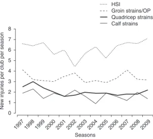
Epidemiological data obtained from Australian football, rugby union and soccer across a number of years indicates that rates of HSIs has not de-clined in recent decades (figure 1).[1,2,9,13,15,17,29] This is particularly worrying when taking into account that HSIs have, for a long time, been a well documented problem that has received con-siderable attention in the literature. Moreover, other injuries, such as ankle sprains in soccer[30] and posterior cruciate ligament injuries in Australian football,[31] have shown reduced incidence rates following the implementation of relatively effec-tive preventaeffec-tive measures. The lack of decline in HSI rates highlights that current practices aimed at preventing them require further scientific in-vestigation. In particular, whilst a number of risk factors for HSIs have been identified, the poten-tial role of the nervous system in strain injury
aetiology has been largely overlooked. Further-more, whilst it is commonly accepted that the aetiology of HSIs is complex and multifactorial in nature,[5,9,16]these factors are often considered in isolation. This review examines the causative and risk factors purportedly associated with HSIs, taking an integrated approach to further under-stand how these factors may interact, and also considers the impact of intervention programmes on these variables. Assimilating this information, we also propose a novel hypothesis as to how previous injury may lead to maladaptation of hamstring structure and function.
2. Literature Search
The articles selected for review were obtained via searches of MEDLINE and SPORTDiscus between 1966 and April 2011. The following key-words were searched in combination: ‘hamstring’, ‘knee flexor’, ‘muscle strain’, ‘injury’, ‘mechanism’, ‘risk factors’ and ‘prevention’. From the abstracts returned, articles were included for review if they related to hamstring injury incidence, causation, risk factor analysis or prevention. Full-text copies of selected articles were then sourced and the
re-ference lists of these articles were hand searched to identify other potential articles.
3. Hamstring Strain Injury (HSI) Incidence and Recurrence Rates
In track and field, one group has reported that HSIs account for 26.0%of all injuries sustained, with most occurring in sprinting events.[19] In comparison, observations from Australian foot-ball and soccer indicate that HSIs are responsible for 13–15%[1,2,4]and 12–14%[13,14,16]of all injuries, respectively. These figures are comparable with reports from American football training camps (12%)[8]and rugby union training (15%).[12]HSIs are also the single largest cause of lost playing time in Australian football[7]and are the predominant injury type responsible for prolonged absence (>28 days) from training and playing in soccer.[13] When compared with earlier epidemiology data from Australian football,[2] rugby union[2] and soccer,[17] recent observations indicate that the incidence of HSIs in sport has trended upwards over the past two decades. Further, data from the Australian Football League Annual Injury Re-port displays an increasing trend in the incidence of HSIs over the past seven competitive seasons, whilst other major injuries, including other pre-valent lower limb muscle strains, have remained largely stagnant (figure 1).[7]
In addition to high incidence rates and signif-icant time lost, HSIs also exhibit a very high rate of recurrence.[2,7,9,15,32-35]Over 13 seasons of ob-servation, 27%of all HSIs in the Australian Foot-ball League are recurrences of previous injuries; however, recent evidence suggests this is trending downwards, arguably, because of a more conser-vative approach in return to play strategies rather than improved rehabilitation practices.[7] Sim-ilarly, high rates of the recurrence of HSIs have also been reported in American football (32%),[32] rugby union (21%)[9]and soccer (16%).[36]
4. Hamstring Function during Running and Potential for Strain Injury
Although kicking, tackling, cutting and slow-speed stretching can result in HSIs,[9,15,17,24,37]
8 7 6 5 4 3 2 1
New injuries per club per season
0 1997 1998 1999 2000 2001 2002 2003 Seasons 2004 2005 2006 2007 2008 2009 HSI Groin strains/OP Quadricep strains Calf strains
Fig. 1.Injury incidence in the Australian Football League over 13 years. An injury is defined as ‘‘any physical or medical condition that prevents a player from participating in a regular season (home and away) match’’ (adapted from Orchard and Seward,[7]with per-mission).HSI=hamstring strain injury;OP=osteitis pubis.
running accounts for the majority of HSIs in soccer[15] and rugby union,[9]which suggests the demands of running give the greatest insight into the causes of HSI.
Studies of running biomechanics have found the hamstrings are active for the entire gait cycle with peaks in activation during the terminal swing and early stance phases.[38,39]During the terminal swing phase the hamstrings are required to contract forcefully whilst lengthening to decel-erate the extending knee and flexing hip.[38,40-43] It is also in terminal swing that the hamstrings reach their maximum length.[38,43] Of the three biarticular hamstring muscles, BF long head (BFL) undergoes the greatest stretch, reaching almost 110% of the length in upright standing during terminal swing, whilst semimembranosus and semitendinosus reach 107.5%and 108.2%, respect-ively.[43]In contrast, the maximum torques for hip extension and knee flexion are found to occur during ground contact in overground sprint-ing.[44]During this phase the hamstrings are act-ing primarily concentrically to extend the hip;[40] however, it has been reported that an eccentric contraction of the hamstrings occurs during the late stance phase of overground sprinting.[38]
The presence of a high force eccentric con-traction during the stance[38]and swing[38,39,41-43] phases likely contributes to the high rates of HSIs during maximal speed running. The terminal swing phase is considered the most hazardous as the hamstring muscle-tendon units are at their longest length of the gait cycle and are most heavily activated.[38,39,41-43]This suspicion has been sup-ported by two independent serendipitous obser-vations of acute HSIs during biomechanical studies of running, the timing of which was consistent with the insult occurring in terminal swing.[45,46] Whilst the stance phase is another possible period of susceptibility to HSIs, due to high hip exten-sion and knee flexion torque,[44,47]it involves much shorter hamstring lengths compared with term-inal swing.[38,41-43]
5. Causes of HSIs
In addition to strain injuries the hamstrings are also affected by tendinopathies[9]and
back-related injuries that referred pain to the posterior thigh.[20]These injuries display varying aetiological characteristics and, as such, the causes of these injuries vary considerably. For the purposes of this section, the focus will be on the cause of HSIs during running.
There is some debate as to whether muscle strain or the magnitude of eccentric force is the causa-tive factor in muscle strain injuries. Observations fromin-situanimal models suggest that the mag-nitude of muscle strain is the primary determin-ing factor in the occurrence of strain injury.[48-50] Many investigators have also suggested thatin vivo muscle strain injuries are associated with high force eccentric contractions,[38,41,42,50-57]where the length-ening demands placed on the muscle exceed the mechanical limits of the tissue.[41]It remains to be seen if both high eccentric force and high muscle strain are necessary conditions for a strain injury or whether each on their own is sufficient to bring about strain injury. Biomechanical observations suggest that eccentric contraction is a necessary condition for an HSI during running[45,46]and this claim is strengthened by the lack of strain injuries in con-centrically biased sports, such as swimming and cycling.[58,59] An argument for muscle strain being a necessary condition is less clear given that HSIs have been reported for both high (i.e. kicking)[9,15] and low (i.e. sprinting)[9,15,19,25]strain tasks. Poten-tially, an inter-relationship exists between eccentric force and muscle strain that dictates whether a muscle is injured. For example, strain injury may be avoided in tasks that involve high levels of strain if the level of eccentric force is low and the same may be true for high eccentric force/low strain activities.
There is also some uncertainty as to whether HSIs most typically occur as a result of accumu-lated microscopic muscle damage,[60]or as a result of a single event that exceeds the mechanical limits of the muscle.[61] It seems feasible, however, that both may contribute. For example, the accumula-tion of microscopic damage may leave the muscle tissue in a vulnerable state and more susceptible to injury in the event of a single traumatic event, such as bending to pick up or catch a ball.
Whilst the potential role of accumulated muscle damage in muscle strain injury aetiology
is not disputed, debate continues as to the physio-logical process responsible for damage. Morgan[60] first proposed the accumulated damage theory when he postulated that microscopic damage caused to individual sarcomeres following eccentric exercise was a result of preferential lengthening of weaker sarcomeres. This theory suggests that dur-ing eccentric contractions, there is non-uniform lengthening of adjacent sarcomeres when muscles are operating on the descending limb of the length-tension curve.[60]This difference in sarco-mere length impacts upon the force-creating capa-bilities of sarcomeres, as per the properties of the length-tension curve, which indicates that sarco-meres extended past their optimum length dis-play a reduction in force-generating capacity.[62] This results in weaker sarcomeres (i.e. sarcomeres longer than optimal length) lengthening uncon-trollably during eccentric contractions and even-tually being excessively stretched so that passive structures take up most of the tension due to the reduction in actin-myosin overlap.[60] The con-sequential damage to individual sarcomeres as a result of this uncontrolled lengthening was termed ‘sarcomere popping’ and was proposed to be the first step towards macroscopic muscle damage, such as muscle strain injury.[60]
Morgan’s hypothesis[60]is not, however, uni-versally accepted. It has been criticized because it is based upon single myofibril stretch studies performedin vitroand in situ that involve fibre strains not considered to be within the physiolo-gical range.[63]Butterfield[63]also argues that the expectation of unstable sarcomere lengthening on the descending limb of the length-tension curve is flawed given that the length-tension curve is de-termined under isometric conditions, whilst muscle lengthening occurs during dynamic eccentric con-traction. Indeed, evidence exists of inherent stability of the length-tension curve during lengthening con-traction,[50,64]which is thought to be attributable to the physiological characteristics of titin.[63]Further evidence[65] also argues against the assertion that sarcomeres at a longer length will lengthen un-controllably when exposed to eccentric contraction. This still remains an area of great controversy.
Our current understanding of HSIs suggest that high levels of eccentric force[38,41,42,45,46,50-57]
and muscle strain[48-50] are implicated in the ae-tiology of strain injury; however, it is not clear whether accumulated microscopic muscle da-mage[60] or the presence of a single injurious event[61]are most typically responsible for injury. Potentially, any one of these factors may be the primary cause of HSIs depending on the injurious activity type. For example, muscle strain may be the predominant mechanism in kicking HSIs whereas forceful eccentric contractions may be the major mechanism in running HSIs.
6. Anatomical Factors that Predispose Hamstrings to Strain Injury
The predominately biarticular nature of the hamstrings allows for simultaneous extension at the hip and flexion at the knee during concentric contraction and significant lengthening during concurrent hip flexion and knee extension, as seen in running[40] and kicking.[66] Such lengthening demands are thought to predispose the hamstrings to strain injury as the lengthening may exceed the mechanical limits of the muscle[41] or lead to the accumulation of microscopic muscle damage.[60,67]
The two heads of the BF muscle are innervated by different nerve branches; BFL by the tibial portion of the sciatic nerve and the BF short head (BFS) by the common peroneal branch of the sciatic nerve, and it has been suggested that this dual innervation is a possible explanation for HSIs because of the potential for uncoordinated contraction of the two heads of BF.[15,32]This, however, remains unsubstantiated and is yet to be the focus of scientific investigation.
Another commonly held belief is that the hamstring muscles possess a high number of type II fibres[68]and this would be expected to increase the risk of strain injury given that fast glycolytic fibres have shown a greater propensity for muscle damage following eccentric contraction in animal models.[69] However, whilst early histochemical analysis suggested that the hamstrings consisted predominately of type II muscle fibres (58%),[68] a more recent study reported that only 51% of fibres in the BFLwere classified as fast twitch.[70] This discrepancy may be due in some part to the difference in the ages of the study participants.
Subjects from the study by Garrett and collea-gues[68]ranged from 37 to 76 years of age, whereas the hamstrings utilized in the study by Dahmane et al.[70]better reflected the ages seen in elite sport (17–40 years). Whilst fibre type distribution may be one factor that impacts upon the strain injury risk of muscles, it’s role in HSIs may have been overstated previously given the fact that the vas-tus lateralis has been shown to have a greater proportion of type II muscle fibres[71]compared with BFL;[70] yet, the hamstrings are more com-monly injured than the quadriceps.[7,11-13,72] In this example, the differing lengthening demands of the muscles may have a greater influence over the propensity for strain injury than fibre type distribution.
Variations in muscle architecture may also explain high rates of muscle-specific HSIs. For example, BFSpossesses much longer fascicles but a much smaller physiological cross-sectional area compared with BFL[73]and this variation of ar-chitecture may predispose the BF; particularly, the long head, to high rates of strain injury. Longer fascicles allow for greater muscle extensibility[63] and reduce the risk of over lengthening during eccentric contraction.[67]However BF
L, which un-dergoes the greatest lengthening of all the ham-strings during sprinting,[43] has shorter fascicles compared with the BFSand this may predispose the BFLto repetitive over lengthening and accu-mulated muscle damage.[60,67] Consideration must be given to the fact that the available hamstring architecture data from this cadaveric study[73]has been performed on muscles from donors aged 68–88 years and the architectural characteristics of these muscles may differ markedly from younger, athletic populations.
The degree of anterior pelvic tilt may also im-pact upon the risk of HSIs given that the common origin for the long hamstrings, the ischial tu-berosity,[74]is found on the posterior aspect of the pelvis. As a result, excessive anterior pelvic tilt will place the hamstring muscle group at longer lengths[75]and some have proposed that this may increase the risk of strain injury.[15,76]
Whilst some commonly held beliefs relating to HSIs risk, such as the importance of fibre type distribution, may now be questioned, the
importance of structure still remains crucial to hamstring muscle function. As such, the anatomy of the hamstrings most likely contributes to its high propensity to injury; however, each of the aforementioned anatomical variables may increase the risk of injury via discrete mechanisms. An un-derstanding of each of these anatomical factors must also be intepreted with an understanding of the causes of HSIs presented in section 5.
7. Risk Factors for HSIs
A number of unalterable and alterable risk factors have been proposed for HSIs, including, but not limited to, increasing age,[6,15,16,20,72,77-79] previous injury,[6,20,72,78,79]ethnicity,[9,15,20]strength imbalances,[5,32,80-87] extremes of flexibility[76,88-92] and fatigue.[32,49,93,94] This section details those prospective studies, which have identified un-alterable and un-alterable factors that elevate the risk of an athlete sustaining an HSI. In addition, both intervention studies and randomized con-trolled trials (RCTs) aimed at preventing HSIs are examined to provide a thorough understanding of the alterable causative factors responsible for HSIs.
7.1 Unalterable Risk Factors
7.1.1 Age
Increasing age has been identified by a number of investigators as an independent risk factor for HSIs in Australian footballers[6,20,77,78] and soc-cer players.[15,16,72,79]Australian footballers older than 23[78]or 24 years[6]and soccer players older than 23 years[15]are at an elevated risk of HSI, with the odds ratios (ORs) as high as 4.4 (95%CI 1.6, 12.5) for the older athlete.[6] Furthermore, each year of age has been reported to increase the risk of sustaining an HSI by as much as 1.3-fold (OR; 95%CI 1.1, 1.5) in Australian footballers[20] and by 1.8-fold (OR; 95%CI 1.2, 2.7) in soccer players.[16] Importantly, all studies that report age as a significant risk factor have utilized re-gression or multivariate analysis to conclude that increasing age increases the risk of sustaining an HSI independently of confounding variables such as previous injury.[6,15,16,20,72,77-79]
One attempt to identify age-related changes that lead to an increased risk of HSIs in Australian football identified increased bodyweight and re-duced hip flexor flexibility as predictors of HSIs in athletes aged 25 years or older.[77] Despite achieving significance, the increase in risk was moderate with risk ratios of 1.07 (95%CI 1.0, 1.2) and 1.15 (95%CI 1.0, 1.3), respectively.[77]Other suggestions are that decreases in muscle mass and strength due to ageing could partially explain the increased risk of HSIs in the older athlete;[6] how-ever, evidence to support this hypothesis[95,96]comes from cross-sectional studies that included non-elite, non-athletic cohorts of significantly greater age ranges than are observed in elite sport. It is, in our view, particularly unlikely that athletes aged 24–30 years are weaker or have less muscle mass than their 18- to 20-year-old counterparts. Other hypotheses are age-related changes to muscle struc-ture[6] and entrapment of L5/S1 nerve root due to hypertrophy of the lumbosacral ligament;[97] however, more evidence is required to test these hypotheses.
Despite the consistent identification of age as a risk for HSIs, no convincing explanation has been given as to why athletes older than 24 years are at significantly greater risk than younger ath-letes. Ideally, long-term longitudinal studies are required to determine the physiological changes that occur across an athlete’s career to further elucidate the relationship between increasing age and increased HSI risk.
7.1.2 Previous Injury
A number of studies have indicated that Australian footballers with previous HSIs are at an elevated risk of sustaining a future HSI.[6,20,78] HSI from the previous season was also a signif-icant risk factor for hamstring injury in elite professional soccer players[79] and has been re-ported to increase the risk of future injury as much as 11.6-fold (OR; 95%CI 3.5, 39.0).[72]
Following an HSI, the primary goal must be to identify the predisposing factor responsible for the injury, which then should be a target for reha-bilitation and/or intervention.[34] If this predis-posing factor is not ameliorated, the athlete will remain at an elevated risk of future HSIs despite
sufficient convalescence. Additionally, a number of suggested post-HSI maladaptations are thought to contribute to the increased risk of future in-jury. These maladaptations include the formation of non-functional scar tissue[34]that is associated with an alteration in muscle tissue lengthening mechanics,[98] reduced flexibility,[89,90,92] persis-tent reductions in eccentric strength,[53,81,90,99] long-term atrophy of the injured muscle,[100] al-terations in the angle of peak knee flexor torque[67] and alterations in lower limb biomechanics.[20] Given the retrospective nature of these observa-tions[34,53,67,81,89,90,92,98-100]it is difficult to ascer-tain if these traits are the cause of or the result of previous injury; however, it is accepted that modifications (or maladaptations) do occur fol-lowing HSIs.[34] From the available literature, persistent reductions in eccentric strength,[53,81,90,99] the alterations in the angle of peak knee flexor torque[60,67,86]and reduced flexibility[89,90,92]have been examined most extensively in the literature and will be discussed in the following sections. The emerging evidence relating to the impact of scar tissue on muscle tissue lengthening mechanics,[98] however, is also worthy of further discussion. Find-ings from Silder and colleagues[98] suggest that previous hamstring injury at the muscle-tendon junction results in a proliferation of scar tissue in this region and ultimately leads to adjacent muscle fibres experiencing greater strain during eccentric contraction. Such an adaptation to muscle tissue lengthening mechanics following injury would imply a greater risk of re-injury given the associa-tion between higher levels of muscle fibre strain and susceptibility to muscle damage.[54]
The high rate of recurrence and the elevated risk associated with previous injury highlights the importance of preventing first-time HSIs and avoiding the vicious injury-reinjury cycle.[34] Fur-thermore, whilst previous injury has been identi-fied as elevating the risk of future injury, much work still needs to be done to determine what maladaptations are responsible for this increased risk.
7.1.3 Ethnicity
Three independent studies have identified Aboriginal[20]and Black African or Caribbean[9,15]
ethnicity as risk factors for HSIs; however, only one study reported the risk to be significantly increased (OR 11.2; 95% CI 2.1, 62.5).[20] Both high proportions of type II fibres[69,101] and ex-cessive anterior pelvic tilt[15,76] have been sug-gested as factors in the incidence of HSIs in these populations; however, these are not substantiated and more objective evidence is required to deter-mine how ethnicity impacts upon HSI risk.
7.2 Alterable Risk Factors
7.2.1 Strength Imbalances
Strength imbalances of the hamstring muscle group have long been suggested as causes of HSIs.[80]For the purposes of this review a strength imbalance can include any of the following: knee flexor weakness, bilateral knee flexor strength asymmetry and low ratios of knee flexor to knee extensor strength, otherwise known as hamstrings to quadriceps (H : Q) ratios.
Strength
Experimental data from animal models has shown that fully stimulated muscles are able to withstand greater amounts of stress before stretch-induced failure compared with partially activated muscles.[48]The authors postulated that stronger muscles would provide greater protection from strain injury and that muscle weakness may be a risk factor for muscle strain injury;[48] however, the evidence linking hamstring weakness to HSIs in humans is mixed.[5,83,85] Whilst one prospec-tive study has found that subsequently injured Australian footballers demonstrated lower peak concentric hamstring torque in preseason iso-kinetic testing,[83]this finding was not replicated in a larger but otherwise similar study a year later.[5] Prospective data on sprinters supports the find-ings of Orchard and colleagues,[83] as isometric knee flexion strength relative to bodyweight was significantly lower in subsequently injured limbs.[85]
Bilateral Asymmetry
Testing to assess unilateral hamstring strength allows for the determination of a weaker limb, if one exists. It has been proposed that a significantly weaker hamstring on one leg compared with the contralateral leg, termed hamstring bilateral
asym-metry, may predispose the weaker hamstring to an elevated risk of injury.[102]The use of a between-leg comparison of strength may be a more mean-ingful marker of weakness for individuals than a comparison with a group average or standardized score.
Early studies suggested that between-leg ham-string strength asymmetry of greater than 10% was a predictor of hamstring injury in American footballers and track and field athletes.[32,80] Later, elite Australian footballers with a bilateral asymmetry of 8%or more were found to have an increased risk of HSIs,[83] whilst soccer players with an asymmetry of more than 15%were at an increased risk.[82] It should be noted, however, that some authors have found no predictive power of bilateral strength imbalances.[5,86]
Whilst some disagreement exists in the litera-ture to date, a number of studies have identified that bilateral hamstring strength asymmetry leads to an increased risk of sustaining an HSI in a number of athletic cohorts.[32,80,82-84]Further ex-ploration of imbalances between the hamstrings and other muscles of the hip joint is warranted, as this may impact upon hamstring loading partic-ularly during the terminal swing phase of run-ning. Any alterations in running biomechanics associated with hamstring strength asymmetry should also be explored to determine if hamstring loading is affected as a result of imbalance.
Hamstrings : Quadriceps Strength Ratio
A lower H : Q ratio suggests a relatively poor capacity for the hamstrings to act as ‘brakes’ at the flexing hip and extending knee joints during the terminal swing phase of running. Thus, forceful contraction of the quadriceps, as occurs during the early swing phase of gait, has the potential to produce angular momentum at the knee joint that exceeds the mechanical limits of the hamstring.[103] Initial research[32,80,83] focused on comparisons of concentric strength imbalances across the knee joint, known as the conventional hamstrings to quadriceps ratio (H : Qconv), but has been criticized, as it neglects the functional role of the hamstrings during the terminal swing phase of gait; that of a forceful eccentric contraction.[38,39,41,43] More recently, the comparison of eccentric hamstrings to
concentric quadriceps strength, known as a func-tional strength ratio (H : Qfunc), has been sug-gested[103]and popularized.[5,81,82,84,86]
One of the earliest studies to examine the re-lationship between H : Qconv ratios and future injury risk found that American footballers with a H : Qconvratio of less than 0.50 were at an ele-vated risk of HSI.[32,80]A later small-scale study in Australian footballers found that an H : Qconv ratio of less than 0.61 put an individual at a sub-stantially increased risk of HSIs,[83]whilst a larger study performed only 1 year later was unable to find an association between H : Qconvor H : Qfunc ratios and future HSIs in Australian footballers.[5] These studies employed athletes at different levels of expertise and professionalism and employed different methodologies, all of which make com-parison of the findings difficult. With respect to sprinters, prospective observations found that neither H : Qconvor H : Qfuncratios displayed any significant differences between athletes who did or did not suffer an HSI.[86]Whilst Cox regres-sion analysis did determine that an H : Qconv ratio below 0.60 led to an increase in the risk of sustaining an HSI by a 17.4-fold hazard ratio (95%CI 1.3, 231.4),[86]the sample size of the in-jured group (n=8) should have precluded the use of this statistical method. Other prospec-tive observations have found that preseason H : Qfunc[84]and an isometric H : Q ratio[85]were sig-nificantly lower in the subsequently injured limbs of sprinters.
Many of these studies are limited due to their small sample sizes, which makes detecting small associations between H : Q ratios and HSI risk difficult.[104]The most powerful study to have ex-amined the association between H : Q ratios and HSIs (n=462) found that uncorrected strength imbalances in soccer players, which included an H : Qconv ratio below 0.45–0.47 (exact cut-off depends on dynamometer brand used) and an H : Qfuncratio below 0.80–0.89 were associated with a significantly greater frequency of HSIs compared with athletes without strength imbalances.[82] Fur-thermore, the correction of strength imbalances, including normalizing H : Q ratios, led to a sig-nificant reduction in HSI frequency compared with athletes who had uncorrected imbalances
(see section 7.3.2).[82]These findings provide the strongest evidence available that sufficient H : Q ratios protect athletes from future HSIs.
Angle of Peak Knee Flexion Torque
Athletes with a greater knee angle at peak con-centric knee flexion torque (those who produce peak knee flexor torque at shorter muscle lengths) are proposed to be at greater risk of HSIs.[67]The hamstrings in these individuals would be expec-ted to work on the descending limb of the length-tension relationship across a greater range of motion, leaving them more prone to damage (see section 5).[105]
Athletes with a history of unilateral hamstring injury display peak knee flexion torque at a greater degree of knee flexion on their injured limb com-pared with the uninjured limb (figure 2);[67] how-ever, it is not known if this is the cause of, or the result of, previous injury given the retrospective nature of these observations. In an attempt to determine a relationship between the angle of peak torque and future HSI occurrence, a recent prospective study in elite and sub-elite Japanese sprinters was performed.[86]This investigation found no association between the angle of peak knee
120 100 80 40 60 Torque (Nm) 20 0 0 20 40
Knee joint angle (°)
60 80 100 Uninjured leg Previously injured leg
Fig. 2.Unpublished concentric knee flexor torque-joint angle re-lationships from a single elite male athlete tested at 60/second in our laboratory. Angle of peak torque is indicated by the downward ar-rows. 0indicates full knee extension, 100indicates 100of knee flexion. The previously injured hamstring produces its peak torque at shorter muscle lengths (greater angle of peak torque), and hence operates to a greater extent along the descending limb of the length-tension curve.
flexor torque and subsequent HSIs during the competitive season.[86] Currently, the evidence pertaining to the usefulness of the angle of peak knee flexor torque to predict previous or future HSIs is too sparse to draw any firm inferences, and more work in this area is required.
7.2.2 Flexibility
Flexibility training has traditionally been pro-posed as a key component of injury prevention in athletes despite a lack of convincing prospective scientific evidence.[94,106]It is proposed that greater flexibility may reduce the risk of strain injury due to a greater ability of the passive components of the muscle-tendon unit to absorb energy as a re-sult of greater compliance;[106,107] although this point is disputed in the literature.[108]
Prospective studies in both American[80] and Australian footballers[6,83,107] have found no re-lationship between the hamstring flexibility from the sit-and-reach or toe-touch test and future HSI risk. In contrast to popular belief, Australian foot-ballers with a history of HSIs, who displayed greater sit-and-reach flexibility were actually more likely to sustain a recurrent HSI.[6]Furthermore, poor hamstring flexibility, as assessed via an active or passive knee extension test or a straight-leg raise, did not increase the risk of HSIs in Australian footballers,[109]soccer players[72]or sprinters.[86] In contrast, some studies have reported relationships between flexibility and hamstring injury.[16,88,91] A study in elite soccer players found that ham-string flexibility of less than 90 in a passive straight-leg raise correlated significantly with fu-ture HSIs.[91] Further studies also identified reduced hamstring flexibility as a significant in-dependent risk factor for HSIs in elite soccer players.[16,88]
Whilst the weight of evidence suggests that there is no protective benefit of greater hamstring flexibility on HSI risk, methodological flaws exist with the measurement techniques employed. Foreman and colleagues[110]suggest that no gold-standard measurement for flexibility has been established and that tests of hamstring length, such as the sit-and-reach, straight-leg raise and toe-touch test can be inaccurate if they do not allow for stabilization at the hip and lumbar spine.
Future studies should employ more objective measures of flexibility, such as the method used by Arnason and colleagues[72] that involves a tension metre to determine the limits of range of motion. This is, as opposed to the subjective as-sessment of the end of range of motion by the investigator or subject, which may display good levels of inter- and intra-tester reliability but may suffer with respect to ecological validity. Even if such subjective measurements are reproducible, there is no means of determining whether a sub-ject has been stretched to their maximal range of motion. The use of a more objective approach would be expected to improve the ecological va-lidity of clinical flexibility tests given that a set level of passive tension is defined as the end of range for all subjects.
7.2.3 Fatigue
Fatigue and its associated performance decre-ments have often been suggested as causative factors for injury.[32,49,94]Indeed, studies of injury incidence have shown that HSIs occur at a greater rate in the latter stages of competitive matches and training.[9,13,15,111,112]
The effect of fatigue on muscle lengthening properties was initially examined in a laboratory setting. In these experiments, muscles that were pre-fatigued via electrical stimulation absorbed less energy before failure when compared with unfatigued muscles.[49] Fatigued and control muscles still failed at the same length, indicating that a fatigued muscle may be more likely to suffer a strain injury due to a reduced capacity to resist over lengthening.[49]
With respect to human muscle function, one group has shown that fatigue of the hamstrings induced by repeated dynamic efforts leads to an increase in the amount of knee extension ob-served during the terminal swing phase of run-ning.[113]This increase in knee extension would be expected to lead to a greater strain on the ham-strings during the terminal swing phase of gait;[43] however, it was matched by a reduction in hip flexion.[113] These alterations in knee- and hip-joint positions suggest that fatigue from dynamic exercise may lead to alterations in propriocep-tion, a phenomenon that has been reported in
response to other experimental models of knee flexor fatigue.[114] In these trials, isokinetic ex-ercise that induced a 30%reduction in knee flexor maximal voluntary contraction force resulted in a reduction in proprioceptive ability, whereby, hamstring length was underestimated in a fatigued state.[114] This could lead to the perception of normal hamstring muscle lengths during running, whilst in reality, repeated over-lengthening of the hamstrings is occurring. Such deficits in pro-prioception when fatigued may elevate the risk of HSIs given the assertions made by Morgan[60]that continual over-lengthening would lead to micro-scopic muscle damage that may accumulate to be-come macroscopic damage (i.e. strain injury).
More recent work has also shown that inter-mittent running designed to mimic the demands of competitive soccer, significantly reduces eccentric hamstring torque with little or no impact on con-centric knee flexion or extension strength.[115,116] In our own unpublished work we have found marked variability in the loss of eccentric ham-string strength. Those who exhibit greater levels of preferential eccentric hamstring fatigue would be expected to be at a greater risk of an HSI with prolonged activity given the link between ec-centric weakness and HSI risk.[82,84]
Other potential factors linking fatigue with elevated risk of muscle strain injuries, such as altered technique, reductions in concentration and other intrinsic physiological changes, such as reduced coordination of muscle recruitment pat-terns, have been suggested[93] but are yet to be rigorously tested.
7.3 Addressing Risk Factors to Reduce the Risk of HSIs
Intervention studies and RCTs are important in determining if reported risk factors are indeed causative factors in injury aetiology. These study designs can determine whether interventions in-tended to improve purported causative factors result in reductions in the risk of sustaining an HSI. In fact, risk factors cannot be considered causative unless there is a reduction in the risk of sustaining an HSI following an intervention aimed at ameliorating them.
7.3.1 Eccentric Strength Training
Nordic Hamstring Exercise
Two RCTs[117,118]and one intervention study[56] have examined the benefits of the Nordic ham-string exercise (NHE) on HSI rates. The NHE is a bodyweight exercise that requires athletes to begin in a kneeling position and to gradually lower their upper bodies towards the ground by extending at the knee while contracting the knee flexors eccen-trically to slow the descent. During the exercise, the athlete’s ankles are typically held down by a part-ner.[119]The NHE has been shown to increase ec-centric hamstring torque[119] and shift the torque-joint angle curve of the hamstrings to longer muscle lengths[120] and both are suggested mechanisms by which the NHE may reduce HSI rates.
The implementation of NHEs failed to reduce rates of HSIs in cohorts of amateur Australian footballers[118] and professional soccer players;[117] however, compliance with both intervention programmes was extremely low. Gabbe and col-leagues[118] reported that approximately half of all participants allocated to their intervention group did not complete the second training ses-sion and that fewer than 10%completed the five planned sessions. Engebretsen and colleagues[117] also reported that only 21%of players performed 20 or more of 30 planned sessions of NHEs. Fur-thermore, the use of extremely high-volume and low-frequency (once per 2–3 weeks) hamstring training in one of these interventions[118]was incon-sistent with conventional conditioning practices.[119] In contrast, elite soccer teams who chose to implement the NHE as part of their preseason and inseason conditioning programmes displayed a 65%reduction in HSIs compared with teams that did not.[56] Furthermore, the teams that utilized the intervention displayed significantly lower rates and severity of HSIs compared with pre-vious seasons.[56]This study was, however, limited by a non-randomized approach as individual teams decided if they were to participate in the interven-tion. Interestingly, the implementation of NHEs did not reduce the rate of HSI recurrence.[56]
Flywheel Training
Training on a flywheel ergometer,[121]which is designed to augment the amount of eccentric
torque required during the performance of a lying-leg curl, has been reported to increase eccentric hamstring strength and reduce HSIs rates.[57] A small-scale RCT performed on two elite soccer teams (n=30 players) found that flywheel ham-string training in the preseason significantly re-duced the number of HSIs compared with the control group. However, the control group dis-played a remarkably high rate of HSI incidence (66%)[57] and this potentially diminishes the sig-nificance of these findings.[122]
Considerations for Exercise Selection
At present, the literature pertaining to the benefits of eccentric strength training on reducing HSI incidence is inconclusive.[122]Whilst a num-ber of factors, including a lack of compliance to eccentric strength training interventions[117,118] may contribute to this, exercise selection may also be a factor. The semimembranosus and semi-tendinosus reportedly exhibit greater activation levels at shorter muscle lengths, whereas the BFL is most powerfully activated at longer lengths during isokinetic knee flexion.[123]
MRI has recently revealed that the BFL and semimembranosus muscles were significantly less active than the semitendinosus and gracilis during a heavily loaded eccentric leg curl, which mimics the knee joint range of motion and ham-string lengths experienced in the NHE and fly-wheel training.[124] It is therefore possible that these exercises may be suboptimal in bringing about adaptation in the BFL, the muscle most fre-quently injured.[22-24]Exercises that better target the BFL, such as the stiff-legged deadlift[125]may prove more effective in hamstring injury preven-tion than those that have so far been employed in RCTs.
7.3.2 Strength Imbalance Correction
A large scale cohort study (n=462) of iso-kinetic hamstring strength in elite soccer players found that correction of strength deficits (either concentric or eccentric asymmetries or low H : Q ratios) lead to similar HSI rates compared with athletes without strength deficits.[82]Participants who had strength deficits but did not undergo isokinetic rehabilitation or who did undergo
iso-kinetic rehabilitation but did not perform post-intervention testing showed significantly higher rates of HSIs.[82]This study is of great significance, as it employed one of the largest sample sizes of any HSI prevention study and suggests that a reduction in the risk of HSIs can be achieved via the detection and subsequent correction of iso-kinetic strength deficits.
7.3.3 Flexibility Training
An intervention study performed on elite soc-cer players found that a prescribed contract-relax flexibility training protocol performed during the warm up did not reduce the rate of HSIs com-pared with teams that did not incorporate flex-ibility training.[56] Similarly, an RCT involving recreational-level runners, who completed a 16-week unsupervised intervention consisting of warm-up and cool-down procedures and stretching, showed no difference in the rate of HSIs compared with a control group.[126]Consideration must, however, be given to the potential that the intervention may have been inadequate to increase flexibility because of the brief duration of stretching ex-ercises (10 seconds).[122] These findings are not totally unexpected given the lack of evidence for poor flexibility being a risk factor for HSIs (see section 7.2.2). However, further work needs to be performed, with greater control over other con-founding variables, such as aerobic and eccentric hamstring conditioning, to fully elucidate the ef-fect of flexibility training on HSI rates.
8. Hamstring Strain Recurrences and Neuromuscular Inhibition
Whilst there is an extensive list of risk factors for HSIs that have been examined through a number of different methodological designs, epidemiological data suggest that first-time and recurrent HSI rates in sport are not in de-cline.[1,2,7,9,13,15,29]This suggests that our current understanding of what increases the risk of a fu-ture HSI has not accounted for all contributing factors or that we are unable to resolve previously identified factors effectively. Despite previous HSIs being consistently identified as one of the primary risk factors for a future HSI[6,20,72,78,79]
maladap-tation associated with HSIs, particularly the ner-vous system function, has been largely overlooked. Potentially, a number of reported maladaptations associated with prior HSIs may be explained by a common neurological mechanism in response to previous injury.
Weakness after painful musculoskeletal injury is typically mediated by both muscular and neural adaptations. For example, following traumatic knee injuries involving anterior cruciate ligament ruptures, maximal voluntary activation of the quad-riceps is significantly reduced, even years after the injury occurred[127,128]and despite restoration of knee stability.[128]In the case of HSIs, however, little attention has been paid to the possibility that prolonged deficits in activation contribute to the high injury recurrence rate. This is surprising, given that the torque-velocity relationships of previously injured hamstrings are characteristic of heightened neuromuscular inhibition in the sense that they show greater deficits in eccentric than concentric strength.[53,90,99]Prolonged neu-romuscular inhibition at long muscle lengths after HSIs could potentially account for observations of preferentially eccentric weakness,[53,90,99] per-sistent atrophy of the previously injured mus-cles[100]and alterations in the angle of peak knee flexor torque,[67]all of which are purported risk factors for HSIs and have been observed in ath-letes following ‘successful’ rehabilitation and the return to full competition and training.
A reduction in the capacity of the nervous system to activate injured muscles presumably constitutes a strategy to unload damaged tissues and thereby reduce pain in the acute recovery period. As the greatest pain after hamstring strain is typically felt at longer muscle lengths, it is not surprising that there is now evidence for a length-specific reduction in hamstring activation.[129] Inhibition, particularly during eccentric actions and at longer muscle lengths, may also impede the rehabilitation process by limiting adaptations within the previously injured muscle(s).
The early and middle stages of treatment for HSIs are characterized by the avoidance of ex-cessive stretching to prevent further scar formation and submaximal exercises performed through a limited range of motion, and with hip-joint
move-ments restrained primarily to the frontal plane.[130] Thus, by the time athletes are in the late stages of rehabilitation, their hamstring muscles might be expected to have shed in-series sarcomeres[131] and to have atrophied considerably. Having fewer in-series sarcomeres would be expected to shift the peak of the knee flexor torque-joint angle curve to shorter muscle lengths and create even greater weakness at longer lengths than atrophy alone.[67]Such hamstring function is detrimental, as running requires strength at relatively long muscle lengths to decelerate hip flexion and knee extension during terminal swing.[41-43]
The return to running at progressively faster speeds and the use of more intense strengthening exercises later in rehabilitation should increase exposure to forceful eccentric actions at relatively long muscle lengths,[38,40-43]and might therefore be expected to return muscles to their original size and fascicles to their pre-injury lengths.[132] How-ever, any lingering neuromuscular inhibition would spare the previously injured hamstring muscle(s) from significant activation during eccentric ac-tions at long length and would therefore limit or prevent hypertrophy and sarcomerogenesis. Evidence of persistent atrophy in the previously injured BFL with simultaneous compensatory hypertrophy of the uninjured BFSin recreational level athletes, 5–23 months after HSIs and after a full return to training and competition,[100]is con-sistent with the hypothesis of prolonged muscle-specific inhibition.
Additional investigation is required to confirm whether previously injured athletes display sig-nificantly greater levels of neuromuscular inhibi-tion within the previously injured leg compared with their contralateral uninjured limb, and whether inhibition is confined specifically to the injured muscle. Ultimately, to identify neuromuscular inhibition as a causative factor in recurrent HSIs, prospective studies and RCTs need to be performed to determine if inhibition following HSIs result in an increased risk of re-injury and whether ameli-orating this neurological deficit reduces the in-cidence of recurrent HSIs. Techniques, such as surface electromyography,[123,129,133-135]twitch inter-polation[136,137] and electrical stimulation,[134,138,139] have been used previously to assess voluntary
muscle activation, and all should be considered for future work in this area. Further work also needs to be carried out to rigorously determine the full extent of physiological maladaptation associated with altered neural function following HSIs.
9. Conclusion
HSIs remain the predominate injury in a num-ber of sports despite concerted efforts to expand scientific knowledge. Additionally, HSIs have shown a high rate of recurrence and the capacity to impact negatively on individual and team per-formance and financial viability of elite sports clubs. Whilst it is widely acknowledged that the causes of HSIs are multifactoral, the interaction between these factors is often overlooked. This review has integrated the role of the hamstrings in running, the specifics of hamstring anatomy and reported risk factors and interventions for HSIs to better understand the causes of this injury.
Sports medicine practitioners and sports injury researchers alike need to appreciate the complex nature of HSIs and understand that no one-single approach can be considered the gold standard for HSI prevention or rehabilitation. For example, a focus solely on markers of performance (i.e. ec-centric strength, flexibility) may neglect the im-portant role that correct running technique may have on injury avoidance. The biomechanical demands of running, the anatomical organization of the hamstrings and a range of unalterable and alterable risk factors, such as age, previous injury, ethnicity, strength imbalances, flexibility and fa-tigue have all been linked to HSIs. All of these factors need to be considered, as does the inter-action between these factors and the impact of reported interventions, by practitioners looking to prevent HSIs. Furthermore, understanding of the exact causes of HSIs remains elusive but muscle strain, high-force eccentric contraction, accumulated muscle damage and/or a single in-jurious event, may all potentially play a role and all should be considered when developing HSI preventative strategies.
Further to this, more work needs to be carried out in the area of assessing maladaptation asso-ciated with previous HSIs. Whilst it is commonly
known that previous HSIs are the primary risk factor for future injury, very little is known about the maladaptations associated with a previous insult. Understanding only that previous injury elevates the risk of injury without an understand-ing as to why, gives little insight into how HSIs should be successfully rehabilitated. We propose a novel integrated framework of how previous injury may lead to persistent neuromuscular in-hibition, which could conceivably result in a cascade of maladaptations that elevate the risk of future HSIs. This area should be a focus of future research given the high levels of HSI recurrence for a number of years in many sports.
Acknowledgements
No funding was used to assist in the preparation of this review. The authors have no conflicts of interest to declare that are directly relevant to the content of the review.
References
1. Orchard J, Seward H. Epidemiology of injuries in the Australian Football League, seasons 1997-2000. Br J Sports Med 2002 Feb; 36 (1): 39-44
2. Seward H, Orchard J, Hazard H, et al. Football injuries in Australia at the elite level. Med J Aust 1993 Sep 6; 159 (5): 298-301
3. Orchard J, Wood T, Seward H, et al. Comparison of in-juries in elite senior and junior Australian football. J Sci Med Sport 1998 Jun; 1 (2): 83-8
4. Gabbe B, Finch C, Wajswelner H, et al. Australian foot-ball: injury profile at the community level. J Sci Med Sport 2002 Jun; 5 (2): 149-60
5. Bennell K, Wajswelner H, Lew P, et al. Isokinetic strength testing does not predict hamstring injury in Australian Rules footballers. Br J Sports Med 1998 Dec; 32 (4): 309-14 6. Gabbe BJ, Bennell KL, Finch CF, et al. Predictors of
hamstring injury at the elite level of Australian football. Scand J Med Sci Sports 2006 Feb; 16 (1): 7-13
7. Orchard J, Seward H. Injury Report 2009: Australian Football League. Sport Health 2010; 28 (2): 10-9 8. Feeley BT, Kennelly S, Barnes RP, et al. Epidemiology of
National Football League training camp injuries from 1998 to 2007. Am J Sports Med 2008 Aug; 36 (8): 1597-603 9. Brooks JH, Fuller CW, Kemp SP, et al. Incidence, risk, and
prevention of hamstring muscle injuries in professional rugby union. Am J Sports Med 2006 Aug; 34 (8): 1297-306 10. Brooks JH, Fuller CW, Kemp SP, et al. A prospective study of injuries and training amongst the England 2003 Rugby World Cup squad. Br J Sports Med 2005 May; 39 (5): 288-93
11. Brooks JH, Fuller CW, Kemp SP, et al. Epidemiology of injuries in English professional rugby union: part 1 match injuries. Br J Sports Med 2005 Oct; 39 (10): 757-66
12. Brooks JH, Fuller CW, Kemp SP, et al. Epidemiology of in-juries in English professional rugby union: part 2 training injuries. Br J Sports Med 2005 Oct; 39 (10): 767-75 13. Ekstrand J, Ha¨gglund M, Walde´n M. Injury incidence and
injury patterns in professional football: the UEFA injury study. Br J Sports Med. Epub 2010 May 29
14. Hawkins RD, Hulse MA, Wilkinson C, et al. The associa-tion football medical research programme: an audit of injuries in professional football. Br J Sports Med 2001 Feb; 35 (1): 43-7
15. Woods C, Hawkins RD, Maltby S, et al. The Football Association Medical Research Programme: an audit of injuries in professional football. Analysis of hamstring injuries. Br J Sports Med 2004 Feb; 38 (1): 36-41 16. Henderson G, Barnes CA, Portas MD. Factors associated
with increased propensity for hamstring injury in English Premier League soccer players. J Sci Med Sport. Epub 2009 Oct 1
17. Ekstrand J, Gillquist J. Soccer injuries and their mechan-isms: a prospective study. Med Sci Sports Exerc 1983; 15 (3): 267-70
18. Bennell KL, Crossley K. Musculoskeletal injuries in track and field: incidence, distribution and risk factors. Aust J Sci Med Sport 1996 Sep; 28 (3): 69-75
19. Drezner J, Ulager J, Sennett MD. Hamstring muscle injuries in track and field athletes: a 3-year study at the Penn Relay Carnival [abstract]. Clin J Sport Med 2005; 15 (5): 386 20. Verrall GM, Slavotinek JP, Barnes PG, et al. Clinical risk
factors for hamstring muscle strain injury: a prospective study with correlation of injury by magnetic resonance imaging. Br J Sports Med 2001 Dec; 35 (6): 435-9 21. Blankenbaker DG, Tuite MJ. Temporal changes of muscle
injury. Semin Musculoskelet Radiol 2010 Jun; 14 (2): 176-93
22. Koulouris G, Connell D. Evaluation of the hamstring muscle complex following acute injury. Skeletal Radiol 2003 Oct; 32 (10): 582-9
23. Koulouris G, Connell DA, Brukner P, et al. Magnetic re-sonance imaging parameters for assessing risk of recur-rent hamstring injuries in elite athletes. Am J Sports Med 2007 Sep; 35 (9): 1500-6
24. Verrall GM, Slavotinek JP, Barnes PG, et al. Diagnostic and prognostic value of clinical findings in 83 athletes with posterior thigh injury: comparison of clinical find-ings with magnetic resonance imaging documentation of hamstring muscle strain. Am J Sports Med 2003 Nov-Dec; 31 (6): 969-73
25. Askling CM, Tengvar M, Saartok T, et al. Acute first-time hamstring strains during high-speed running: a longitudinal study including clinical and magnetic resonance imaging findings. Am J Sports Med 2007 Feb; 35 (2): 197-206 26. Engebretsen AH, Myklebust G, Holme I, et al. Intrinsic
risk factors for hamstring injuries among male soccer players: a prospective cohort study. Am J Sports Med 2010 Jun; 38 (6): 1147-53
27. Woods C, Hawkins R, Hulse M, et al. The Football Asso-ciation Medical Research Programme: an audit of injuries in professional football-analysis of preseason injuries. Br J Sports Med 2002 Dec; 36 (6): 436-41
28. Verrall GM, Kalairajah Y, Slavotinek JP, et al. Assessment of player performance following return to sport after hamstring muscle strain injury. J Sci Med Sport 2006 May; 9 (1-2): 87-90
29. Ekstrand J, Gillquist J, Mo¨ller M, et al. Incidence of soccer injuries and their relation to training and team success. Am J Sports Med 1983 Mar-Apr; 11 (2): 63-7
30. Ekstrand J, Gillquist J, Liljedahl SO. Prevention of soccer injuries. Supervision by doctor and physiotherapist. Am J Sports Med 1983 May-Jun; 11 (3): 116-20
31. Orchard JW, Seward H. Decreased incidence of knee pos-terior cruciate ligament injury in Australian Football League after ruck rule change. Br J Sports Med 2009 Dec; 43 (13): 1026-30
32. Heiser TM, Weber J, Sullivan G, et al. Prophylaxis and man-agement of hamstring muscle injuries in intercollegiate foot-ball players. Am J Sports Med 1984 Sep-Oct; 12 (5): 368-70 33. Orchard J, James T, Alcott E, et al. Injuries in Australian
cricket at first class level 1995/1996 to 2000/2001. Br J Sports Med 2002 Aug; 36 (4): 270-4
34. Croisier JL. Factors associated with recurrent hamstring injuries. Sports Med 2004; 34 (10): 681-95
35. Drezner JA. Practical management: hamstring muscle in-juries. Clin J Sport Med 2003 Jan; 13 (1): 48-52 36. Ekstrand J, Ha¨gglund M, Walde´n M. Epidemiology of
muscle injuries in professional football (soccer). Am J Sports Med. Epub 2011 Feb 18
37. Askling C, Lund H, Saartok T, et al. Self-reported ham-string injuries in student-dancers. Scand J Med Sci Sports 2002 Aug; 12 (4): 230-5
38. Yu B, Queen RM, Abbey AN, et al. Hamstring muscle kinematics and activation during overground sprinting. J Biomech 2008 Nov 14; 41 (15): 3121-6
39. Chumanov ES, Heiderscheit BC, Thelen DG. Hamstring musculotendon dynamics during stance and swing phases of high speed running. Med Sci Sports Exerc 2011 Mar; 43 (3): 525-32
40. Novacheck TF. The biomechanics of running. Gait Posture 1998 Jan 1; 7 (1): 77-95
41. Chumanov ES, Heiderscheit BC, Thelen DG. The effect of speed and influence of individual muscles on hamstring mechanics during the swing phase of sprinting. J Biomech 2007; 40 (16): 3555-62
42. Thelen DG, Chumanov ES, Best TM, et al. Simulation of biceps femoris musculotendon mechanics during the swing phase of sprinting. Med Sci Sports Exerc 2005 Nov; 37 (11): 1931-8
43. Thelen DG, Chumanov ES, Hoerth DM, et al. Hamstring muscle kinematics during treadmill sprinting. Med Sci Sports Exerc 2005 Jan; 37 (1): 108-14
44. Mann RV. A kinetic analysis of sprinting. Med Sci Sports Exerc 1981; 13 (5): 325-8
45. Heiderscheit BC, Hoerth DM, Chumanov ES, et al. Iden-tifying the time of occurrence of a hamstring strain injury during treadmill running: a case study. Clin Biomech (Bristol, Avon) 2005 Dec; 20 (10): 1072-8
46. Schache AG, Wrigley TV, Baker R, et al. Biomechanical response to hamstring muscle strain injury. Gait Posture 2009 Feb; 29 (2): 332-8
47. Mann R, Sprague P. A kinetic analysis of the ground leg during sprint running. Res Q Exerc Sport 1980 May; 51 (2): 334-48 48. Garrett Jr WE, Safran MR, Seaber AV, et al. Biomechanical
comparison of stimulated and nonstimulated skeletal muscle pulled to failure. Am J Sports Med 1987 Sep-Oct; 15 (5): 448-54
49. Mair SD, Seaber AV, Glisson RR, et al. The role of fatigue in susceptibility to acute muscle strain injury. Am J Sports Med 1996 Mar-Apr; 24 (2): 137-43
50. Garrett Jr WE. Muscle strain injuries: clinical and basic aspects. Med Sci Sports Exerc 1990 Aug; 22 (4): 436-43 51. Stanton P, Purdham C. Hamstring injuries in sprinting: the
role of eccentric exercise. J Orthop Sports Phys Ther 1989; 10 (9): 343-9
52. Kujala UM, Orava S, Ja¨rvinen M. Hamstring injuries. Current trends in treatment and prevention. Sports Med 1997 Jun; 23 (6): 397-404
53. Lee MJ, Reid SL, Elliott BC, et al. Running biomechanics and lower limb strength associated with prior hamstring injury. Med Sci Sports Exerc. Epub 2009 Sep 2 54. Lieber RL, Fride´n J. Muscle damage is not a function of
muscle force but active muscle strain. J Appl Physiol 1993 Feb; 74 (2): 520-6
55. Lieber RL, Fride´n J. Mechanisms of muscle injury gleaned from animal models. Am J Phys Med Rehabil 2002 Nov; 81 (11 Suppl.): 70S-9S
56. Arnason A, Andersen TE, Holme I, et al. Prevention of hamstring strains in elite soccer: an intervention study. Scand J Med Sci Sports 2008 Feb; 18 (1): 40-8
57. Askling C, Karlsson J, Thorstensson A. Hamstring injury occurrence in elite soccer players after preseason strength training with eccentric overload. Scand J Med Sci Sports 2003 Aug; 13 (4): 244-50
58. Johnson JN. Competitive swimming illness and injury: common conditions limiting participation. Curr Sports Med Rep 2003 Oct; 2 (5): 267-71
59. Mellion MB. Common cycling injuries: management and prevention. Sports Med 1991 Jan; 11 (1): 52-70 60. Morgan DL. New insights into the behavior of muscle
during active lengthening. Biophys J 1990 Feb; 57 (2): 209-21
61. Verrall GM, Slavotinek JP, Barnes PG. The effect of sports specific training on reducing the incidence of hamstring injuries in professional Australian Rules football players. Br J Sports Med 2005 Jun; 39 (6): 363-8
62. Gordon AM, Huxley AF, Julian FJ. The variation in iso-metric tension with sarcomere length in vertebrate muscle fibres. J Physiol 1966 May; 184 (1): 170-92
63. Butterfield TA. Eccentric exercise in vivo: strain-induced muscle damage and adaptation in a stable system. Exerc Sport Sci Rev 2010 Apr; 38 (2): 51-60
64. Rassier DE, Herzog W, Pollack GH. Stretch-induced force enhancement and stability of skeletal muscle myofibrils. Adv Exp Med Biol 2003; 538: 501-15
65. Telley I, Stehle R, Ranatunga K, et al. Dynamic behaviour of half-sarcomeres during and after stretch in activated rabbit psoas myofibrils: sarcomere asymmetry but no ‘sarcomere popping’. J Physiol 2006; 573 (1): 173-85
66. Orchard J, Walt S, McIntosh A, et al. Muscle activation during the drop punt kick. J Sports Sci 1999; 17 (10): 837-8 67. Brockett CL, Morgan DL, Proske U. Predicting hamstring strain injury in elite athletes. Med Sci Sports Exerc 2004 Mar; 36 (3): 379-87
68. Garrett Jr WE, Califf JC, Bassett 3rd FH. Histochemical correlates of hamstring injuries. Am J Sports Med 1984 Mar-Apr; 12 (2): 98-103
69. Lieber RL, Fride´n J. Selective damage of fast glycolytic muscle fibres with eccentric contraction of the rabbit ti-bialis anterior. Acta Physiol Scand 1988 Aug; 133 (4): 587-8 70. Dahmane R, Djordjevic S, Smerdu V. Adaptive potential of human biceps femoris muscle demonstrated by histo-chemical, immunohistochemical and mechanomyographical methods. Med Biol Eng Comput 2006 Nov; 44 (11): 999-1006
71. Pernus F, Erzen I. Arrangement of fiber types within fas-cicles of human vastus lateralis muscle. Muscle Nerve 1991 Apr; 14 (4): 304-9
72. Arnason A, Sigurdsson SB, Gudmundsson A, et al. Risk factors for injuries in football. Am J Sports Med 2004 Jan-Feb; 32 (1 Suppl.): 5S-16S
73. Woodley SJ, Mercer SR. Hamstring muscles: architecture and innervation. Cells Tissues Organs 2005; 179 (3): 125-41 74. Abebe E, Moorman C, Garrett Jr W. Proximal hamstring
avulsion injuries: injury mechanism, diagnosis and disease course. Oper Tech Sports Med 2009; 17 (4): 205-9 75. Sherry MA, Best TM. A comparison of 2 rehabilitation
programs in the treatment of acute hamstring strains. J Orthop Sports Phys Ther 2004 Mar; 34 (3): 116-25 76. Hennessey L, Watson AW. Flexibility and posture
assess-ment in relation to hamstring injury. Br J Sports Med 1993 Dec; 27 (4): 243-6
77. Gabbe BJ, Bennell KL, Finch CF. Why are older Australian football players at greater risk of hamstring injury? J Sci Med Sport 2006 Aug; 9 (4): 327-33
78. Orchard JW. Intrinsic and extrinsic risk factors for muscle strains in Australian football. Am J Sports Med 2001 May-Jun; 29 (3): 300-3
79. Ha¨gglund M, Walde´n M, Ekstrand J. Previous injury as a risk factor for injury in elite football: a prospective study over two consecutive seasons. Br J Sports Med 2006 Sep; 40 (9): 767-72
80. Burkett LN. Causative factors in hamstring strains. Med Sci Sports 1970; 2 (1): 39-42
81. Croisier JL, Forthomme B, Namurois MH, et al. Hamstring muscle strain recurrence and strength performance disorders. Am J Sports Med 2002 Mar-Apr; 30 (2): 199-203 82. Croisier JL, Ganteaume S, Binet J, et al. Strength
im-balances and prevention of hamstring injury in profes-sional soccer players: a prospective study. Am J Sports Med 2008 Aug; 36 (8): 1469-75
83. Orchard J, Marsden J, Lord S, et al. Preseason hamstring muscle weakness associated with hamstring muscle injury in Australian footballers. Am J Sports Med 1997 Jan-Feb; 25 (1): 81-5
84. Sugiura Y, Saito T, Sakuraba K, et al. Strength deficits identified with concentric action of the hip extensors and eccentric action of the hamstrings predispose to hamstring