Western University Western University
Scholarship@Western
Scholarship@Western
Electronic Thesis and Dissertation Repository
7-22-2011 12:00 AM
Walk Softly and Carry a Big Stick: Strategies to Decrease Dynamic
Walk Softly and Carry a Big Stick: Strategies to Decrease Dynamic
Knee Joint Loading
Knee Joint Loading
Daniel J. Bechard
The University of Western Ontario
Supervisor
Dr. Trevor Birmingham
The University of Western Ontario Joint Supervisor Dr. Thomas Jenkyn
The University of Western Ontario
Graduate Program in Health and Rehabilitation Sciences
A thesis submitted in partial fulfillment of the requirements for the degree in Doctor of Philosophy
© Daniel J. Bechard 2011
Follow this and additional works at: https://ir.lib.uwo.ca/etd
Part of the Other Rehabilitation and Therapy Commons, and the Physical Therapy Commons
Recommended Citation Recommended Citation
Bechard, Daniel J., "Walk Softly and Carry a Big Stick: Strategies to Decrease Dynamic Knee Joint Loading" (2011). Electronic Thesis and Dissertation Repository. 209.
https://ir.lib.uwo.ca/etd/209
This Dissertation/Thesis is brought to you for free and open access by Scholarship@Western. It has been accepted for inclusion in Electronic Thesis and Dissertation Repository by an authorized administrator of
WALK SOFTLY AND CARRY A BIG STICK: STRATEGIES TO DECREASE DYNAMIC KNEE JOINT LOADING
(Spine title: Strategies to Decrease Knee Joint Loading)
(Thesis format: Integrated Article)
by
Daniel J. Bechard
Graduate Program in Health and Rehabilitation Sciences
A thesis submitted in partial fulfillment
of the requirements for the degree of Doctor of Philosophy
The School of Graduate and Postdoctoral Studies The University of Western Ontario
London, Ontario, Canada
ii
THE UNIVERSITY OF WESTERN ONTARIO School of Graduate and Postdoctoral Studies
CERTIFICATE OF EXAMINATION
Co-Supervisors
______________________________ Dr. Trevor Birmingham
______________________________ Dr. Thomas Jenkyn
Examiners
______________________________ Dr. Tim Wilson
______________________________ Dr. Michael Hunt
______________________________ Dr. Bert Chesworth
______________________________ Dr. Kara Patterson
The thesis by
Daniel J. Bechard
entitled:
Walk Softly and Carry a Big Stick: Strategies to Decrease Dynamic Knee
Joint Loading
is accepted in partial fulfillment of the requirements for the degree of
Doctor of Philosophy
______________________ _______________________________
iii
Abstract
Excessive dynamic loading of the knee joint, quantified indirectly during three
dimensional gait analysis, is a risk factor for the progression of knee osteoarthritis (OA). The
overall objective of this thesis was to explore the effects of prolonged walking and the use of
Nordic walking poles on selected gait characteristics indicative of knee joint load. The first
study evaluated the time-varying behaviour, reliability, and validity of selected gait
kinematics during 60 minutes of treadmill walking in 20 healthy adults. Maximum lateral
trunk lean angle and maximum toe-out angle did not change over time, were consistent from
day to day and were consistent with values assessed during over-ground gait analysis,
suggesting that these measures are appropriate for use in studying potential adaptive gait
mechanisms. The second study compared the time-varying behaviour of selected gait
kinematics during 30 minutes of treadmill walking in 20 participants with, and 20
participants without, medial compartment knee OA, and explored correlations between these
gait kinematics and pain intensity. Trunk lean, toe-out, and pelvic rise were different between
those with and without knee OA, but did not systematically change over time in either group.
Trunk lean and contralateral pelvic drop were significantly correlated to pain intensity. The
third study was a technical report describing the use of three dimensional gait analysis and a
Nordic walking pole instrumented with a compression load cell. This methodology was then
used in the fourth study to evaluate the effect of walking poles used by 34 patients with
medial compartment knee OA. Despite small reductions in the vertical ground reaction force,
walking with poles increased the frontal plane lever arm, and therefore the knee adduction
moment. The pole force in the vertical direction was inversely related to the increase in first
iv
plausible, patients with knee OA do not naturally adopt gait characteristics or use walking
poles in a way to decrease knee joint loads. These findings support the need for future efforts
directed at explicitly teaching walking techniques, including the optimal use of assistive
devices, to decrease knee joint loading.
Keywords: compensatory gait biomechanics, human locomotion, measurement properties,
gait compensation, pain, pelvic tilt, prolonged gait, knee adduction moment, lever arm,
v
Co-Authorship Statement
This thesis contains materials from one published manuscript (Chapter 2), one
manuscript accepted for publication (Chapter 3), and two manuscripts in preparation to be
submitted for peer review (Chapter 4 and 5). Daniel Bechard was primarily responsible for
collection, analysis, and interpretation of data for all studies in this thesis. Dr. Trevor
Birmingham and Dr. Thomas Jenkyn were co-authors for all four studies (Chapter 2 to 5) and
assisted in study design, data analysis, and manuscript preparation. Dr. Aleksandra Zecevic
was co-author for Chapters 1, 2, 3, and 5 and assisted in study design, and manuscript
preparation. Dr. Robert Giffin and Mr. Ian Jones were co-authors on Chapters 3 and 5. They
assisted in study design, patient recruitment, and data collection. Ms. Kristyn Leitch was a
co-author for Chapter 5 and assisted with data processing and manuscript writing. Daniel
vi
Acknowledgments
First and foremost I would like to thank my co-supervisors, Dr. Trevor Birmingham
and Dr. Thomas Jenkyn. They provided guidance and friendship throughout this process
which I will never forget. I will always look to their examples of mentorship as a framework
to help me guide future students. I would also like to thank the other prominent individuals
that made this dissertation a success. My advisory committee: Dr. Alexandra Zecevic and Dr.
Robert Giffin, and my comprehensive committee: Dr. Monica Maly, and Dr. Philip Doyle.
These individuals insured my breadth of knowledge, attention to detail, and ultimate success.
I thank all of you for your truly unique perspective and expertise. I would also like to thanks
those individuals that I worked with on a daily basis. These individuals were the cause of the
good days and helped me persevere through the bad. These individuals were my family over
the past five years. This group consists of our Research Associate and Lab Manager Ian
Jones. Ian gave me the hands on tools that I needed to collect data, trouble shoot, and
provided daily guidance which was seldom regarding academics. I would also like to thank
the rest of the students that passed through the Wolf Orthopaedic Biomechanics Laboratory
over the past five years. WOBL was always a welcoming place where students worked to
develop their skills together rather than compete against each other. I will always remember
sharing a beer and the occasional ‘cheap shot’ with Dr. Shawn Robbins and Angelo
Boulougouris. This group also consisted of Dr. Volker Nolte, Chris McCully and the rest of
the UWO Rowing Team and Michelle Darvill and the Canadian Under 23 Women’s Rowing
Team. These groups shed light on what it truly means to work towards excellence. Most of
all, I want to thank my parents Jerome and Zuzanna, my sister Nicole and brother-in-law
vii
unconditional love and support. Finally, to my wife Natalie. She provided me unwavering
viii
Table of Contents
Certificate of Examination ... ii
Abstract ... iii
Co-Authorship Statement………...………..v
Acknowledgments... vi
List of Tables ... xii
List of Figures ... xiv
List of Appendices ... xivvi
List of Abbreviations, Symbols, Nomenclature ... xvii
Chapter 1 : Introduction and Background ... 1
1.1 Overview ... 1
1.2 Articular Cartilage ... 1
1.3 Osteoarthritis ... 2
1.4 The Impact of Osteoarthritis ... 4
1.5 3-D Gait Analysis and the Knee Adduction Moment ... 6
1.6 Methods of Reducing the Knee Adduction Moment ... 12
1.6.1 Trunk lean and Toe-out ... 14
1.6.2 Walking Poles and Canes ... 24
1.7 Thesis Outline ... 30
1.8 References ... 31
Chapter 2 : Time-varying Behaviour, Test-retest Reliability and Concurrent Validity of Lateral Trunk Lean and Toe-out During Prolonged Treadmill Walking ... 44
2.1 Overview ... 44
ix
2.3 Methods... 47
2.3.1 Study Design ... 47
2.3.2 Participants ... 48
2.3.3 Over-ground Gait Analysis ... 48
2.3.4 Treadmill Gait Analysis ... 49
2.3.5 Data Reduction ... 49
2.3.6 Statistical Analysis ... 50
2.4 Results ... 51
2.5 Discussion ... 55
2.6 Conclusions ... 59
2.7 References ... 59
Chapter 3: Toe-out, Lateral Trunk Lean and Pelvic Obliquity During Prolonged Walking in Patients with Medial Compartment Knee Osteoarthritis and Healthy Controls ... 64
3.1 Overview ... 64
3.2 Introduction ... 65
3.3 Methods... 68
3.3.1 Participants ... 68
3.3.2 Gait Analysis ... 69
3.3.3 Data Reduction ... 70
3.3.4 Statistical Analysis ... 71
3.4 Results ... 72
3.5 Discussion ... 79
3.6 Conclusions ... 84
x
Chapter 4: The Effects of an Instrumented Walking Pole on Knee Joint Loading During
Gait: A Technical Note ... 91
4.1 Overview ... 91
4.2 Introduction ... 92
4.3 Methods... 94
4.4 Results ... 97
4.5 Discussion ... 101
4.7 References ... 103
Chapter 5: The Effects of Nordic Walking Poles on the Knee Adduction Moment in Patients with Medial Compartment Knee Osteoarthritis ... 107
5.1 Overview ... 107
5.2 Introduction ... 108
5.3 Methods... 110
5.3.1 Participants ... 110
5.3.2 Gait Analysis ... 111
5.3.3 Statistical Analysis ... 115
5.4 Results ... 115
5.5 Discussion ... 121
5.6 Conclusions ... 124
5.7 References ... 125
Chapter 6: Discussion ... 130
6.1 Overview ... 130
6.2 Implications and Future Research ... 132
xi
6.4 Limitations ... 134
6.5 References ... 136
Appendices ... 141
xii
List of Tables
Table 1.1: Summary of findings from articles investigating trunk lean ... 21
Table 1.2:Summary of findings from articles investigating toe-out ... 22
Table 1.3: Summary of findings from studies investigating walking poles and canes ... 28
Table 2.1: Demographics of the study participants (n=20). ... 48
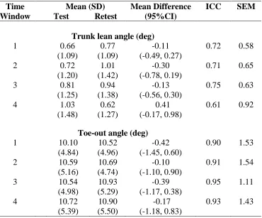
Table 2.2: Agreement between testing days and standard error of measurement for lateral trunk lean and toe-out measures (in degrees) during prolonged treadmill walking divided into four time intervals.. ... 52
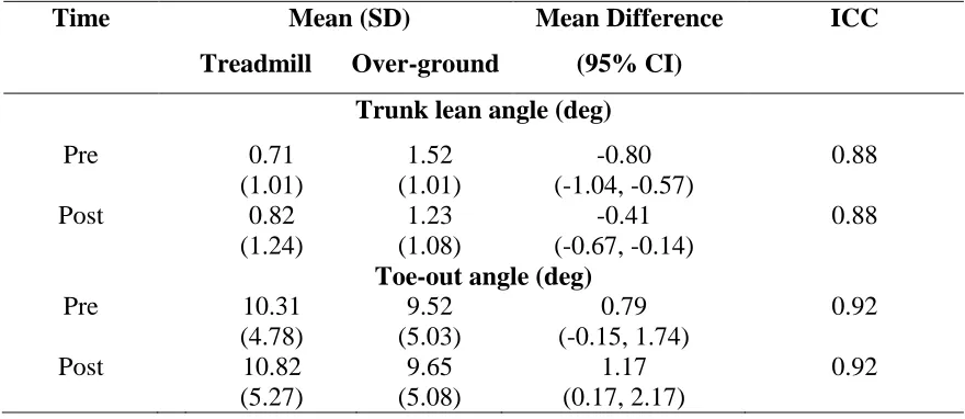
Table 2.3: Agreement between treadmill and over-ground walking measures. Trunk lean and toe-out (in degrees) values obtained during the first over-ground walking session (pre) are compared to window one of treadmill walking. Values obtained during window four of treadmill walking are compared to the second over-ground walking session (post). ... 53
Table 3.1: Demographics of the study participants (n=40), listing mean (standard deviation). ... 69
Table 3.2: Summary statistics, mean difference and 95% confidence intervals for toe-out, trunk lean and pelvic obliquity measures during 30 minutes of treadmill walking for both healthy adults and patients with knee osteoarthritis. ... 75
Table 4.1: Subject demographics including sex, age (y), height (m), mass (kg), and body mass index (kg/m2) ... 94
Table 4.2: Values for all kinetic and kinematic outcome measures at first and second peak knee adduction moment for both with and without poles conditions. Means (standard deviations) were calculated from ten trials, five from each participant ... 99
xiii
Table 5.2: Sample means (SD) for all kinetic and kinematic outcome measures at the time of
first peak knee adduction moment for both with poles and without poles conditions and mean
difference with 95% confidence intervals ... 117
Table 5.3: Sample means (SD) for all kinetic and kinematic outcome measures at the time of
second peak knee adduction moment for both with poles and without poles conditions and
xiv
List of Figures
Figure 1.1. An illustration of the knee adduction moment (A) and mechanical axis angle (B).
The knee adduction moment consists of the line of action of the ground reaction force and the
lever arm extending from the centre of the knee to the line of the ground reaction force. The
mechanical axis angle is a measure of limb alignment and consists of the angle created by
lines drawn from the centre of the femoral head and centre of the ankle to the centre of the
knee. Negative values indicate varus alignment (bow legs) and positive values indicate
valgus alignment (knock knees)... 10
Figure 1.2. A schematic diagram of the degenerative cycle that occurs in patients with knee
osteoarthritis that includes a cyclic process of cartilage degeneration and joint space
narrowing, increased mechanical axis angle, and increased knee joint load ... 11
Figure 1.3. An illustration of trunk lean angle measured from the midpoint of a line
connecting the anterior superior iliac spines and the midpoint of the line connecting the
acromion processes with respect to the vertical. Frontal plane ground reaction force is also
shown. Positive values represent a lean towards the stance limb while negative values
represent a lean towards the swing limb ... 17
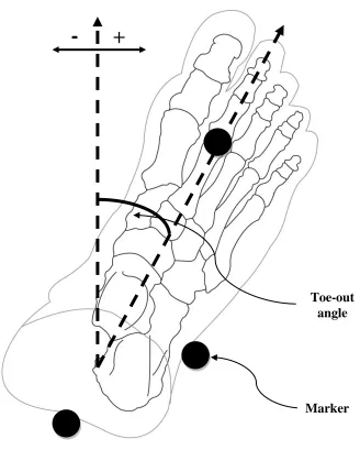
Figure 1.4. An illustration of toe-out angle measured as a line connecting the middle of the
ankle to the head of the second metatarsal with respect to the forward progression of the
body. Positive values indicate toeing out while negative values indicate toeing in ... 20
Figure 1.5. An illustration of the knee adduction moment and the moment generated by the
walking pole during gait ... 27
Figure 2.1. Mean lateral trunk lean angle with standard deviation bars plotted over the entire
60 minutes of treadmill walking in 5 minute intervals for both the first and second test
xv
Figure 2.2. Mean toe-out angle with standard deviation bars plotted over the 60 minutes of
treadmill walking in 5 minute intervals for the first and second test sessions ... 55
Figure 3.1. Ensemble averages for toe-out angle, lateral trunk lean angle and pelvic obliquity
angle. Averages were calculated by normalizing each trial to 100% of stance for each in both
patients with osteoarthritis and healthy adults. Ninety-five percent confidence intervals are
indicated where data were sampled ... 76
Figure 3.2. Time-varying behaviour of toe-out, trunk lean, pelvic drop and pelvic rise over 30
minutes of prolonged treadmill walking for groups with and without knee osteoarthritis. Data
points include sample means at each time point +/- 95% confidence intervals ... 77
Figure 3.3. Time-varying behaviour of pain intensity of 30 minutes of prolonged treadmill
walking for individuals with knee OA. All healthy participants reported no knee pain
throughout testing. Data points include sample means at each time point +/- 95% confidence
intervals ... 79
Figure 4.1. An ensemble curve (n=2) of the moment generated by the pole about the knee,
and knee adduction moment, for with and without poles over 100% stance ... 100
Figure 5.1. An ensemble curve (n=34) of the knee adduction moment, frontal plane lever
arm, and vertical ground reaction force over 100 percent stance with and without walking
poles. 95% confidence intervals are shown for all measures at the time of first and second
peak knee adduction moment. * p<0.05 ... 119
Figure 5.2. A scatter plot of the pole force in the vertical direction versus the difference in
first peak knee adduction moment with and without poles. Negative values along the X axis
xvi
List of Appendices
Appendix A: Letter of Information and Censent Forms ... 141
Appendix B: Ethics Approval ... 152
Appendix C: Permission to Use Copyright Material ... 156
xvii
List of Abbreviations, Symbols, Nomenclature
ANOVA – analysis of variance
AP - Anteroposterior
BMI – body mass index
BW – body weight
CI – confidence interval
GRF – ground reaction force
ICC – intraclass correlation coefficient
KAM – knee adduction moment
KL Grade – Kellgren and Lawrence grade of OA severity
MAA – mechanical axis angle
ML - mediolateral
OA – osteoarthritis
SD – standard deviation
SEM – standard error of measurement
1
Chapter 1:
Introduction and Background
1.1 Overview
Excessive dynamic loading of the knee joint, quantified indirectly during three
dimensional gait analysis, is a risk factor for the progression of knee osteoarthritis (OA) –
a leading cause of physical disability and health care use. The purpose of the present
chapter was to provide an introduction to knee OA, gait analysis, and knee joint loading.
Selected gait patterns (i.e. kinematics) and interventions (i.e. therapeutic devices such as
canes and walking poles) proposed to reduce knee joint loading were described to provide
relevant background and rationale for the studies in this thesis.
1.2 Articular Cartilage
Articular cartilage can be described as a thin, dense, connective tissue that covers
the epiphyses of synovial joints. This tissue has two main functions, first, to better
distribute loads across weight bearing surfaces and secondly, to reduce friction and wear
that is associated with moving loaded joint surfaces.
The structural components of articular cartilage are chondrocytes, collagen,
proteoglycans, and water. Chondrocytes are small in population, but responsible for the
maintenance of the extracellular matrix within cartilage. Within the matrix, collagen
provides tensile strength, while proteoglycans retain water and other ions. A healthy,
2
adaptable to dynamic motion and variable loads through the movement of extracellular
fluid.
The viscoelastic properties of articular cartilage allow tissue to adapt during gait
through a ‘creep’ and ‘stress relaxation’ response to loading. In a ‘creep’ response,
interstitial fluid will exude from the tissue when a constant load is applied. This will
cause the tissue to deform in a first rapid response that will slow until solid matrix take up
a sufficient amount of the load. When equilibrium is achieved the tissue deformation will
then stabilize. In a ‘stress relaxation’ response, a stress is applied in which fluid exudation
takes place. After a maximum strain, deformation due to fluid exiting the tissue occurs,
much like in the creep response. Remaining fluid will then redistribute within the tissue
allowing a relaxation of the tissue until equilibrium is achieved (Mow & Hung, 2001).
Through proper function of these principles, healthy cartilage can be maintained, however
damage to the cartilage can result from high or rapid loads leading to a ‘flushing’ of
essential proteoglycan and collagen compounds. OA occurs, when the repair cannot keep
up with the destructive process.
1.3 Osteoarthritis
Osteoarthritis is a progressive, degenerative condition of the articular cartilage that
involves a complex interaction between biochemical and biomechanical pathologies
(Lawrence et al., 2008). OA usually occurs in weight bearing joints, of which the medial
compartment of the knee is most commonly affected. The onset of OA is thought to be
triggered by progressive ‘wear and tear’ due to cyclic joint loading during regular,
3
anterior cruciate ligament tear which compromises joint stability (Poole, 1999). In each
case, the damage of the articular cartilage exceeds the repairing capacity. Risk factors for
OA development include genetic predisposition, age, sex, obesity, and previous joint
injury. Women tend to have a higher prevalence and severity of knee OA; however,
symptoms present similarly in both sexes (O’Connor, 2006).
Initially arthritis is identified through symptoms such as weight bearing pain,
crepitus (cracking or popping sounds or sensations), stiffness, and loss of range of motion
that contribute to activity limitations and compensations (Altman et al., 1986; Hunt et al.,
2008; Mundermann, Dyrby, & Andriacchi, 2005). Diagnosis is usually completed by
radiograph in conjunction with Altman’s criteria (1986) and grading of severity through
the Kellgren and Lawrence scale (1957). A positive assessment through Altman’s criteria
includes knee pain, age greater than 18 years, radiographic evidence of osteophytes (bone
spurs), and at least one of: age greater than 50 years, morning knee stiffness lasting longer
than 30 minutes, or crepitus associated with motion of the knee. Kellgren and Lawrence
devised a commonly used classification system in order to rate the severity of disease on
a five point scale. No indication of OA is represented by zero while severe OA which
includes joint space narrowing, presence of osteophytes and severe subchondral sclerosis
(thickening of bone under the cartilage) is given a rating of four. Individuals often seek
care due to pain attributing to activity limitations; however, pain is not the deciding factor
4
1.4 The Impact of Osteoarthritis
Conditions, such as OA, where acute and long term activity limitations are
prevalent are costly in today’s society. Sixteen percent of Canadians over the age of 15
(Health Canada, 2003) and 21.5% of Americans over 18 (Bitton, 2009) are affected by
arthritis, of which OA is the most common form. This is projected to increase to 21% by
2026 for Canadians and 25% of Americans by 2030, primarily due to the rapidly
increasing rate of obesity and aging of the population.The cost of arthritis in developed
nations is staggering. In 1997, costs of arthritis were estimated to be between 1 and 2.5%
of the gross domestic product of five nations including the US and Canada (Reginster,
2002). During this year, the total medical expenditures for arthritis and other rheumatic
disabilities in the US was $233.5 billion, but by 2003, costs had increased to $321.8
billion (Yelin, 2007).
The individual impact is equally significant. In Canada, 50% of those with arthritis
under the age of 75 report limitations in activities that take place at home, work, school,
or other settings. Of those with limitations, 40% require assistance with activities of daily
living (Health Canada, 2003). Individual costs that result from these limitations range
from increased care required, more days spent seeking medical treatment, to lost wages
(Bitton, 2009).
Depending on the severity of symptoms, the individual will reassess daily
activities and community participation in an incremental process. Valued activities are the
first to be reassessed and modified when symptoms begin. Community mobility,
5
the severity of the symptoms progress, independence is continuously challenged (Cott &
Gignac, 1999). Individuals with conditions that affect their mobility will initially modify
valued activities such as sport and recreation. If a task cannot be completed with a
perception of safety and confidence, participation is likely reduced or stopped completely
as a final resort (Maly & Krupa, 2007). Community mobility is altered next. It is described
as doing what you want when you want (leaving the home to go shopping, or driving a
car). This stage could result in avoidance of leaving home due to the perceived risk to
safety, and pain (Charmez, 1995). As severity of symptoms progress, independence in
household activities is threatened (general home care and meal preparation). Individuals will modify their daily tasks in order to become more time and effort efficient because of
increasing pain and mobility disruption. At this stage, the attention is only given to the
basic activities needed to take care of themselves and their household. When in-home
mobility is challenged, tasks such as walking from room to room and up stairs can be
threatened. Finally, the most severe limitation affects personal care. This includes
difficulty or inability to complete fundamental daily tasks such as dressing, shaving, and
bathing (Charmez, 1995; Cott & Gignac, 1999; Maly & Krupa, 2007).
At each stage of reassessment, symptoms of the condition provide reason for a
perceived risk of pain, further damage, and inability to complete otherwise standard tasks,
therefore these activities are constantly modified or removed from daily life. Individuals
that live with knee OA experience increasing frustration of limitations to independence.
This frustration often leads to depression, decreasing self efficacy, and attempts to engage
6
Cott, & Kontos, 2002; Maly & Krupa, 2007). With limited independence, compromised
safety, and frustration, coupled with increased mobility restrictions, a sedentary lifestyle
and social isolation become an increasing risk to general well being. For these reasons,
research that analyses the causes of disease initiation and progression and offers deeper
insight into potential solutions is essential.
1.5 3-D Gait Analysis and the Knee Adduction Moment
Today, 3D motion capture systems are frequently used to examine human
mobility. Most systems are based on a system of multiple high resolution digital cameras
that identify and record the positions of reflective markers placed on the skin of the
patient, and force plates that detect ground reaction forces acting on the body in three
dimensions.
Traditionally, these systems are used for analysis of over-ground gait. The
treadmill however provides the advantage of removing the constraints of the traditional
over-ground walkway allowing for a longer, uninterrupted walking path. Despite its
advantages, treadmill walking is different from over-ground walking (Marsh et al., 2006;
Matsas, Taylor, & McBurney, 2000; Riley, Paolini, Croce, Paylo, & Kerrigan, 2006;
Wass, Taylor, & Matsas, 2005). This may be a result of the increased level of
coordination required and difference in visual perception during gait. These differences
may also be exacerbated depending on the level of experience, and age of the individual.
Differences can however, be mitigated through proper familiarization techniques (Matsas
et al., 2000; Wass et al., 2005). Walking that occurs on a treadmill could therefore better
7
Regardless of any drawbacks, use of the treadmill provides a suitable method for
measuring gait kinematics over prolonged periods of time.
Whether over-ground or on a treadmill, 3D motion capture systems do not directly
measure internal joint forces, however inverse dynamics calculations enable accurate
estimates of forces acting on joints (Winter, 1990). It has been proposed that interventions
anchored on objective, accurate data that motion capture provides can lead to more
successful treatment resulting in fewer hospital visits for secondary invasive procedures
and faster rehabilitation resulting in a greater quality of life (Hailey, & Tomie, 2000;
Wren, Woolf, & Kay, 2005).
Investigation of knee joint load in patients with knee OA is an example of how
motion capture can reveal the mechanisms that contribute to mobility limitations
(McGibbon & Krebs, 2002; Andriacchi et al., 2004; Mundermann et al., 2004; Sharma et
al., 2008; Rutherford, Hubley-Kozey, Deluzio, & Stanish, 2008; Ramsey,
Snyder-Mackler, Lewek, Newcomb, & Rudolph, 2007). Knee joint load is commonly described
using the external knee adduction moment. This moment is composed of the frontal plane
ground reaction force acting on the body, lever arm that extends from the ground reaction
force line of action to the frontal plane centre of rotation of the knee, and inertial
properties of the lower limb (Figure 1.1 A). During gait, the line of action from the
ground reaction force passes medially to the centre of rotation of the knee. This creates a
torque tending to adduct the tibia with respect to the femur. The knee adduction moment
usually follows a ‘double hump’ pattern, with first peak occurring in the first half of
8
has been found to have a positive correlation with compressive forces in the medial
compartment (Schipplein & Andriacchi, 1991) and is considered a valid (Hurwitz,
Sumner, Case, Block, & Andriacchi, 1998; Jackson et al., 2004; Thorp et al., 2006; Wada
et al., 2001) and reliable (Birmingham, Hunt, Jones, Jenkyn, & Giffin, 2008) proxy for
knee joint loading of the medial compartment.
Mechanical axis angle continues to be the best predictor of the knee adduction
moment (Hurwitz, Ryals, Case, Block, & Andriacchi, 2002; Hunt et al., 2008) and
consists of a line drawn in the frontal plane from the centre of the ankle and centre of the
femoral head to the centre of the knee (Figure 1.1 B). This creates an angle of deviation
that represents a quantification of alignment. Individuals with neutral (mechanical axis
angle) alignment experience 75% of the load passing through their knee in the medial
compartment (Hsu, Himeno, Coventry, & Chao, 1990). Individuals with valgus alignment
(positive mechanical axis angle) will experience a greater than normal load on the lateral
compartment, whereas more common in individuals with knee OA a varus alignment
(negative mechanical axis angle) will result in increased loading on the medial
compartment. High knee adduction moments are considered a major risk factor for the
initiation, severity, and progressions of OA. Miyazaki et al. (2002) showed that an
increase in the peak knee adduction moment of 1% body weight times height (BW*Ht)
(approximately 25% increase of absolute load) resulted in a 6.5 fold greater risk of
progression of radiographic knee OA over a six year period, increasing the risk of further
mobility limitations. The altering of joint biomechanics therefore has the potential to
9
patients with medial compartment knee OA. As cartilage degeneration and joint space
narrowing occurs, malalignment increases and leads to increased loading thereafter
10
Figure 1.1. An illustration of the knee adduction moment (A) and mechanical axis angle
(B). The knee adduction moment consists of the line of action of the ground reaction
force and the lever arm extending from the centre of the knee to the line of the ground
reaction force. The mechanical axis angle is a measure of limb alignment and consists of
the angle created by lines drawn from the centre of the femoral head and centre of the
ankle to the centre of the knee. Negative values indicate varus alignment (bow legs) and
positive values indicate valgus alignment (knock knees).
A
B
+
-
Mechanical Axis Angle Ground
Reaction Force
Lever Arm
Knee Adduction
11
Figure 1.2. A schematic diagram of the degenerative cycle that occurs in patients with
knee osteoarthritis that includes a cyclic process of cartilage degeneration and joint space
narrowing, increased mechanical axis angle, and increased knee joint load.
Cartilage
Degeneration
and Joint
Space
Narrowing
Increased
Mechanical
Axis Angle
Increased
12
1.6 Methods of Reducing the Knee Adduction Moment
The primary goal of any treatment for symptomatic knee OA is to increase
function and reduce pain. Patients with knee OA are encouraged to participate in physical
activity, yet the potential for further degeneration and increased knee loading is
considerable. However, strategies can be developed to mitigate this potential.
During quiet standing the mass of the individual is shared between limbs, however
during gait, this force regularly reaches 1.2 times bodyweight acting through a single
limb. The line of action of the ground reaction force acting further away from the centre
of rotation of the knee causes a greater knee adduction moment. A common strategy to
address the symptoms of knee OA has focused on the reduction of the knee adduction
moment and the transfer of a portion of load, borne by the medial compartment, to the
lateral. A well established invasive treatment option for those with unicompartmental
knee OA is a medial opening wedge high tibial osteotomy (HTO). The purpose of this
surgery is to realign the affected limb from a varus alignment to neutral (mechanical axis
approximately zero). By moving the shank laterally, the intention is to move the ground
reaction force vector closer to the knee’s centre of rotation during the stance phase of gait
thereby reducing the knee adduction moment. This redistributes the load travelling
through the knee, thereby relieving the affected compartment (Giffin & Shannon, 2007).
This corrective surgery is designed for those with varus alignment and unicompartmental
knee OA (genu varum) and therefore, tolerate a higher than normal load in their medial
13
technique that result in lower knee adduction moment, improved quality of life and daily
function (Birmingham et al., 2009).
Invasive treatment strategies are often considered a last resort. The more common
methods by which individuals manage the symptoms associated with knee OA are
through conservative techniques such as corrective devices. Two common non-invasive
methods of reducing knee adduction moment are medial unloader braces and lateral heel
wedges.
Several exhaustive literature reviews advocate the benefits of medial unloader
braces for those with knee OA (Gravlee & Van Durme, 2007; Gross & Hillstrom, 2008;
Pollo & Jackson, 2006; Ramsey, Briem, Axe, & Snyder-Mackler 2007). The purpose of
the medial unloader brace is to correct malalignment and improve medial lateral stability
in patients with medial compartment knee OA. Unloader braces improve alignment
during quiet standing and maintain that alignment during gait (Komistek et al., 1999).
These devices have been shown to reduce the knee adduction moment from between 10 to
13% (Lindenfeld, Hewett, & Andriacchi, 1997; Pollo, Otis, Backus, Warren, &
Wickiewicz, 2002) while increasing function and decreasing levels of pain during daily
activity (Gaasbeek, Groen, Hampsink, van Heerwaarden, & Duysens, 2007; Kirkley,
Webster-Bogaert, & Litchfield, 1999).
The use of a lateral heel wedge is another non-invasive method in the treatment of
knee OA. The lateral heel wedge adjusts the position of stance thereby changing the
location of the centre of pressure of the affected limb. Limited but growing evidence
14
(2002) and Crenshaw, Pollo, and Calton (2000) used lateral wedges between 5 and 10
degrees and reported reductions of less than 10% knee adduction moment, however the
effects on pain and function are currently mixed. Maillefert et al. (2001) and Baker et al.
(2007) reported no difference in pain or function scores whereas other studies (Rodrigues
et al. 2008; Rubin & Menz, 2005) advocate the device’s role in improving these same
variables.
1.6.1 Trunk lean and Toe-out
Various gait kinematics, identified using 3-D motion analysis, have been
associated with decreased knee joint load during gait. In particular, increased lateral trunk
lean over the stance limb (Andriacchi & Mundermann, 2006; Hunt et al., 2008; Tanaka et
al., 2008; Mundermann et al., 2005; Mundermann, Asay, Mundermann, & Andriacchi,
2008) and increased toe-out (foot progression) angle (Andrews, Noyes, Hewett, &
Andriacchi, 1996; Chang et al. 2007; Guo, Axe, & Manal, 2007; Hurwitz et al., 2002;
Jenkyn, Hunt, Jones, Giffin, & Birmingham, 2008; Rutherford et al., 2008) have been
consistently reported to decrease the knee adduction moment. Hunt et al. (2008)
investigated the role of gait kinematics in the variation of the knee adduction moment in
120 patients with knee OA. They determined that trunk lean and toe out explained 13 and
12% of the variation in knee adduction moment respectively. These gait kinematics have
been frequently discussed as potential adaptive gait patterns adopted by patients with
knee OA in an attempt to lessen the load and symptoms on the affected medial
compartment (Andrews et al., 1996; Andriacchi & Mundermann, 2006; Hunt et al., 2008;
15
2008). Lateral trunk lean and toe-out have also been discussed as a possible therapeutic
intervention for patients with knee OA (Chang et al., 2007; Gou et al., 2007;
Mundermann et al., 2008).
Deviating the trunk towards the stance limb reduces the knee adduction moment
by moving the line of action of the ground reaction force closer to the centre of rotation of
the knee, thereby decreasing the frontal plane lever arm and knee adduction moment.
Previous investigations have focused on the role of lateral trunk lean in both
healthy adults (Mundermann et al., 2008) and patients with knee OA (Tanaka et al., 2008;
Hunt et al., 2008; Hunt, Wrigley, Hinman & Bennell, 2010). Mundermann et al. (2008)
investigated the effects of increased lateral trunk lean on the knee adduction moment in
19 healthy adults. Increasing trunk lean by 10 degrees resulted in a reduction of knee
adduction moment by 65% on average (Mundermann et al., 2008).
In patients with knee OA, trunk lean angles of two to five degrees are more
common nevertheless, the effects are still apparent (Andriacchi & Mundermann, 2006;
Hunt et al., 2008; Mundermann et al., 2005; Tanaka et al., 2008;). For example, Hunt et
al. (2010) investigated proximal segment walking mechanics in 75 patients with knee OA
of varying severities and 20 healthy adults. They reported an average trunk lean angle of
5.0 degrees in patients with severe knee OA compared to 1.6 degrees in individuals with
no knee pain.
This thesis defines lateral trunk lean as the angle of a line drawn from the
16
of the acromion processes with respect to the vertical (Figure 1.3). Positive values
indicate a trunk lean towards the stance limb while a negative value indicates a lean
17
Figure 1.3. An illustration of trunk lean angle measured from the midpoint of a line
connecting the anterior superior iliac spines and the midpoint of the line connecting the
acromion processes with respect to the vertical. Frontal plane ground reaction force is
also shown. Positive values represent a lean towards the stance limb while negative
values represent a lean towards the swing limb.
+
+
+
-
18
Toeing-out during gait is also a commonly proposed compensation strategy
suggested to reduce knee joint loads (Andrews et al., 1996; Chang et al., 2007; Guo et al.,
2007; Hurwitz et al., 2002; Jenkyn et al., 2008; Rutherford et al., 2008). Due to the centre
of pressure following the lateral border of the foot throughout stance, decreases in the
knee’s frontal plane lever arm and adduction moment are most pronounced during later
stance (Hurwitz et al., 2002). Variability for toe-out is quite high (-2.2 degrees toe in
(Jenkyn et al., 2008) to almost 40 degrees toe-out (Chang et al., 2007)), however toe-out
has been consistently shown to have a negative correlation with the knee adduction
moment in both patients with knee OA (Chang et al., 2007; Gou et al., 2007; Hurwitz et
al., 2002; Jenkyn et al., 2008; Rutherford et al., 2008) and healthy adults (Andrews et al.,
1996; Teichtahl, Cicuttini, Janakiramanan, Davis, & Wluka, 2006).
The effectiveness of reducing the knee adduction moment through increases in
toe-out has been demonstrated in the past. Gou et al. (2007) investigated voluntary
implementation of increased toe-out angle during gait in 10 patients with knee OA. An
average increase in toe-out angle of 16.6 degrees resulted in a 38% reduction in second
peak knee adduction moment. This is of importance since a smaller toe-out angle has
been associated with a greater likelihood of OA progression after 18 months (Chang et
al., 2007). The toe-out angle has been defined in this thesis as the angle between a line
drawn from the centre of the ankle to the head of the second metatarsal and the forward
progression of the body (Figure 1.4). Positive values indicate a toe-out while negative
19
In summary, although lateral trunk lean and toe-out clearly affect knee joint
loading, and disease progression, their roles as adaptive, compensatory mechanisms are
currently unclear. Specifically, it is not known if magnitudes of trunk lean and toe-out
20
Figure 1.4. An illustration of toe-out angle measured as a line connecting the middle of
the ankle to the head of the second metatarsal with respect to the forward progression of
the body. Positive values indicate toeing out while negative values indicate toeing in.
+
-
Toe-out angle
21
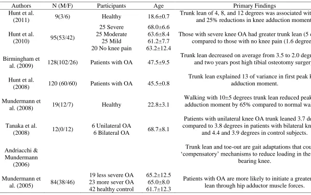
Table 1.1: Summary of findings from articles investigating trunk lean
Authors N (M/F) Participants Age Primary Findings
Hunt et al.
(2011) 9(3/6) Healthy 18.6±0.7
Trunk lean of 4, 8, and 12 degrees was associated with 7, 21, and 25% reductions in knee adduction moment.
Hunt et al.
(2010) 95(53/42)
25 Severe 25 Moderate
25 Mild 20 No knee pain
68.0±6.6 63.6±8.4 61.2±7.7 63.2±12.4
Those with severe knee OA had greater trunk lean (5 degrees) compared to those with no knee pain (1.6 degrees).
Birmingham et
al. (2009) 128(102/26) Patients with OA 47.5±9.5
Trunk lean decreased on average from 3.5 to 2.0 degrees pre and two years post high tibial osteotomy surgery.
Hunt et al.
(2008) 120 (60/60) Patients with OA 45.5±0.8
Trunk lean explained 13 of variance in first peak knee adduction moment.
Mundermann et
al. (2008) 19(12/7) Healthy 22.8±3.1
Walking with 10±5 degrees trunk lean reduced peak knee adduction moment by 65% compared to normal walking.
Tanaka et al.
(2008) 12(0/12)
6 Unilateral OA
6 Bilateral OA 68.7±8.1
Patients with unilateral knee OA trunk leaned 3.7 degrees compared to 3.8 degrees in patients with bilateral knee OA
and 4.4 and 3.9 degrees in control subjects.
Andriacchi & Mundermann
(2006)
Trunk lean and toe-out are gait adaptations that could be ‘compensatory’ mechanisms to reduce loading in the weight
bearing knee.
Mundermann et
al. (2005) 84(38/46)
19 less severe OA 23 more sever OA 42 healthy control
65.2±12.5 65.0±8.0 61.7±12.3
22
Table 1.2:Summary of findings from articles investigating toe-out
Birmingham
et al. (2009) 128(102/26) Patients with OA 47.5±9.5
Toe-out increased from 12.0 to 13.2degrees pre and two years post high tibial osteotomy surgery.
Hunt et al.
(2008) 120 (60/60) Patients with OA 45.5±0.8
Toe-out angle explained 12% of variance in first peak knee adduction moment.
Authors N (M/F) Participants Age Primary Findings
Fregly et al.
(2008) 1(1/0) Patient with OA 41
Increasing toe-out by 15 degrees reduced second peak knee adduction moment by 38%.
Jenkyn et al.
(2008) 180(141/39)
Medial compartment
OA 48.1(21-76)
Toe-out results in significant reductions in first and second peak knee adduction moment (11.7 and 34.4% respectively).
Lynn et al.
(2008) 11(6/5) Healthy 22.9±1.8
Increasing toe-out angle from 19 to 40 degrees resulted in a 12% increase and a 93% decrease in first and second peak knee adduction moment. Ten degrees toe in resulted in 64%
increase in second peak knee adduction moment.
Lynn et al.
(2008) 24(12/12) 12 Healthy
12 Knee OA
68.7±8.4 67.4±10
Exaggerated toe-out by 9.6 and 11 degrees resulted in reductions in second peak knee adduction moment in both
healthy adults (22.5%) and patients with knee OA (42%) respectively.
Reinbolt et al.
(2008) 1(1/0)
Medial compartment
OA 41
Increased toe-out resulted in a 6% increase and 31% decrease in first and second peak knee adduction moment respectively.
Rutherford et
al. (2008) 140(72/68)
50 Healthy 46 Mild to Moderate
44 Severe
53±10 60±9 67±8
Toe-out was associated with the knee adduction moment in only healthy and mild groups at second peak. Second peak is
23
with mild OA.
Schache et al.
(2008) 1(1/0) Healthy 26
Increasing toe-out by 11 degrees reduced the knee adduction moment by 23% during second peak.
Chang et al.
(2007) 56(23/33) Patients with OA 66.6
Greater toe-out was associated with a lower likelihood of knee OA progression (Odds Ratio = 0.60).
Guo et al.
(2007) 10(6/4) Patients with OA 64±8
A 16.6 degree increase in toe-out translated to a 40% reduction in second peak knee adduction moment.
Hurwitz et al.
(2002) 111(56/55)
62 with OA 49 healthy
62±10 59±10
Toe-out was significantly correlated with second peak knee adduction moment (r=-0.452).
Lin et al.
(2001) 44 Healthy children 11-13
Increased toe-out from 10 to 20 degrees resulted in a 55% reduction in first peak knee adduction moment and 700%
increase in second peak. Andrews et al.
(1996) 11(5/6) Healthy 23-42
24
1.6.2 Walking poles and Canes
Assistive walking devices such as canes are a common technique for individuals
with disability to maintain independence, improve function, enhance safety, and protect
joints (Van der Esch, Heijmans, & Dekker, 2003).
For patients with knee OA, the purpose of a cane, is in part to reduce knee joint
loading and symptoms related to OA. Patients with symptomatic knee OA who use a
cane, will carry the device on the contralateral side of the affected limb. By pressing
down on the device, a moment is generated about the knee in the frontal plane that acts to
abduct the femur relative to the tibia. Through the frontal plane force applied by the user
and the long lever arm, the moment provided by the cane resists the knee adduction
moment of the stance limb (Figure 1.5). This moment has been identified to be an
important factor in estimating the effectiveness of walking devices on reducing knee joint
load (Gross & Hillstrom, 2009). Kemp, Crossley, Wrigley, Metcalf, and Hinman (2008)
investigated the effects of contralateral cane use on knee joint loading in forty patients
with knee OA. They determined a 10% reduction in the knee adduction moment
compared to walking unassisted. Chan, Smith, Kirtley, and Tsang (2005) evaluated the
effects of cane placement in 14 patients with knee OA. They determined a 7% reduction
in knee adduction moment with contralateral cane placement compared to unaided gait.
Despite the positive effects of cane use, significant drawbacks have been identified such
as reduced walking speed (Chan et al., 2005).
Nordic walking poles may act similarly to canes as users are encouraged to apply
25
suggested as an option for individuals with disability to maintain a reasonable level of
physical activity (Fregly et al., 2009; Oakley et al., 2008). Manufacturers have since
incorporated populations with disability into marketing audiences as they are promoted to
improve fitness while reducing stress on the joints of the lower limb (Urban Poling Inc.,
2011). Walking poles however, could offer more benefit to patients with knee OA than
other assistive walking devices. Walking with poles has been shown to decrease ground
reaction force acting on the stance limb, increase walking speed, stride length, and
cadence (Willson, Torry, Decker, Kernozek, & Steadman, 2001) while achieving a higher
workload as measured by VO2 depending on surface type in healthy adults (Schiffer,
Knicker, Dannohl, & Struder, 2009; Hansen & Smith, 2009). Investigations focusing on
individuals with disability also report benefits. Oakley et al. (2008) investigated the
effects of walking with poles on 20 patients with Intermittent Claudication, a circulatory
disease of the lower extremities. They found patients walked further, with less pain, and
higher workload without an increase in perceived exertion compared to walking without
poles.
Findings from studies evaluating the effects of walking poles on knee joint
loading vary substantially. Fregly, D’Lima, and Colwell (2009) investigated medial
compartment direct contact force in one patient with an instrumented total knee
replacement. They observed a 27% decrease in medial compartment contact force late in
stance in one individual with an instrumented total knee replacement. Walter, D’Lima,
Colwell, and Fregly (2010) also evaluated knee joint loading in one individual with an
instrumented total knee replacement. They found a 33% and 47% decrease for first and
26
Conversely, Stief et al. (2008) investigated walking with poles on the lower extremities
during gait in 15 healthy adults. They found a 15% increase in first peak knee adduction
moment compared to unaided gait. Jensen et al. (2010) evaluated knee joint loading with
different magnitudes of pole force in 10 healthy adults. They found no change in either
first or second peak knee adduction moment between self-selected pole force and when
force applied to the pole was increased by 2.4 times.
In summary, the benefits associated with walking poles may make them desirable
to those dealing with the symptoms associated with knee OA. Their potential however, to
27
Figure 1.5. An illustration of the knee adduction moment and the moment generated by
the walking pole during gait. Knee
Adduction Moment Abduction
28
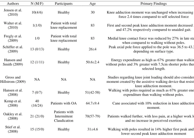
Table 1.3: Summary of findings from studies investigating walking poles and canes
Authors N (M/F) Participants Age Primary Findings
Jensen et al.
(2010) 10(4/6) Healthy 30 Knee adduction moment was unchanged when increasing pole
force 2.4 times compared to self selected force
Walter et al.
(2010) 1(1/0)
Patient with total
knee replacement 83 First and second peak knee adduction moment decreased 33.1
and 47.2% respectively compared to unaided gait.
Fregly et al.
(2009) 1/0
Patient with total
knee replacement 83 Medial knee contact force was reduced by 27% in late stance
when compared to walking without poles. Schiffer et al.
(2009) 13 (0/13) Healthy 26±4
Peak axial pole force applied to the pole was 36.5 to 43.3 N depending on surface type.
Hansen and
Smith (2009) 12 (1/11) Healthy 50.6±2.4
Energy expenditure as high as 67% greater than walking without poles and 3% greater with 7.5cm shorter poles then self
selected length.
Gross and
Hillstrom (2009) NA NA NA
Studies regarding knee joint loading should also consider the moment created by the assistive walking device that resists the
knee adduction moment. Hansen et al.
(2008) 7 (0/7) Healthy 51(42-58)
Walking with poles required as much as 67% greater energy expenditure than walking without poles.
Kemp et al. (2008)
40
(16/24) Patients with OA 64.7±9.4 Cane associated with 10% reduction in knee adduction
moment.
Oakley et al.
(2008) 21 (21/0)
Patients with Intermittent Claudication
70(57-79) Patients walked further, with less pain, at a higher work load,
and no increase in perceived exertion.
Stief et al.
(2008) 15 (15/0) Healthy 31±4.6 Walking with poles resulted in 14% higher first peak and 2%
29
Bohne et al.
(2007) 15 (15/0) Healthy (20-49)
Walking poles reduced sagittal plane moments for the ankle, knee, and hip while walking on a decline by 16.4, 10.6, and
9.7% respectively.
Chan et al.
(2005) 14(0/14) Patients with OA NA
The use of canes caused slower walking speeds, and greater knee adduction moment when held on the ipsilateral side (40%)
compared to an 8% decrease on the contralateral side.
Willson et al.
(2000) 13 (8/5) Healthy 29.5±5.1
Using walking poles increased gait speed, stride length, and cadence while decreasing ground reaction force compared to
30
1.7 Thesis Outline
The overall objective of this thesis was to explore the effects of prolonged
walking and the use of Nordic walking poles on selected gait characteristics indicative of
knee joint load. The thesis consists of a series of four studies. Data collection methods
included both over-ground and treadmill three-dimensional gait analysis. Healthy
participants had no disability or disease related to mobility, and were recruited from the
surrounding community. Patients with knee OA were assessed and diagnosed by
orthopaedic surgeons at the Fowler Kennedy Sport Medicine Clinic at the University of
Western Ontario. Patients were recruited from the pool of potential participants being
screened for an ongoing study investigating the effects of medial opening wedge high
tibial osteotomy surgery. Individuals with knee OA were referred to this clinic mostly
due to prolonged periods of unresolved pain localized to the knee. Data collected from
healthy adults in study one (Chapter 2) were shared with study two (Chapter 3).
The objectives of study one and two were to investigate the effects of prolonged
walking on gait patterns (selected kinematics) previously suggested to reduce knee joint
load. The first study (Chapter 2) evaluated the time-varying behaviour, reliability, and
validity of lateral trunk lean and toe-out angles during prolonged (60 min) treadmill
walking in healthy adults. Study 2 (Chapter 3) used the methodology evaluated in Study
1 to compare the time-varying behaviour of lateral trunk lean and toe-out during
prolonged (30 min) treadmill walking in healthy adults and patients with knee OA, and
31
reduced for patients with knee OA to mimic a realistic length of activity for patients with
painful mobility.
The objectives of the third and fourth studies were to analyze the effects of a
walking device (Nordic walking poles) on dynamic knee joint loading. The third study
(Chapter 4) was a technical report that quantified frontal plane forces and moments
generated by the pole about the knee in healthy adults. The fourth study (Chapter 5) used
the methodology developed in the third study to evaluate the effects of walking poles on
dynamic knee joint loading and related gait kinematics in patients with knee OA. The
final chapter (Chapter 6) summarized the overall findings and offered direction for future
investigations.
1.8 References
Altman, R., Asch, E., Bloch, D., Bole, G., Borenstein, D., Brandt, K., et al. (1986).
Development of criteria for the classification and reporting of osteoarthritis.
Arthritis and Rheumatism, 29(8), 1039-1049.
Andrews, M., Noyes, F. R., Hewett, T. E., & Andriacchi, T. P. (1996). Lower limb
alignment and foot angle are related to stance phase knee adduction in normal
subjects: A critical analysis of the reliability of gait analysis data. Journal of
Orthopaedic Research, 14, 289-295.
Andriacchi, T. P., & Mundermann, A. (2006). The role of ambulatory mechanics in the
initiation and progression of knee osteoarthritis. Current Opinion in Rheumatology,
32
Andriacchi, T., Mundermann, A., Smith, L., Alexander, E., Dyrby, C., & Koo, S. (2004).
A framework for the in vivo pathomechanics of osteoarthritis at the knee. Annals of
Biomedical Engineering, 32(3), 447-457.
Baker, K., Goggins, J., Xie, H., Szumowski, K., LaValley, M., Hunter, D. J. et al. (2007).
A randomized crossover trial of a wedged insole for treatment of knee
osteoarthritis. Arthritis and Rheumatism, 56(4), 1198-1203.
Birmingham, T. B., Hunt, M. A., Jones, I. C., Jenkyn, T. R., & Giffin, R. J. (2008).
Test-retest reliability of the peak knee adduction moment during walking in patients with
medial compartment knee osteoarthritis. Arthritis and Rheumatism, 57(6),
1012-1017.
Birmingham, T. B., Giffin, R. J., Chesworth, B. M., Bryant, D. M., Litchfield, R. B.,
Willits, K., et al. (2009). Medial opening wedge high tibial osteotomy: A
prospective cohort study of gait, radiographic, and patient-reported outcomes.
Arthritis and Rheumatism, 61(5), 648-657.
Bitton, R. (2009). The economic burden of osteoarthritis. The American Journal of
Managed Care, 15(8), S230-S235
Bohne, M., & Abendroth-Smith, J. (2007). Effects of hiking downhill using trekking
poles while carrying external loads. Medicine and Science in Sport and Exercise,
33
Bourret, E. M., Bernick, L. G., Cott, C. A., & Kontos, P. C. (2002). The meaning of
mobility for residents and staff in long term care facilities. Journal of Advanced
Nursing, 37(4), 338-345.
Chan, G. N. Y., Smith, A. W., Kirtley, C., & Tsang, W. (2005). Changes in knee
moments with contralateral versus ipsilateral cane usage in females with knee
osteoarthritis. Clinical Biomechanics, 20, 396-404.
Chang, A., Hurwitz, D., Dunlop, D., Song, J., Cahue, S., Hayes, K., et al. (2007). The
relationship between toe-out angle during gait and progression of medial
tibiofemoral osteoarthritis. Annals of Rheumatic Disease, 66, 1271-1275.
Charmez, K. (1995). The body, identity, and self: adapting to impairment. The
Sociological Quarterly, 36(4), 656-680.
Cott, C., & Gignac, M. (1999). Independence and dependence for older adults with
osteoarthritis of osteoporosis. Canadian Journal on Aging, 18(1), 1-25.
Crenshaw, S. J., Pollo, F. E., & Calton, E. F. (2000). Effects of lateral-wedged insoles on
the thrust of osteoarthritic knees. Clinical Orthopaedic Related Research, 375,
185-192.
Fregly, B. J., D’Lima, D. D., & Colwell, C. W. Jr. (2009). Effective gait patterns for
offloading the medial compartment of the knee. Journal of Orthopaedic Research,
34
Fregly, B. J., Reinbolt, J. A., & Chmielewski, T. L. (2008). Evaluation of a
patient-specific cost function to predict the influence of foot path on the knee adduction
torque during gait. Computer Methods in Biomechanics and Biomedical
Engineering, 11(1), 63-71.
Gaasbeek, R. D. A., Groen, B. E., Hampsink, B., Heerwaarden, R. J., & Duysens, J.
(2007). Valgus bracing in patients with medial compartment osteoarthritis of the
knee: A gait analysis study of a new brace. Gait and Posture, 26, 3-10.
Giffin, R. J., & Shannon, F., J. (2007). The role of the high tibial osteotomy in the
unstable knee. Sports Medicine Arthroscopy Review, 15(1), 23-31.
Gravlee, J. R., & Van Durme, M. D. (2007). Braces and splints for musculoskeletal
conditions. American Family Physician, 75(3), 342-348.
Gross, D. K., & Hillstrom, H. (2009). Primary care using noninvasive devices and
biomechanical principles. Medical Clinics of North America, 34(3), 179-200.
Guo, M., Axe, M. J., & Manal, K. (2007). The influence of foot progression angle on the
knee adduction moment during walking and stair climbing in pain free individuals
with knee osteoarthritis. Gait and Posture, 26, 436-441.
Hailey, D., & Tomie J. An assessment of gait analysis in the rehabilitation of children
35
Hansen, L., Henriksen, M., Larsen, P., & Alkjaer, T. (2008). Nordic walking does not
reduce the loading of the knee joint. Scandinavian Journal of Medicine Science and
Sports, 18, 436-441.
Hansen, E., & Smith, G. (2009). Energy expenditure and comfort during Nordic walking
with different pole lengths. Journal of Strength and Conditioning Research, 23(4),
1187-1194.
Health Canada (2003). Arthritis in Canada. An ongoing challenge. Health Canada,
Ottawa.
Hsu, R. W. W., Himeno, S., Coventry, M. B., & Chao, E. Y. S. (1990). Normal axial
alignment of the lower extremity and load-bearing distribution at the knee. Clinical
Orthopaedics and Related Research, 255, 215-227.
Hunt, M. A., Birmingham, T. B., Bryant, D., Jones, I., Giffin, J. R., Jenkyn, T. R. et al.
(2008). Lateral trunk lean explains variation in dynamic knee joint load in patients
with medial compartment knee osteoarthritis. Osteoarthritis and Cartilage, 16,
591-599.
Hunt, M. A., Simic, M., Hinman, R. S., Bennell, K. L., & Wrigley, T. V. (2011).
Feasibility of a gait retraining strategy for reducing knee joint loading: Increased
trunk lean guided by real-time biofeedback. Journal of Biomechanics,44(5),
943-947.
Hunt, M. A., Wrigley, T. V., Hinman, R. S., & Bennell, K. L. (2010). Individuals with
36
compared with individuals with less severe OA and those without knee pain.
Arthritis Care and Research, 62(10), 1426-1432.
Hurwitz, D. E., Sumner, Andriacchi, T. P., & Sugar (1998). Dynamic knee loads during
gait predict proximal tibial bone distribution. Journal of Biomechanics, 31,
423-440.
Hurwitz, D. E., Ryals, A. R., Case, J. P., Block, J. A., & Andriacchi, T. P. (2002). The
knee adduction moment during gait in subjects with knee osteoarthritis is more
closely correlated with static alignment than radiographic disease severity, toe out
angle and pain. Journal of Orthopaedic Research, 20, 101-107.
Jackson, B. D., Teichtahl, A. J., Morris, M. E., Wluka, A. E., Davis, S. R., & Cicuttini, F.
M. (2004). The effects of knee adduction moment on tibial cartilage volume and
bone size in healthy women. Rheumatology, 43, 311-314.
Jenkyn, T. R., Hunt, M. A., Jones, I. C., Giffin, R. J., & Birmingham, T. B. (2008).
Toe-out gait in patients with knee osteoarthritis partially transforms external knee
adduction moment into flexion moment during early stance phase of gait: A
tri-planar kinetic mechanism. Journal of Biomechanics, 41, 276-283.
Jensen, S. B., Henriksen, M., Aaboe, J., Hansen L., Simonsen, E. B., & Alkjaer, T.
(2010). Is it possible to reduce the knee joint compression force during level
walking with hiking poles? Scandinavian Journal of Medicine and Science in
37
Kellgren, J. H., & Lawrence, J. S. (1957). Radiological assessment of osteoarthritis.
Annals of the Rheumatic Diseases, 16, 494-502.
Kemp, G., Crossley, K. M., Wrigley, T. V., Metcalf, B. R., & Hinman, R. S. (2008).
Reducing joint loading in medial knee osteoarthritis: Shoes and canes. Arthritis and
Rheumatism, 59(5), 609-614.
Kerrigan, D. C., Lelas, J. L., Goggins, J., Merriman, G. J., Kaplan, R. J., & Felson, D. T.
(2002). Effectiveness of a lateral-wedge insole on knee varus torque in patients
with knee osteoarthritis. Archives of Physical Medicine and Rehabilitation, 83,
889-893.
Kirkley, A., Webster-Bogaert, S., Litchfield, R., Amendola, A., MacDonald, S.,
McCalden, R. et al. (1999). The effect of bracing on varus gonarthrosis. Journal of
Bone and Joint Surgery, 81, 539-548.
Komistek, R. D., Dennis, D. A., Northcut, E. J., Wood, A., Parker, A. W., & Traina, S.
M., (1999). An invivo analysis of the effectiveness of the osteoarthritic knee brace
during heel-strike of gait. Journal of Arthroplasty, 14, 738-742.
Lawrence, R., Felson, D., Helmick, C., Arnold, L., Choi, H., Deyo, R. et al. (2008).
Estimates of the prevalence of arthritis and other rheumatic conditions in the united
states. Arthritis and Rheumatism, 58(1), 26-35.
Lin, C., Lai, K., Chou, Y., & Ho, C. (2001). The effect of changing the foot progression
angle on the knee adduction moment in normal teenagers. Gait and Posture, 14,
38
Lindenfeld, T. N., Hewett, T. E., & Andriacchi, T. P. (1997). Joint loading with valgus
bracing in patients with varus gonarthrosis. Clinical Orthopaedics Related
Research, 344, 290-297.
Lynn, S. K., & Costigan, P. A. (2008). Effect of foot rotation on knee kinetics and
hamstring activation in older adults with and without signs of knee osteoarthritis.
Clinical Biomechanics, 23, 779-786.
Lynn, S. K., Kajaks, T., & Costigan. P. A. (2008). The effect of internal and external foot
rotation on the adduction moment and lateral-medial shear force at the knee during
gait. Journal of Science and Medicine in Sport, 11, 444-451.
Maillefert, J. F., Hudry, C., Baron, G., Baron, G., Kieffert, P., Bourgeois, P. et al. (2001).
Laterally elevated wedged insoles in the treatment of medial knee osteoarthritis: a
prospective randomized controlled study. Osteoarthritis Cartilage, 9(8), 738-745
Maly, M., & Krupa, T. (2007). Personal experience of living with knee osteoarthritis
among older adults. Disability and Rehabilitation, 29(18), 1423-1433.
Marsh, A. P., Katula, J. A., Pacchia C. F., Johnson, L. C., Koury K. L., & Rejeski W. J.
(2006) Effect of treadmill and overground walking on function and attitudes in
older adults. Medicine and Science in Sport and Exercise, 38(6), 1157-1164.
Matsas, A., Taylor, N., & McBurney, H. (2000) Knee joint kinematics from familiarized
treadmill walking can be generalized to overground walking in young unimpaired