Western University Western University
Scholarship@Western
Scholarship@Western
Electronic Thesis and Dissertation Repository
8-21-2019 3:00 PM
Scaffold Design Considerations for Soft Tissue Regeneration
Scaffold Design Considerations for Soft Tissue Regeneration
Madeleine M. Di Gregorio The University of Western Ontario
Supervisor
Hamilton, Douglas W.
The University of Western Ontario
Graduate Program in Biomedical Engineering
A thesis submitted in partial fulfillment of the requirements for the degree in Master of Engineering Science
© Madeleine M. Di Gregorio 2019
Follow this and additional works at: https://ir.lib.uwo.ca/etd
Part of the Biomaterials Commons
Recommended Citation Recommended Citation
Di Gregorio, Madeleine M., "Scaffold Design Considerations for Soft Tissue Regeneration" (2019). Electronic Thesis and Dissertation Repository. 6532.
https://ir.lib.uwo.ca/etd/6532
This Dissertation/Thesis is brought to you for free and open access by Scholarship@Western. It has been accepted for inclusion in Electronic Thesis and Dissertation Repository by an authorized administrator of
ii
Abstract
Tissue engineering has emerged as a promising strategy for the replacement of
degenerating or damaged tissues in vivo. Also known as regenerative medicine, integral to this therapeutic strategy is biomimetic scaffolds and the biomaterial structural components used to form them. In this study, three different biomaterial scaffolds for tissue engineering applications were fabricated: three-dimensional reverse embedded collagen scaffolds, polymer fusion printed polycaprolactone (PCL) scaffolds, and electrospun gelatin scaffolds. Three-dimensional collagen and PCL scaffolds promoted human adipose-derived stem/stromal cell (ASC) spreading,
proliferation, and fibronectin deposition in vitro. Secondly, this study investigated the efficacy of exogenous galectin-3 delivery as a therapeutic in skin healing, given that galectin-3 has been implicated in several wound healing processes. Gelatin polymer blended with recombinant galectin-3 was electrospun into a protein delivery scaffold and employed in a murine model of cutaneous wound healing. Treatment of wounds with the galectin-3/gelatin scaffolds, or with topical galectin-3, did not enhance wound closure, re-epithelialization, or influence macrophage phenotypes in vivo.
Keywords
iii
Summary for Lay Audience
Following injury, the body’s natural healing mechanisms mount a defense against
iv
Co-Authorship Statement
This thesis was written by M.M. Di Gregorio with input, suggestions, and revisions from Dr. D.W. Hamilton. Experiments were designed by Dr. D.W. Hamilton. All studies were
v
Acknowledgements
I am sincerely thankful to Dr. Douglas Hamilton for his supervision of my research, mentorship, and support of my academic goals. Thank you for sharing your immense knowledge and showing patience and kindness during my graduate work. Production of this thesis would not have been possible without your guidance and encouragement. Thank you to my advisory
committee members, Dr. Elizabeth Gillies and Dr. Amin Rizkalla, for valuable input regarding my project’s organization and goals. This collaboration has been incredibly rewarding.
Thank you to all members of the Hamilton lab. It has been a pleasure to meet and work alongside an amazing group of scientists; I am thrilled to see what the future holds for each of you. JT and Georgia taught me animal studies. Adam taught me the fundamentals of
electrospinning and was an excellent resource when I had questions. Sarah helped edit this thesis; Sanduni and Alex offered unlimited positivity and friendship. Thank you, Dr. John de Bruyn, for teaching me rheological testing. Thank you also to Dr. Rizkalla and Khalid for your collaboration and introducing me to polymer fusion 3D printing.
Thank you to my family especially; Dad, Oma and Nonno, for your endless
vi
Table of Contents
Abstract ... ii
Keywords... ii
Summary for Lay Audience ... iii
Co-Authorship Statement ... iv
Acknowledgements ... v
Table of Contents ... vi
List of Tables ... ix
List of Figures ... x
List of Appendices ... xi
List of Abbreviations ... xii
1 Introduction ... 1
1.1 Cutaneous Wound Healing ... 1
1.1.1 Anatomy and Physiology of Skin ... 1
1.1.2 Acute Wound Healing ... 5
1.1.3 Chronic Wounds ... 9
1.1.4 Clinical Treatment of Chronic Wounds ... 11
1.2 Tissue Engineering and Regenerative Medicine ... 17
1.2.1 Biomaterial Scaffold Design Requirements ... 17
1.2.2 Three-Dimensional Bioprinting ... 18
1.2.3 Collagen-Based Biomaterials ... 19
1.2.4 Polycaprolactone-Based Biomaterials ... 20
1.2.5 Electrospinning ... 21
1.2.6 Stem Cell Therapy in Wound Healing ... 24
1.3 Galectin-3 as a Therapeutic for Chronic Wounds ... 26
1.3.1 Galectin-3 Overview ... 26
1.3.2 Roles in Inflammation ... 26
1.3.3 Roles in Angiogenesis ... 27
1.3.4 Roles in Re-Epithelialization ... 28
1.4 Hypothesis and Objectives ... 28
1.4.1 Rationale ... 28
1.4.2 Hypothesis ... 29
1.4.3 Objectives ... 29
1.5 References ... 31
2 Design and Validation of 3D Printed Scaffolds for Adipose Derived Stromal Cell Tissue Engineering ... 46
2.1 Introduction ... 46
2.2 Materials and Methods ... 47
vii
2.2.2 Gelatin Support Slurry Rheology ... 48
2.2.3 Gelatin Support Slurry Microparticle Morphology ... 49
2.2.4 Scaffold Computer-Aided Design ... 49
2.2.5 Printing of Porous Collagen Scaffolds ... 50
2.2.6 Printing of Porous Polycaprolactone Scaffolds ... 50
2.2.7 3D Printed Scaffold Morphology ... 51
2.2.8 Collagen Scaffold Hydrophilicity ... 51
2.2.9 Cell Isolation and Culture ... 51
2.2.10 In Vitro Proliferation Assay ... 52
2.2.11 In Vitro Cytoskeleton Morphology ... 52
2.2.12 In Vitro Fibronectin Detection ... 53
2.2.13 In Vitro Phenotype Study ... 53
2.3 Results ... 57
2.3.1 Preparation of 3D Printed Scaffolds ... 57
2.3.2 In Vitro Validation of 3D Printed Scaffolds as a Support for ASC Cell Growth ... 71
2.4 Discussion and Conclusion ... 87
2.5 References ... 90
3 Investigating the Effects of Exogenous Galectin-3 in Murine Wound Healing ... 93
3.1 Introduction ... 93
3.2 Materials and Methods ... 94
3.2.1 Electrospinning Galectin-3-Loaded Fibrous Gelatin Scaffolds ... 94
3.2.2 Scanning Electron Microscopy ... 95
3.2.3 Effect of Galectin-3 on Murine Wound Healing In Vivo ... 95
3.2.4 Murine Wound Healing Kinetics ... 96
3.2.5 Effect of Galectin-3 on Arginase I Enrichment During Wound Healing ... 97
3.3 Results ... 97
3.3.1 Preparation of Electrospun Galectin-3/Gelatin Scaffolds ... 97
3.3.2 In Vivo Effects of the Electrospun Galectin-3/Gelatin Scaffold ... 102
3.4 Discussion and Conclusion ... 118
3.5 References ... 121
4 General Discussion ... 124
4.1 Summary of Thesis Objectives ... 124
4.2 Summary of Fabricated Biomaterial Scaffolds ... 126
4.3 Galectin-3 to Modulate Inflammation In Vivo ... 128
4.4 Future Directions ... 130
4.4.1 Improving 3D Printed Scaffolds ... 130
4.4.2 Improving Electrospun Scaffolds ... 131
4.4.3 ASC Differentiation ... 132
4.4.4 Establishing In Vitro and In Vivo Effects of Galectin-3 ... 132
4.5 Limitations of Results ... 134
4.5.1 Human Adipose Derived Stem/Stromal Cells ... 134
4.5.2 Murine Model of Wound Healing ... 135
4.6 Conclusion ... 136
viii
5 Appendices ... 144
Appendix A: Supplementary Rheological Results ... 144
Appendix B: G-Code and FFF Files for Scaffolds ... 145
Appendix C: Summary of Early Embedded Printing Attempts ... 163
Appendix D: Autofluorescence of PCL Scaffolds ... 165
Appendix E: Example of the Appearance of Individual Z Slices for Scaffold Confocal Microscopy . 166 Appendix F: Arginase-I Immunofluorescent Staining of Day 9 Murine Wound Sections No Primary Antibody Negative Control ... 168
Appendix G: Research Ethics Approvals ... 169
Curriculum Vitae ... 171
ix
List of Tables
Table 1. A summary of cutaneous cell types within the epidermis, dermis, and
hypodermis 4
Table 2. Spatiotemporal coordination of normal wound healing 8
x
List of Figures
Figure 2.1. Preparation of the Sacrificial Support Slurry 55
Figure 2.2. Justification for Reverse Embedded Printing 59
Figure 2.3. Rheological Properties of the Gelatin Support Slurry 61
Figure 2.4. Effect of Blend Duration on Gelatin Microparticle Morphology 63
Figure 2.5. Collagen Scaffold Computer-Aided Design 65
Figure 2.6. Porous Collagen Scaffold Morphology 67
Figure 2.7. Polycaprolactone (PCL) Scaffold Morphology 69
Figure 2.8. ASC Proliferative Potential 73
Figure 2.9. ASC Cytoskeletal Morphology 75
Figure 2.10. ASC Cytoskeletal Morphology after 14 Days 78
Figure 2.11. Fibronectin Detection After 3 Days 80
Figure 2.12. Fibronectin Detection After 14 Days 83
Figure 2.13. ASC Phenotype Analysis 85
Figure 3.1. Effect of Gelatin on Electrospun Fiber Morphology 98
Figure 3.2. Refined Electrospun Fiber Morphology 100
Figure 3.3. Representative Images of Murine Full-Thickness Dermal Wounds 104
Figure 3.4. Quantification of Murine Wound Healing Kinetics 106
Figure 3.5. Masson’s Trichrome Staining Following In Vivo Full-Thickness Wounding 108 Figure 3.6. Re-epithelialization and Epithelial Thickness Following In Vivo
Full-Thickness Wounding 110
Figure 3.7. Example of Biological Variability in Masson’s Trichrome Staining of
Full-Thickness Wounds 112
Figure 3.8. Arginase I Enrichment of Full-Thickness Wounds 114
xi
List of Appendices
Appendix A: Supplementary Rheological Results ... 144
Appendix B: G-Code and FFF Files for Scaffolds ... 145
Appendix C: Summary of Early Embedded Printing Attempts ... 163
Appendix D: Autofluorescence of PCL Scaffolds ... 165
Appendix E: Example of the Appearance of Individual Z Slices for Scaffold Confocal Microscopy . 166 Appendix F: Arginase-I Immunofluorescent Staining of Day 9 Murine Wound Sections No Primary Antibody Negative Control ... 168
xii
List of Abbreviations
2D Two-dimensional
3D Three-dimensional
AMM Amniotic mesenchymal stem cell
ANOVA Analysis of variance
ADSC Adipose-derived stem cell
AD-MSC Adipose-derived mesenchymal stem cell
AMSC Adipose MSC
ASC Adipose-derived stem/stromal cell
Asc 2-P Ascorbic acid 2-phosphate
AM Additive manufacturing
BGP β-glycerophosphate, glycerol 2-phosphate
BM Bone marrow
BMDM Bone marrow derived macrophage
BO Bio-Oss
BSA Bovine serum albumin
CAD Computer-aided design
CCN2 Connective tissue growth factor 2
CKI Cyclin-dependent kinase inhibitor
CSR Controlled shear rate
CXCL Chemokine ligand
c-Abl Abelson tyrosine kinase
DCB Decellularized bone
DFU Diabetic foot ulcer
DMEM Dulbecco's modified eagle medium
DMSO Dimethyl sulfoxide
DNA Deoxyribonucleic acid
ECM Extracellular matrix
EGF Epidermal growth factor
FBS Fetal bovine serum
xiii
FGF Fibroblast growth factor
FRESH Freeform reversible embedding of suspended hydrogel
GAG Glycosaminoglycan
GSK-3β Glycogen synthase kinase-3 beta
G’ Storage modulus
G” Loss modulus
HBOT Hyperbaric oxygen therapy
HF Hair follicle
HFSC Hair follicle stem cell
IBMX Isobutylmethylxanthine
IFN-γ Interferon gamma
IFE Interfollicular epidermis
IL Interleukin
iNOS Inducible nitric oxide synthase
LPS Lipopolysaccharide
M1 Classical activated macrophage
M2/a Alternative activated macrophage
M2b Type 2 macrophage
M2c Deactivated macrophage
M2d M2-like macrophage
MCR Modular compact rheometer
MMP Matrix metalloproteinase
MRWD Moisture-retentive wound dressing
MSC Mesenchymal stem cell
NO Nitric oxide
NPWT Negative-pressure wound therapy
PBS Phosphate buffered solution
PC Progenitor cell
PCL Poly(ϵ-caprolactone)
PCLDMA Polycaprolactone dimethylacrylate
xiv
PEGDA Poly(ethylene glycol) diacrylate
PEO Poly ethylene oxide
PI Propidium Iodide
PLA Polylactic acid
PLGA Poly(lactic-co-glycolic)acid
PPARγ Peroxisome proliferator-activated receptor gamma
PSA Prostate-specific antigen
PU Pressure ulcer
PVA Polyvinyl alcohol
RM Regenerative medicine
RNA Ribonucleic acid
ROI Reactive oxygen intermediate
RPM Revolutions per minute
RUNX2 Runt-related transcription factor 2
SC Stem cell
SG Sebaceous gland
SMA Smooth muscle actin
SMC Smooth muscle cell
STL Stereolithography
SVF Stromal vascular fraction
T3 Triiodothyronine
TCP Tricalcium phosphate
TCPS Tissue culture polystyrene
TBS Tris-buffered saline
TE Tissue engineering
TGF Transforming growth factor
TIMP Tissue inhibitor of matrix metalloproteinase
TNF Tumor necrosis factor
TRM Tissue resident memory
UV Ultraviolet
xv
VEGFR Vascular endothelial growth factor receptor
VLU Venous leg ulcer
V/V Volume per volume
WT Wild type
𝜂 Viscosity
|η*| Complex viscosity
1
1
Introduction
1.1 Cutaneous Wound Healing
1.1.1 Anatomy and Physiology of Skin
Skin is part of the integumentary system and represents the largest organ of the human body.1,2 Skin is multifunction: it acts as a selectively permeable barrier to protect the body from
harmful pathogens in the external environment; thermoregulation is achieved through blood vessel control and perspiration via sweat glands; cutaneous sensory receptors, including
mechanoreceptors, nociceptors (pain) and thermoreceptors, mediate the body’s interactions with the external environment.2,3 The epidermis, dermis, and subcutaneous hypodermis of skin are
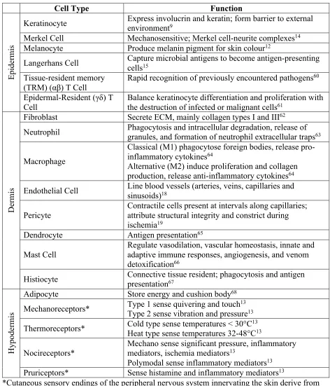
comprised of a variety of cell types with specific biological roles (Table 1).2
The epidermis is subdivided into five layers: the stratum basale, stratum spinosum, stratum granulosum, stratum lucidum, and stratum corneum.4–6 The epidermis is the outermost
layer of skin and provides a cell-dense barrier between the body and the external environment. Keratin, the main structural protein found in the epidermis, is water insoluble and mechanically protects epithelial cells from damage or stress while preventing the invasion of foreign bodies.7
This water-resistant barrier is synthesized by keratinocytes, which are the dominant cell type within the epidermis, accounting for 95% of the cells located in this layer.8 Cornification, or
keratinization, is the process by which proliferating keratinocytes of the basal layer undergo terminal differentiation into highly specialized corneocytes.9,10 In this process, keratinocytes
migrate superficially, become flat, polyhedral, anucleated, and lose 55% of their water volume.3
Other cell types present in the epidermis include melanocytes, which produce melanosomes containing melanin.11,12 This protein provides skin with pigmentation and protects skin from
ultraviolet radiation that can cause sunburn injury to skin, damage to nucleic DNA, and
accelerated aging of skin.3 Merkel cells, also known as Merkel-Ranvier cells or tactile epithelial
cells, are oval-shaped mechanoreceptors essential for light touch sensation in hairless skin of vertebrates.13,14 Langerhans cells also occupy the epidermis and act as antigen-presenting cells.15
The dermal layer confers pliability, elasticity, and tensile strength to skin.1–3 These
mechanical properties are achieved through a diverse arrangement of connective tissues, cell types, and structural components. The extracellular matrix (ECM) is a three-dimensional
2
and collagen, and hydrated gels of glycosaminoglycans (GAGs) linked to proteoglycans.2,3,16 The
ECM occupies the interstitial space, providing structural support and biochemical cues to cells, while also acting as a compression buffer. The most abundant ECM protein is collagen (types I and III), which is synthesized by dermal fibroblasts and conveys structural integrity to the skin by resisting stress.3 In addition to fibroblasts, various leukocyte populations (including
neutrophils and macrophages) can enter the dermis through the vascular networks in response to different stimuli. Blood vessels, lined with squamous endothelial cells, can become permeable allowing leukocytes and clotting proteins access to connective tissue.17,18 On the outside of blood
vessels, pericytes wrap around capillaries and confer structural integrity to the vessel wall.19
Other components of the dermal layer include epidermally derived appendages such as hair follicles, nails, and sebaceous, sweat and mammary glands; also, dermal dendrocytes, mast cells, histiocytes, blood vessels, nerves, and lymphatics. The dermis is subdivided into the upper papillary dermis and lower reticular dermis, with distinctions in the structure and organization of collagen and elastin fibers between these sublayers. The dermal vasculature provides nutrition to the skin and assists the body in thermoregulation. Dilation and constriction of blood vessels leads to heat dissipation or conservation, respectively.20 Thermoregulation is also controlled by
arrector pili muscles (attached to hair follicles) and sweat glands.3 Piloerection, the erection or
bristling of hairs, traps air close to the skin for an additional layer of insulation. Endothermic evaporation of sweat cools the surface of the skin.
The subcutaneous hypodermis consists primarily of fatty adipocytes but also
contains fibrous septa of loose connective tissue, nerves, and blood vessels. Hypodermal fat lobules insulate and cushion the body, provide buoyancy, and store energy.3 In addition to
adipocytes, fibroblasts and macrophages are also found in the hypodermis. Together, the epidermis, dermis, and hypodermis allow skin to maintain a physical barrier to the external environment; if this barrier is disrupted via injury to the skin, a spatiotemporally coordinated process is initialed in order to restore barrier function, maintain internal homeostasis and guard sterility.21
Skin cells are continually shed and replenished through desquamation, the shedding of the outermost layer of the stratum corneum. In this process, individual corneocytes are shed following degradation of cell-cell junctions known as corneodesmosomes.22 The rate of
3
maintained by permanently residing stem cells that sustain principal differentiated epidermal lineages, the interfollicular epidermis (IFE), sebaceous gland (SG), hair follicle (HF),24,25 and
Merkel cell mechanoreceptors.26 Through radiation dose-survival studies, it was proposed that
stem cells comprise about 2-7% of basal layer cells27 while another study of murine basal layer
cells suggested a larger stem cell population, 10-12% of cells in the basal layer.28 Maintenance of
skin homeostasis is dependent on the ability of stem cells to replenish the turnover of apoptosing epithelial lineages.
Epithelial stem cells (SCs) reside in a specific microenvironment called the niche, where stem cell behaviour is influenced by cell-cell communication, cell-ECM interactions, and growth factors.29 Cutaneous mesenchymal stem/stromal cells (MSCs) include dermal papilla cells (DPC)
involved in hair follicle cycling,30,31 and the dermal sheath cells (DSC), which are capable of
differentiating into wound healing fibroblasts that repair the dermis.32 Human FH-derived DPCs
and DSCs have been shown to differentiate into adipogenic and osteogenic lineages in vitro, suggesting multi-lineage potential of these MSCs.33 Hair follicle stem cells (HFSCs) reside in the
follicle bulge and have been shown to give rise to IFE, HF, and SG lineages after
transplantation.34–39 Under normal physiological conditions, however, lineage tracing in mice has
shown that HFSCs only contribute to HF regeneration and not the SG, IFE, or infundibulum.40–45
In the IFE, a single basal layer of proliferative cells replenishes suprabasal terminally differentiated cells. This basal layer consists of a heterogeneous proliferative population of quiescent, long-lived IFE SCs and short-lived, differentiation-fated progenitor cells (PC).46–52
Between the bulge and SG is the isthmus, where another pool of resident SCs maintain the isthmus, SG, SG ducts, and infundibulum.53–56 Epithelial SCs are normally confined to
4
Table 1. A summary of cutaneous cell types within the epidermis, dermis, and hypodermis.
Cell Type Function
E
pi
de
rm
is
Keratinocyte Express involucrin and keratin; form barrier to external environment9
Merkel Cell Mechanosensitive; Merkel cell-neurite complexes14
Melanocyte Produce melanin pigment for skin colour12
Langerhans Cell Capture microbial antigens to become antigen-presenting
cells15
Tissue-resident memory (TRM) (αβ) T Cell
Rapid recognition of previously encountered pathogens60
Epidermal-Resident (γδ) T Cell
Balance keratinocyte differentiation and proliferation with the destruction of infected or malignant cells61
D
erm
is
Fibroblast Secrete ECM, mainly collagen types I and III62
Neutrophil Phagocytosis and intracellular degradation, release of granules, and formation of neutrophil extracellular traps63
Macrophage
Classical (M1) phagocytose foreign bodies, release pro-inflammatory cytokines64
Alternative (M2) induce proliferation and collagen production, release anti-inflammatory cytokines64
Endothelial Cell Line blood vessels (arteries, veins, capillaries and sinusoids)18
Pericyte
Contractile cells present at intervals along capillaries; attribute structural integrity and constrict during ischemia19
Dendrocyte Antigen presentation65
Mast Cell Regulate vasodilation, vascular homeostasis, innate and adaptive immune responses, angiogenesis, and venom detoxification66
Histiocyte Connective tissue resident; phagocytosis and antigen presentation67
H
ypode
rm
is
Adipocyte Store energy and cushion body68
Mechanoreceptors* Type 1 sense quivering and touch13
Type 2 sense vibration and pressure13
Thermoreceptors* Cold type sense temperatures < 30°CHeat type sense temperatures 32-48°C1313
Nocireceptors* Mechano sense significant pressure, inflammatory mediators, ischemia mediators13
Polymodal sense inflammatory mediators13
Pruriceptors* Sense histamine and inflammatory mediators13
*Cutaneous sensory endings of the peripheral nervous system innervating the skin derive from the dorsal root ganglia and the trigeminal ganglia. Nerves in the skin form epidermal plexus from which some fibres cross the dermo-epidermal junction.13
5 1.1.2 Acute Wound Healing
Following injury, normal wound healing is achieved via four overlapping phases: hemostasis, inflammation, proliferation, and remodeling (Table 2). Thrombin, the principal enzyme involved in hemostasis, is produced primarily in the liver and circulates systemically in the blood plasma. This serine protease cleaves fibrinogen to create a polymerized fibrin matrix that captures circulating platelets to form a hemostatic plug and establish hemostasis.69 Within
the first hour post-injury, inflammatory cytokines such as interleukin (IL) -1α, IL-1β, IL-6, IL-8, tumor necrosis factor (TNF) -α, platelet derived growth factor (PDGF), and transforming growth factor (TGF) -β, are released by aggregated platelets to recruit early inflammatory cells and initiate healing.70 When injury first occurs, vasoconstriction is induced locally to reduce blood
loss. However, this is subsequently reversed to allow changes in blood flow, an increase in permeability of blood vessels, and the infiltration of fluid, proteins, and leukocytes from the circulatory system into the site of tissue damage.
Recruited leukocytes produce a chemotactic gradient that stimulates the migration of neutrophils, monocytes, smooth muscle cells (SMCs), and fibroblasts. Neutrophils, which
comprise 60-70% of the leukocyte population, are part of the innate immune response and one of the first cell types to respond to injury. Neutrophils are granulocytes and lack immune memory; they function by releasing reactive oxygen species to produce toxic metabolites (hydrogen peroxide) that kill invading bacteria and certain fungal species, secreting serine proteases and matrix metalloproteinases (MMPs) to debride necrotic tissue, and phagocytosing dead bacteria and wound debris.70 Neutrophils cannot renew their lysosomes and are short-lived, resulting in
pro-6
regenerative wound healing and tissue repair. Following exhaustion of inflammatory cells, exocytotic release of a second wave of signaling molecules, including TGF-α, TGF-β, fibroblast growth factor (FGF) 2/basic FGF (bFGF), PDGF, and vascular endothelial growth factor
(VEGF), recruits keratinocytes, fibroblasts, endothelial cells, and pericytes to initiate the overlap of late-stage inflammation with the proliferative stage of healing.71
During fibroplasia, fibroblasts proliferate and migrate into the wound bed, depositing collagen-rich ECM to slowly replace the fibrin clot with a permanent matrix.72 This ECM
improves structural integrity of the damaged site and provides a supportive scaffold for neovascular growth (angiogenesis) and re-epithelialization.73 Through cadherin-11-mediated
adhesion between macrophages and fibroblastic cells, β-producing macrophages and TGF-β-activating myofibroblasts are held in close proximity, facilitating efficient myofibroblast activation and stimulating fibrosis.74 Persistent fibroplasia leads to the accumulation of a dense
fibrotic scar tissue.72 As ECM is deposited, re-epithelializing keratinocytes upregulate
anti-fibrotic urokinase-type plasminogen activator, MMP-1 and MMP-3, and downregulate pro-fibrotic connective tissue growth factor 2 (CCN2), collagen I and II, fibronectin, plasminogen activator inhibitor-1, α-smooth muscle actin (α-SMA), and tissue inhibitor of matrix
metalloproteinase (TIMP)-2/3.75 Maturation of the granulation tissue, composed of the newly
deposited collagen network and migrating endothelial cells and macrophages, is enhanced by angiogenesis and re-epithelialization.76
Angiogenesis, vascular growth from pre-existing vasculature via endothelial cell migration, proliferation and vessel formation, re-establishes normoxia and nutrient supply to tissues.71,77 Post-injury hypoxic conditions promote the release of inflammatory mediators to
increase vascular permeability and dilation, facilitating endothelial cell migration into the wounded tissue.78,79 Angiogenic factors, particularly VEGF-A and FGF-2/bFGF, promote
endothelial cell proliferation and the formation of new capillary tubules in the developing granulation tissue to restore vascularization.80–83 To prevent excessive scar formation, wound
progression through fibroplasia and angiogenesis must be tightly regulated. Proteolytic ECM remodeling decreases ECM density, decreases cell-ECM interactions (by cleaving matrix components such as collagen, fibronectin and laminin84), and releases matrix-bound angiogenic
7
During re-epithelialization, epithelial cell proliferation and migration is initiated.
Fibroblasts and macrophages release epidermal growth factor (EGF), TGF-α and FGF, initiating re-epithelialization.86 Hemidesmosome links between the epidermis and the basement membrane
separate, releasing cell-ECM linkages. Keratinocyte migration over the granulation tissue separates the eschar from viable tissue. Behind the migrating epithelial tongue, keratinocytes proliferate and mature, ultimately restoring the barrier function.87 Macrophages secrete TGF-β
that signals fibroblasts to migrate into the granulation tissue and produce new ECM
components.88 Macrophage-activated myofibroblasts, highly contractile cells, anchor to the ECM
and contract the wound, pulling opposing edges together.86,87 Myofibroblast contractility is
dependent on a positive feedback loop initiated by endogenous TGF-β and tensile forces, which increases the density of α-SMA enrichment in stress fibers, increases force production and tension development, and consequently upregulates TGF-β. Re-modeling of granulation tissue occurs as fibroblasts upregulate expression of collagen I, and MMPs degrade the temporary collagen III matrix. In the final stages of remodeling, the scar tissue consists of mainly an acellular matrix of parallel collagen I fiber bundles.87,89,90 The repaired tissue which has the
composition of a scar does successfully restore barrier function, but possesses approximately 80% of the mechanical elasticity of unwounded skin.87,91 However, in certain instances, skin can
8
Table 2. Spatiotemporal coordination of normal wound healing.
Post-Injury Period
Within Minutes
Within
Hours ~3 Days ~1 Week Weeks Years
Wound Healing Stage
Hemostasis Inflammation Proliferation Remodeling
Active Cell Types
-Platelets -Neutrophils
-M1
macrophages -Mast cells
-Fibroblasts -M2 macrophages -Endothelial cells -Keratinocytes
-Myofibroblasts
Major Events
-Vasoconstriction -Hemostatic plug formation
-Chemotactic recruitment of inflammatory cells
-Inflammatory cell migration into the wound bed
-Defense against foreign infection
-Granulation tissue -Fibroplasia
-Randomly oriented ECM
-Angiogenesis -Re-epithelialization
-Parallel collagen ECM
-Contraction of wound bed -Scar
9 1.1.3 Chronic Wounds
Reduced ability to heal is often the result of a variety of morbidities including vascular insufficiency, diabetes, malnutrition, patient age, pressure, infection, and edema.70 A persistent
tissue injury that fails to proceed through the reparative process after 3 months is classified as a chronic (non-healing) wound.92 Such wounds become a significant burden to the patient, causing
pain, impaired limb function, sepsis, hospitalization and the need for amputation. Worldwide, chronic wounds account for approximately 4% of the global healthcare expenditure.93–96 Within
Canada, chronic wounds pose a significant burden to the Canadian healthcare system, costing on average $10,000 to treat a single wound.97 The most common chronic wound classifications are
venous leg ulcers (VLUs), arterial ulcers, diabetic foot ulcers (DFUs) and pressure ulcers (PUs). The largest class by occurrence is VLUs, caused by chronic venous insufficiency as a result of trauma, aging, obesity, pregnancy, phlebitis, deep vein thrombosis, congestive heart failure, and a history of ulcers.70,89 Arterial ulcers, formed following arterial disease and
inadequate blood supply to the skin, are most commonly the manifestation of systemic atherosclerosis.98,99 DFUs affect approximately 10% of diabetics annually.89
Hyperglycemia-induced impaired nerve function reduces sensation, causing foot deformity, limited joint
mobility, and structurally compromised peripheries.98 DFUs most frequently appear on the sole
of the foot where peripheral neuropathy of the foot increases the risk of ulceration from repeated mechanical stress.100 Finally, PUs are localized areas of necrotic tissue formed as a result of
extended periods of soft tissue compression, causing localized hypoxia and ischemic-reperfusion injury. PUs, also called bedsores or decubitus ulcers, are associated with impaired mobility, decreased level of consciousness, diabetes mellitus, peripheral vascular disease, malnutrition, and fecal/urinary incontinence.101 With increasing prevalence of obesity, diabetes and
atherosclerosis, as well as an aging population, the impact of chronic wounds on the healthcare system will continue to rise.
At the molecular level, chronic wounds are characterized by dysfunctional proteolytic activity and prolonged inflammation, bacterial infection, cellular senescence and constrained cell proliferation.102 Enrichment of proteases and reactive oxygen species leads to a deficient ECM
10
populations of neutrophils and macrophages are hallmarks of chronic wounds. Abundance of these inflammatory cells cause accumulation of proteolytic factors such as MMP-9, neutrophil elastase, and proteinase-3 within chronic wounds, indicating a disturbed balance between proteases and their inhibitors.105 This imbalance can lead to the excessive breakdown of matrix,
ultimately hindering re-epithelialization and angiogenesis.
During prolonged inflammation, neutrophils and macrophages release pro-inflammatory cytokines such as IL-1β and TNF-α which advances further MMP and TIMP imbalance. Under normal conditions, pro-fibrotic macrophages release soluble factors (such as TGF-β, PDGF-β and galectin-3) to counteract the degradation of extracellular matrix by MMPs.106 TGF-β,
PDGF-β and galectin-3 increase the deposition of matrix and the release of TIMPs by myofibroblasts. Chronic wounds however represent a function of significantly increased neutrophil and pro-inflammatory macrophage populations. Thus, the switch from inflammation to proliferation, requiring downregulation of anti-inflammatory factors and initiation of fibroplasia,
neoangiogenesis, and remodeling, is severely compromised in chronic wounds. In particular, the expression of IL-1β and TNF-α is upregulated during pathological wound healing.86,107,108 These
cytokines signal macrophages to release MMPs, and also suppress the synthesis of ECM proteins and TIMPs. A structurally immature ECM reduces or inhibits cellular migration, prolonging the wound healing process.86,107,108 This characteristic unbalanced proteolytic activity results in
extreme degradation of ECM components, protease inhibitors, growth factors, and other wound repair mediators, creating a harsh microenvironment with abnormal biochemical and physical cues. Wound healing cell types rely on biochemical and physical signals in order for normal cellular activity to take place, which again delays the final stages of wound healing. Bacterial colonization of the affected tissue is an additional challenge resulting from compromised barrier function. This continuous bacterial presence contributes to a feedforward mechanism which elevates the proinflammatory response, preventing inflammatory resolution even further.
Substantial bacterial presence generates a biofilm, a slippery buildup of bacteria, which impedes wound closure and blocks topical application of antibiotic treatments.105,109–111
Angiogenesis normally ensures adequate nutrition and oxygenation during tissue healing. However, in the chronic wound microenvironment, antiangiogenic factors, for example
11
pathological angiogenesis are possible mechanisms that lead to the development of inadequate capillary growth witnessed in chronic venous leg ulcers.113–116
1.1.4 Clinical Treatment of Chronic Wounds
Medical intervention for a chronic wound includes debriding (cleaning) the affected region, removal of necrotic tissue, management of localized and systemic infection, application of bandages, mechanical off-loading to reduce shear forces on the skin and injury recurrence, and restoration of blood flow to the wounded tissue.117 Furthermore, the application of advanced
multidisciplinary wound care strategies, such as combining pressure off-loading and
revascularization with biomedical engineering approaches, has under certain circumstances significantly improved chronic wound healing outcomes.118–120
Debridement involves the removal of unwanted calluses, necrotic tissue, foreign debris, and pathogens from the wound in order to minimize infection and expose underlying healthy tissue to the wound edge.90 Surgical debridement is the preferred method and involves excising
tissue with scissors or a scalpel. However, this procedure can be painful and may damage viable tissue depending on the surgeon’s ability to distinguish regions of affected tissues from
unaffected. Thus, a more accurate means of targeting only the nonviable tissue is by autolytic debridement, which degrades nonviable tissue via the catabolic action of endogenous enzymes. DuoDerm, a clinically available hydrocolloid dressing with autolytic properties, reduced pericapillary fibrin cuffs that can cause venous disease and chronic venous ulcers.121 Similarly,
enzymatic debridement involves exogenous enzymes added exogenous to topical ointments to degrade and remove necrotic material from the wound bed.122 Collagenase, the sole enzymatic
debriding agent approved by the Federal Drug Association (FDA), degrades bioactive fragments within the wound bed in order to increase endothelial cell and keratinocyte migration.123 Lastly,
biosurgical debridement involves the usage of medical grade maggots (Lucilia sericata, Phaenicia sericata, Lucilia cuprina) that ingest necrotic tissue.122,124–126
12
tissues, leading to necrotizing infection, gangrene, or deep abscesses with the need for amputation.127
Covering a wound with dressings provides protection from infection, maintains moisture, absorbs exudate, and promotes tissue regeneration.89,122 Specifically, moisture-retentive wound
dressings (MRWDs) limit moisture vapor transmission from the wound to less than 35 g/m2/hr,
which promotes keratinocyte migration and wound healing.128–130 MRWDs can be fabricated
from films, foams, hydrocolloids, alginates, and hydrogels.
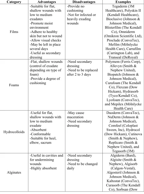
MRWDs differ in their efficacy, advantages and disadvantages (Table 3). Films are comprised of thin, elastic sheets of polyurethane applied mainly to the site of acute surgical wounds.90,122 While films are gas permeable, transparent for visual inspection of wound
progression, and exclude bacteria from entering the wound, they offer limited liquid drainage and can also damage tissue when removed. MRWD foams are moisture absorbent, comfortable to wear, and drape over tissue, but may require application of additional dressings to facilitate occlusion or drainage.90,122,131 Hydrocolloid dressings combine a foam or polyurethane film
dressing with a hydrocolloid matrix that, when applied to wound exudate, forms a gel that promotes autolytic debridement and granulation tissue formation.90,122,131 However,
hydrocolloids should not be employed for highly exudative or infected wounds as they may cause skin maceration.132,133 Alginate dressings are composed of sodium and calcium salts of
alginic acid. Alginates have haemostatic properties, thus are highly absorbent and ideal for highly exudative wounds, but not for dry wounds.90,122,131 Lastly, hydrogels are macromolecular
polymer gels that absorb wound exudate and stimulates autolytic debridement.90,122 Hydrogels
may be applied to a range of wounds from mildly to highly exudative, but maceration can
occur.131 These MRWDs are largely passive dressings, and the ideal clinical treatment of chronic
wounds would also possess an ability to actively stimulate biological healing responses. Bioengineered skin substitutes, in addition to retaining moisture, stimulate healing and are of interest in tissue engineering and regenerative medicine. Epidermis substitutes, including Epicel®, Epidex®, Myskin®, Bioseed®, and Cellspray®, are predominantly autogenic
keratinocyte expansions formulated into delivery dressings or as suspensions that can be topically ejected or sprayed onto the wound site.134 Dermal substitutes are 3D biomaterial
13
mechanically robust, and can be used off the shelf; however, these substitutes depend on host cell infiltration to initiate angiogenesis and remodeling.134 In contrast, collagen-GAG cellular
matrices such as Dermagraft® and OrCel® are pre-seeded with neonatal human foreskin fibroblasts and/or keratinocytes and have been shown to reduce wound contractility to allow re-epithelialization to restore a more natural tissue barrier.135,136 Bilayer skin substitutes contain
both epidermal and dermal components. Apligraf® is an FDA-approved skin substitute
consisting of an epidermal neonatal human foreskin keratinocyte layer and an underlying dermal bovine type I collagen layer seeded with neonatal human fetal fibroblasts.90,122 Apligraf®
dressings already contain within them the cells responsible for the production and delivery of cytokines, growth factors and ECM components to the wound bed, however these bilayer substitutes are expensive and have a limited shelf life of 5-10 days.137,138
Healing outcomes can also be influenced by employing adjuvant therapees in conjunction with standard medical practice. Negative-pressure wound therapy (NPWT) or vacuum-assisted closure therapy applies sub-atmospheric pressure to a local area of tissue damage through a specialized pump.139 NPWT reduces tissue edema, improves circulation, promotes granulation
tissue formation and inhibits bacterial growth.140 Hyperbaric oxygen therapy (HBOT) involves
briefly inhaling 100% oxygen while inside a pressurized chamber.141 Heightened blood and
tissue oxygenation by HBOT improves oxygen delivery to hypoxic tissues and vasoconstricts surrounding healthy tissue, alleviating edema and permitting innate biological combating of infection and ischemia to ensue.142 Topical addition of growth factor formulations have also been
used in chronic wound healing. For instance, a gel composition of recombinant PDGF (Regranex®/Becaplermin) has been employed to mediate cell division, migration and
proliferation. Daily Becaplermin application significantly aided wound closure in PUs and in DFUs.143,144 However, there is increased risk of cancer mortality associated with excessive use
(three or more dispensaries) of Becaplermin.145 Notably, a significant limitation of topical
growth factors is their rapid proteolytic degradation.146
Current clinical strategies, including wound debridement, moisture-retentive dressings, and adjuvant therapies, have various disadvantages and do little to resolve a chronic wound. In Canada, 3.5 million individuals live with diabetes and DFUs will affect one quarter of these patients.95,147 DFUs are the most common cause of non-traumatic lower limb amputations, with
14
a high of mortality rate; in Canada, 30% of patients with diabetes die within one year of amputation and 69% of patients within five years.96,148 Despite the multitude of wound healing
15
Table 3. Examples of clinically available moisture-retentive wound dressings.90,122,149,150
Category Advantages Disadvantages Examples
Films
-Suitable for flat, shallow wounds with low to medium exudates -Promote moist environment -Adhere to healthy skin but not to wound -Allow visual checks -May be left in place several days
-Useful as secondary dressing
-Provide no cushioning
-Not for infected or heavily exuding wounds
Tegaderm (3M Healthcare), Polyskin II
(Kendall Healthcare), Bioclusive (Johnson &
Johnson Medical), Blisterfilm (The Kendall
Co), Omniderm (Omikron Scientific Ltd),
Proclude (ConvaTec), Mefilm (Mölnlycke Health Care), Carrafilm
(Carrington Lab), and Transeal (DeRoyal)
Foams
-Flat, shallow wounds (control of exudate depending on type of foam)
-Provide a degree of cushioning
-Need secondary dressing
-Need to be replaced after 2 to 3 days
Polymem (Ferris Corp), Allevyn (Smith &
Nephew United), Biopatch (Johnson &
Johnson Medical), Curafoam (The Kendall
Co), Flexzan (Dow Hickam), Hydrasorb
(Tyco/Kendall Co), Lyofoam (ConvaTec), and Mepilex (Mölnlycke
Health Care)
Hydrocolloids
-Useful for flat, shallow wounds with low to medium exudate
-Absorbent -Conformable -Suitable for heel, elbow, sacrum -May cause maceration -Need secondary dressing Duoderm (ConvaTec), NuDerm (Johnson &
Johnson Medical), Comfeel (Coloplast Sween, Inc), Hydrocol (Dow Hickam), Cutinova
(Smith & Nephew), Replicare (Smith & Nephew United), and
Tegasorb (3M)
Alginates
-Useful in cavities and for undermining wounds
-Highly absorbent
-Need secondary dressing
-Need to be changed daily
Algiderm (Bard), Algisite (Smith & Nephew), Algisorb
(Calgon-Vestal), Algosteril (Johnson &
Johnson Medical), Kaltostat (ConvaTec), Curasorb (The Kendall
16
Hickam), Melgisorb (Mölnlycke Health Care),
SeaSorb (Coloplast), and Kalginate (DeRoyal)
Hydrogels
-Supply moisture to wounds with low to medium exudate -Suitable for sloughy or necrotic wounds -Useful in flat wounds and cavities
-May be left in place several days
-Need secondary dressing
-May cause maceration
Vigilon (CR Bard), Nu-gel (Johnson & Johnson Medical), Tegagel (3M),
FlexiGel (Smith & Nephew), Curagel (The
Kendall Co), Clearsite (Conmed Corp), Curafil
(The Kendall Co), Curasol (The Kendall
Co), Carrasyn (Carrington Laboratories), Elasto-Gel
(SW Technologies), Hypergel (Scott Health
Care), Normgel (SCA Hygiene Products), 2nd
17
1.2 Tissue Engineering and Regenerative Medicine
Tissue engineering is an area of research that is focused on the combination of natural and synthetic scaffolds, cells, and biologically active molecules in an attempt to form
biologically functional tissues. Similarly, regenerative medicine describes the use of engineered biological materials in conjunction with the body’s innate proliferative mechanisms to expand cell populations and reconstruct tissues in vivo. Focuses of tissue engineering research include the development of bioengineered materials, skin substitutes, biomolecule delivery systems, and stem cell therapies.
1.2.1 Biomaterial Scaffold Design Requirements
Biomaterials can be considered natural or synthetic materials that are designed to interact with biological tissues for augmenting or replacing a native tissue. Tissue engineering scaffolds have the potential to mimic the native extracellular matrix at the nanoscale level, which is important for matrix production, neoangiogenesis and cellular ingrowth, leading to regeneration of tissues.151 Biomaterials must be biocompatible, nonimmunogenic, and if desirable,
biodegradable; that is, a biomaterial should perform safely within the biological environment and any degradation products should be nontoxic. Scaffold structures should ideally mimic native ECM and display a fiber diameter of 50 to 500 nm, 90% porosity, and average pore size of 100 μm to facilitate mass transfer and cell infiltration.152–156 Various fabrication methods for
construction of three-dimensional biomimetic scaffolds have been investigated, including
electrospinning, phase-separation, freeze drying, and self-assembly.157 However, the challenge of
fabricating complex and functional tissues still exists. Current biomaterial fabrication methods are limited in the ability to control hierarchical architecture of scaffolds, formed by nanofibers and nanopores. The insufficient vascularization systems of biological substitutes results in limited diffusion properties of these biomimetic scaffolds.157 As a consequence, biomaterial
scaffolds contain a necrotic center where oxygen transport, nutrient deposit, and waste removal are limited, and viable cells do not penetrate. As such, a scaffold manufacturing technique with controlled material deposition is desirable in order to create complex architectures with
18 1.2.2 Three-Dimensional Bioprinting
Additive manufacturing (AM), also known as three-dimensional (3D) printing, is a mechanized fabrication method that builds objects by repeatedly depositing material layer-by-layer. Adjacent layers fuse together during the process until the final object has been achieved. This technique first requires the creation of a 3D digital model that is generated using computer-aided design (CAD) software. Since building takes place layer-by-layer, the 3D model must be sliced into horizontal planes in order to generate a G-code, or programming language that will control the printer’s extrusion path.158 A basic 3D printer typically includes a movable print bed
that can displace in the vertical (Z) direction and supports the print object, an ejector nozzle that moves in the horizontal (X, Y) plane, and the print material, which can be a wide range of materials, from plastics and metals to soft materials. G-code is sent to the printer and the object can be 3D printed.158,159 Advantages of 3D printing include the availability of low-cost hardware
that is easily customizable, free, open-source software, and limited waste of print materials.160
In 3D bioprinting, the print material is often referred to as a “bioink.” Material properties to consider when choosing a bioink include the desired print resolution, ability for the printed device to be easily sterilized, shear strength and ability to recover scaffold shape, and the material stiffness. As is required for all biomaterials, bioinks need to display biological compatibility with cells and the body such that the biomaterial can adequately function in the biological environment while minimizing any foreign-body response mounted by the host immune system.161 Furthermore, optimized biomaterials should also exhibit the ability to be
remodeled, biodegraded and bioadsorbed by cells, producing nontoxic degradation products.161
Commercially available bioinks include native structural extracellular matrix proteins such as collagen, gelatin, hyaluronic acid, and calcium phosphate.162
Three-dimensional printing of soft materials, such as photocurable resins, polymer powders, or thermoplastic monofilaments, creates the additional challenge of apparent ink viscosity, which can cause the print material to flow out of its extruded shape.163 One approach
to 3D printing of soft materials is to eject the ink layer-by-layer into a secondary yield-stress support bath, embedding the print object in a granular medium. Embedding scaffold structures into a temporary, sacrificial support material is advantageous in that this technique allows for the printing of soft natural and synthetic materials that exhibit low viscosities prior to
19
and porous regions. Freeform reversible embedding of suspended hydrogels (FRESH), is a type of 3D printing that utilizes a support bath with reversible gelation properties, such that the bath can be liquified in order to liberate the 3D printed object post-printing.164–166
1.2.3 Collagen-Based Biomaterials
Twenty-nine collagen types have been identified which account for 33% of the protein in humans and 66% of the dry weight of skin.167 Collagen types I-III, V, and XI have fibrillar
quaternary structures. Collagen molecules are comprised of three polypeptide chains aligned in a parallel manner and coiled in a left‐handed polyproline II‐type (PPII) helix.168 These chains
arrange themselves further into a right‐handed triple helix that is stabilized by interstrand
hydrogen bonds and intrastrand non-covalent interactions.169 Animal collagens arranged in triple
helices, known as tropocollagen, form macroscopic fibers and extracellular matrix networks in tissue, bone, and basement membrane. Ninety percent of the total collagen content in human skin is type I collagen, and this type is most frequently employed for biomedical applications.170
Biomaterial scaffolds fabricated using collagen have been applied extensively as a major component in dermal skin substitutes due to collagen’s high biocompatibility, biodegradability, and molecular composition.171 Collagen extraction for biomedical application is commonly from
bovine skin and tendons; porcine skin, intestine, or bladder mucosa; and rat tail sources.172 The
quality and properties of extracted collagen depends on the source species and the tissue from which it was harvested. One risk associated with collagen derived from animal sources is allergic reaction and pathogen transmission.173,174 To overcome this complication, recombinant collagen
can be produced via heterologous expression in mammalian, insect, yeast, or bacterial cells.175– 177 Biorecognition of collagen is necessary since may cell-surface portions interact and bind to
collagen. Cell-collagen communications are mediated by four different kinds of proteins: Pro‐ Hyp‐Gly-recognizing receptors (for example, glycoprotein VI); integrin family receptors; integrin‐type receptors that recognize cryptic motifs within collagen; and receptors with affinity for non-collagenous domains.178,179 Proteins containing Arg-Gly-Asp or similar integrin‐
recognition sequences, such as decorin and laminin, can bind to both collagen and integrins, promoting cell adhesion and proliferation.180
20
ideal for use in biomedical applications. Exogenous degradation products of collagen types I-III have been shown to induce chemotaxis of human fibroblasts and is thought to promote
regeneration of tissue structure and functionality.181,182 Collagen can be cross-linked via chemical
cross-linking agents including formaldehyde, glutaraldehyde, carbodiimides, polyepoxy compounds, acyl azides, and hexamethylene diisocyanate;167 physical cross-linking methods
using ultraviolet light or dehydrothermal treatment;183 and enzymatic cross-linking such as via
tissue transglutaminase.184 Although chemical cross‐linking can enhance biomaterial stability,
residual electrophilic reagents and compounds produced upon degradation in vivo can be
cytotoxic. An advantage of cross-linking with enzymes is this method is benign and generates no cytotoxic byproducts.
Despite limitations such as immunogenicity of xenogeneic sources and high costs, the use of natural biological materials is of interest in biomaterial fabrication.For instance, collagen-based biomaterials have been demonstrated for a variety of applications, including neural stem cell scaffolds,185 cartilage,186,187 osteochondral,188 and skin.189 Tissue substitutes comprised of
natural biomaterials have garnered interest for applications in transdermal and topical formulation discovery, dermal toxicity studies, and autologous grafts for wound healing.
Collagen-based biomaterials can be classified as either decellularized collagen matrices or more refined scaffolds based on the extent of their purification. Decellularized collagen matrices maintain the natural tissue properties and ECM structure; cellular matter is removed from collagen matrix by physical methods such as snap freezing or high pressure, chemical treatment with acid or alkali treatment, chelation with EDTA, or treatment with detergents or solutions of high osmolarity, and trypsin enzymatic digestion to produce the biomaterial.190 In contrast, more
refined scaffolds are fabricated via collagen protein extraction, purification, and polymerization. Natural collagen can dissolve in aqueous solutions depending on the extant cross-linking. Refined scaffolds require collagen to first be dissolved in aqueous solution; the most common solvent systems include a neutral NaCl solution, dilute acetic acid, or a solution of proteolytic enzymes.167
1.2.4 Polycaprolactone-Based Biomaterials
21
consistent quality, and tunable material properties.191 One such example of a widely employed
synthetic material is polycaprolactone (poly(ϵ-caprolactone), PCL), a biocompatible polyester. PCL is a semi-crystalline, aliphatic polymer, having highly ordered molecular structure.192 The
structure comprises of a repeating unit of one ester group and five methylene groups. Its melting temperature is above body temperature, thus within the body the semi-crystalline structure of PCL results in high toughness.193 Advantages of PCL include its strength and durability, which
are tunable, and its ease of fabrication while allowing for precise control of product architecture.194
PCL is degraded in the body under physiological conditions via hydrolytic mechanisms. Due to its high molecular weight, the polyester has a slow degradation rate of approximately two years in the biological environment.195–197 PCL is biocompatible and non-toxic, while its
durability means that PCL has less chance to induce immunological effects.198 Physical
properties of PCL are easily manipulated by compounding the polymer with secondary
constituents. By using a copolymer of PCL with dl-lactide, a more flexible material with a faster degradation rate than the homopolymer can be achieved.195 Moreover, the high degree of
permeability has made PCL an important candidate for the development of drug delivery systems and in bone tissue regeneration.199–203
PCL has been established as an important biomaterial. It has been approved by the Food and Drug Administration (FDA) for several medical applications, including suture materials and subdermal contraceptive implants.192,204,205 In dentistry, PCL has been employed as a root
canal-filling material; PCL-filled roots demonstrated proper seals to protect against the aqueous environment.206 In wound healing, PCL was employed as wound-dressing material and delivery
system for chemical antiseptic; PCL fibers were shown to exhibit desirable tensile properties following compounding with chlorhexidine diacetate while the antiseptic conferred antimicrobial properties, even at concentrations as low as 1% (w/w).207 Furthermore, PCL composites have
been widely studied for applications in tissue engineering scaffolds that regenerate bone, ligament, cartilage, skin, nerve and vascular tissues.204
1.2.5 Electrospinning
22
solution passes such that as each droplet accumulates at the tip of the needle, it experiences electrostatic repulsion between the surface charges of the droplet and Columbic force exerted by the applied electric field.208 As charge accumulates on the surface of the droplet, a Taylor cone is
formed. Once the electrostatic charge exceeds the surface tension of the polymer solution, a polymer jet is expelled and travels towards a grounded mandrel.208 The solvent evaporates,
leaving a porous, nonwoven fiber mat deposited on the collector. Electrospinning allows for customization of scaffold morphology by varying parameters known to affect fiber shape and size, for instance the concentration and resultant viscosity of the polymer solution, solvent identity, distance from needle to collecting surface, applied voltage, flow rate, temperature, and humidity.209,210
Electrospun nanofibrous scaffolds have been proposed as a novel alternate strategy to conventional wound dressings for management of chronic skin wounds.211,212 Electrospinning
polymer solutions can also be supplemented with bioactive agents to improve scaffold
biocompatibility, biodegradability, biorecognition, sterilizability, and mechanical properties.213
Due to the high surface-area-to-volume ratio, nanoscale diameter, and highly
porous structure of electrospun fibers, even distribution and controlled release of bioactive molecules is possible.214 Immediate burst release with subsequent prolonged release of bioactive
factors via passive diffusion occurs during degradation of polymer fibers.215 Antimicrobial
peptides, cytokines, and growth factors, are absent in chronic wounds, but could be delivered exogenously using a biomolecule delivery system.216 For instance, electrospun polyvinyl alcohol
(PVA)-silk nanofibers have previously been supplemented with EGF, bFGF, and the
antimicrobial peptide LL-37. Biological gradients of these bioactive factors were established and tested using full-thickness excisional wounds on the dorsal surface of diabetic rabbits,
demonstrating this combination of factors accelerated wound healing, lowered MMP expression, regulated ECM secretion, and reduced biofilm or bacterial colonization.217
Several limitations of electrospun scaffolds have been identified however, including 1) the structure which is densely packed nanofibers, resulting in small pore sizes, 2) potential toxicity of residual solvents or cross-linking agents, and 3) difficulty with industrial
upscaling.218–221 Small pore sizes and densely packed fibers result from the fabrication process
23
regeneration.222 Pore size can be increased however, by changing the polymer solution flow rate,
increasing the concentration of the polymer solution to produce thicker fibers, or selective and controlled heating of the electrospinning environment to facilitate solvent evaporation. 214,223,224
However, while thicker fibers can counteract the limitation of pore size, increasing fiber diameter inhibits cellular adhesion and migration.225 Moreover, the use of organic solvents or
chemical cross-linking agents is a common concern associated with blended electrospinning due to cytotoxicity220. Safe crosslinking options include enzymatic crosslinking, electrostatic
crosslinking, or hydrogen bonding with sugars or polyphenols.226 However, these alternative
cross-linking methods are associated with more complicated fabrication procedures and
excessive degradation of bioactive factors loaded into the scaffold for slow release.226 Lastly, the
soluble nature of most biomolecules often results in rapid release from the nanofibrous scaffolds, as fast as 70% released within 30 minutes, followed by degradation or aggregation due to the instability of these proteins against proteolysis, acidity, and heat in the wound
microenvironment.227–229 Despite these challenges, electrospinning is a viable biomaterials
technique due to the great potential for customization of scaffold morphology and material composition.
Electrospun nanofibrous scaffolds can be produced using natural and synthetic polymers.230 Natural polymers such as collagen, gelatin, silk, fibrinogen and chitin are
biocompatible, biodegradable and abundant in the natural environment. However, natural polymers exhibit fragile structural properties, complicated processability, vulnerability to enzymatic degradation and potential immunogenicity. Weak mechanical properties and rapid degradation of natural polymers are overcome using cross-linking to improve scaffold stability. Chemical crosslinkers such as glutaraldehyde, formaldehyde, polyether oxide, hexamethylene diisocyanate and polyurethane, acyl azide and carbodiimides, and glycerol, and physical
crosslinkers including drying, heating, and UV/gamma radiation, covalently bond amino acids on adjacent scaffold structures.
Gelatin is a natural polymer alternative to collagen for use in electrospun biomaterial scaffolds.231 Gelatin, a derivative of collagen, is obtained by the denaturation of collagen in
either acidic or basic processing. Type A gelatin is produced by acid pre-treatment of animal samples while type B gelatin is produced by alkaline pre-treatment.232 Gelatin maintains
24
Moreover, gelatin scaffolds have been shown to stimulate cellular adhesion, migration and proliferation, and accelerate wound healing.233–235
1.2.6 Stem Cell Therapy in Wound Healing
Characteristics of stem cells (SCs) include the ability to self-renew, maintain long-term viability, and multipotent differentiation.236 SC therapies for the treatment of chronic wounds
involves delivering adult stem and progenitor cells to the site of injury. Autologous SC transplant uses a person’s own SCs while an allogeneic transplant uses stem cells from a donor. In wound healing, exogenous progenitor and stem cells have been employed to improve healing and scarring outcomes through SC differentiation and secretory activities. For instance, transplanted human amniotic mesenchymal stem cells (AMMs) into a diabetic murine excisional skin wound showed that AMMs promoted wound healing and increased re-epithelialization and cellularity; AMMs demonstrated engraftment and expression of keratinocyte-specific proteins in vivo, while the secretome was rich in angiogenic factors IGF-1, EGF and IL-8.237 A clinical study of
autologous bone marrow (BM)-derived MSC intramuscular injection into the affected limb of 24 patients with non-healing ulcers demonstrated enhanced wound healing and improved clinical parameters such as painless walking.238 In a study of 41 type 2 diabetic patients with critical limb
ischemia and foot ulcers, ulcer healing, limb perfusion, and painless walking were improved in groups injected intramuscularly with BM-MSCs compared to those that received BM-derived mononuclear cells or normal saline.239 Thus far, SC therapies for wound healing applications are
limited to animal models and small clinical trials, but promising results warrant further investigation into the effective delivery of autologous and allogeneic stem and progenitor cells.240
Adipose-derived stem/stromal cells (ASCs) are MSCs derived from fat tissue and have garnered interest in SC therapy. ASCs have been shown to proliferate rapidly and differentiate in vitro toward the osteogenic, adipogenic, myogenic, and chondrogenic lineages.236,241,242
Moreover, ASCs are resistant to mechanical damage243 and easily harvested from the body by
mechanical liposuction, manual aspiration (Coleman technique), or direct surgical excision.244
The stromal vascular fraction (SVF) derived from adipose tissue is heterogeneous, comprised of MSCs, pre-adipocytes, endothelial cells, pericytes, T cells, and alternative M2 macrophages.245
25
surface markers including CD44, CD90, CD105, CD166, and Stro-1.246 Notably, ASC phenotype
is controversial and under speculation; the International Federation for Adipose Therapeutics and Science and the International Society for Cellular Therapy include ASC positivity for CD13, CD 36, CD73, and CD10, and negativity for CD45, CD31 and CD106.247 Despite the need for a
distinct set of quality control criteria that can define the standard ASC phenotype, ASCs are a promising focus for cell therapy.
In wound healing studies, ACSs injected locally into excisional wounds in diabetic rats with compromised healing abilities resulted in rescue of wound healing rates in diabetic rats almost equivalent to those of wild type rats.248 Moreover, ASCs appear to promote wound
healing under ischemic conditions. Ischemia of rabbit ears was created by ligating two of three main arteries of the ear and subsequently wounding the ear; ASC therapy enhanced wound granulation in rabbit ears under ischemia.249 Radiation models of chronic wounds in rats and
mice have demonstrated that ASCs administered directly into the irradiated region,
intramuscularly into the irradiated limbs, or intravenously resulted in enhanced wound repair.250– 252 ASCs within the subcutaneous adipose tissue niche are in close proximity to cutaneous
wounds and are implemented in the wound healing process.253 ASCs exhibit great migration
potential and may infiltrate the wound to foster wound repair. For instance, ASCs transplanted into the subretinal space of injured rat retinas migrated into the retinal pigmented epithelium after 4 weeks.254
ASCs are capable of affecting other cells through the release of hormones, cytokines, growth factors, and micro RNAs. Extracellular vesicles released by ASCs transport gene
regulatory information that in turn affects angiogenesis, adipogenesis, and other cell pathways in recipient cells.255 Hormones released from ASCs have been shown to affect cancer cell
proliferation.256 In diabetic rats, ASCs injected subcutaneously into full-thickness skin wounds
stimulated angiogenesis and enhanced tissue regeneration after 8 weeks; ASCs amassed in the subdermal layer of the wound boundary and amplified angiogenesis via expression of von Willebrand factor and VEGF.257 Through co-culturing it was demonstrated that ASCs have an
anti-inflammatory effect on monocyte-derived dendritic cells in vitro.258 Moreover, proliferation