ISSN: 2319-8753
I
nternational
J
ournal of
I
nnovative
R
esearch in
S
cience,
E
ngineering and
T
echnology
(An ISO 3297: 2007 Certified Organization)
Vol. 2, Issue 11, November 2013
6235
Release Pattern of Ag
+
ions from
Silver-Loaded Zeolite X and its Subsequent Effect on
Fatty Acid Composition of Bacterial Cells
Dr. B. Kwakye-Awuah
1,*, Dr. A, Mrozik,
2Prof. Z. Piotrowska
-
Seget
2, I. Nkrumah
1Prof. C. Williams
3, Dr. I.
Radecka
3Lecturer, Department of Physics (Materials Science group), KNUST, Kumasi, Ghana1 Professor, Department of Biochemistry, University of Silesia, 28, 40-032 Katowice, Poland2 Principal Lecturer, Research Centre in Applied Science, University of Wolverhampton, WV1 1SB, UK3
*Corresponding author: Tel. +233(0)545207159, Email: [email protected]
Abstract: The kinetics of the release profile of silver ions from zeolite X framework and its effect on fatty acid composition of two gram-negative: Escherichia coli K12 W-T and Pseudomonas aeruginosa NCIMB 8295 and one gram-positive Staphylococcus aureus NCIMB 6571. Silver ions were released from the zeolite framework in an anomalous manner with time. The fatty acid composition was significantly altered when they were exposed to silver-loaded zeolite X. Interaction of silver ions with fatty acid of bacterial cells is likely to affect the survival of the cell
Keywords: Silver-loaded zeolite X, fatty acid, bacteria, silver ions
I. INTRODUCTION
ISSN: 2319-8753
I
nternational
J
ournal of
I
nnovative
R
esearch in
S
cience,
E
ngineering and
T
echnology
(An ISO 3297: 2007 Certified Organization)
Vol. 2, Issue 11, November 2013
6236
analysed as well as variations in the saturated/unsaturated components. In addition, the release profile of silver ions from the zeolite X framework was investigated.II. MATERIALSANDMETHOD
An easy way to comply with the conference paper formatting requirements is to use this document as a template and simply type your text into it.
A. Reagents, bacteria, media, zeolite X and silver-zeolite X
Sodium hydroxide and sodium aluminate were purchased from Aldrich Chemicals, UK. Sodium silicate was purchased from Fischer Chemicals, UK. (TSA) and Tryptone soya broth (TSB) were obtained from Lab M, UK. De-ionized water used in this investigation was supplied by University of Wolverhampton. Zeolite X (batch composition: NaAlO2:4SiO2:16NaOH:325H2O) without silver and silver-loaded zeolite X were synthesized and characterize by
Kwakye-Awuah (2008). Bacterial strains; E. coli K12 W-T, S. aureus NCIMB 6571 and P. aeruginosa NCIMB 8295 used in this study were obtained from the University of Wolverhampton culture collection. The stock cultures were freeze-dried and kept at –20 oC overnight. Before use, cultures were resuscitated and grown on Tryptone soya agar (TSA) overnight at 37 oC.
B. Extraction and analysis
Before the extraction process, extraction reagents (saponification, methylation, extraction solvent and base wash) were first prepared. Saponification reagent was prepared by first diluting 150 ml of methanol (HPLC grade; Aldrich Chemicals, UK) with 150 ml of de-ionized water and adding 45 g of sodium hydroxide. The resulting solution was stirred until the sodium hydroxide was completely dissolved to obtain a methanolic solution. To prepare the methylation reagent, 325 ml of 6.0 N HCl was added to ethanol with constant stirring until a uniform solution was obtained. Extraction solvent was prepared by adding 200 ml of methyl tert-butyl ether (MTBE) to 200 ml hexane (HPLC grade) with uniform stirring for about 5 minutes to ensure a homogeneous solution. Finally base wash reagent was prepared by dissolving 10.8 g of sodium hydroxide in 900 ml de-ionized water. A single bacterial colony of each microorganism was used to inoculate a 100 ml starter culture, which were grown aerobically in TSB (pH 7.2) overnight at 37 oC in a rotary shaker (150 rev min-1) (Bellantone et al., 2000; Kwakye-Awuah et al 2008a). After overnight incubation, a 0.1 ml of each culture ( ) was inoculated into a 200 ml sterile TSB (pH 7.2) to give a final concentration of cells ml-1 for E. coli K12 W-T, cell ml
-1
ISSN: 2319-8753
I
nternational
J
ournal of
I
nnovative
R
esearch in
S
cience,
E
ngineering and
T
echnology
(An ISO 3297: 2007 Certified Organization)
Vol. 2, Issue 11, November 2013
6237
reagent was added to the tube, vortexed for 10 minutes and allowed to settle. Portion of the top phase in the tube was transferred into vials, capped and analysed by chromatography (GC) with a Hewlett Packard 5890 series II (GMI Inc. USA) equipped with flame ionization detectors auto-sampler, integrator and computer. The oven, detector and injection port temperatures were set at 200, 300 and 275 oC respectively and the Helium carrier gas flow was kept at 100 ml/min. Before starting the first run, the whole system was checked to ensure that pressure in helium, hydrogen, nitrogen and compressed air cylinder was enough and the screen displayed ready. Major fatty acids were identified by comparison of retention times with those of pure bacterial fatty acid methyl esters standard (Microbial Identification System; TSBA library version 3.9; MIDI, USA).III.RESULTS AND DISCUSSION
Methyl esters fatty acids detected by gas chromatography (GC) for E. coli K12W-T (Table 1), for S. aureus NCIMB 6571 (Table 2) and P. aeruginosa NCIMB8295 (Table 3) are presented. The results are presented as percentage concentrations with standard errors. MIDI-FAME analysis produced isolate profiles that were characteristic of the assigned genus as reported in literature [15] – [17]. The nomenclature of the fatty acid profiles obtained is as follows: the number of carbon atoms, followed by a colon, the number of double bonds and then by a position of the first double bond from the methyl end (
) end of the molecule. The prefix c or t indicate cis or trans configuration of the double bond, cy – cyclopropane fatty acids, Me – the position of the position of the methyl group from the acid end, and -OH indicates the position of the hydroxyl group from the acid end of the molecule. Branched fatty acids were designed as iso or anteiso, if the methyl branch is one or two carbons from the
end of the acyl chain Sajbidor [29] and Mrozik et al [16]. Peaks that did not display equivalent chain length values of known fatty acids were left unnamed.Without silver-exchanged zeolite Silver-exchanged zeolite X
Fatty acid type Exp 1 Exp 2 Average
composition
Exp 1 Exp 2 Average
Changes in
composition
(%)
Saturated
12:0 3.96 4.12 4.04 3.44 4.55 4.00 0
14:0 7.91 8.06 7.99 8.04 8.70 8.37 0
unknown 0.91 1.91 1.41 0.73 0.00 - 0
14:0 3OH 8.18 8.30 8.24 7.29 7.81 7.55 0
16:0 36.70 36.30 36.50 39.53 38.66 39.10 -6
17:0 cy 15.43 16.15 15.79 23.35 21.56 22.46 + 42
19:0 cy ω8c 2.98 2.84 2.91 6.56 5.51 6.04 + 107
Unsaturated % of total fatty acids
18:1ω7c/9t/12t 12.77 12.69 12.73 7.34 8.92 8.13 - 36
16:1 ω7c 10.29 10.41 10.35 3.06 4.26 3.66 - 65
Table 1, Profile of fatty acids obtained from Escherichia coli K12 W-T grown without silver-exchanged zeolite X
ISSN: 2319-8753
I
nternational
J
ournal of
I
nnovative
R
esearch in
S
cience,
E
ngineering and
T
echnology
(An ISO 3297: 2007 Certified Organization)
Vol. 2, Issue 11, November 2013
6238
For the interpretation of the obtained results, all isolated fatty acids were grouped into; saturated and unsaturated fatty acids [15, 16]. The first group included four subgroups: straight chain, hydroxyl, cyclopropane and branched fatty acids (iso or anteiso). When cells were challenged with zeolite X (Table 1), the fatty acid composition of E. coli K12 W-T exhibited low proportion of an unknown fatty acid, 19:0 cy 8c of the saturated fatty acid obtained, a result similar to that of fatty acids extracted from the cells. These compositions were unaffected upon addition of silver-loaded zeolite X. High proportion of 16:0 fatty acids was obtained when cells were challenged with zeolite X which increased marginally upon addition of silver-loaded zeolite X. The total level of saturated fatty acids in E. coli with zeolite X was 76.9 % whereas it was 88.5 % for silver-loaded zeolite X. The unsaturated fatty acids detected in E. coli K12 W-T were 18:1 7c/9t/12t and 16:1 7c at levels of 12.7 % and 10.4 % respectively. The levels however, dropped to 8.1 % and 3.67 % respectively when challenged with silver-loaded zeolite X.Saturation/unsaturation ratio in E. coli K12 W-T was 3.3 when cells were challenged with zeolite X and 7.4 when cells were challenged with silver-loaded zeolite X. For S. aureus NCIMB 6571 only saturated (branched and unbranched) fatty acids were detected with the characteristics: low proportion of 19:0 iso,16:0, 19:0 anteiso and 17:0 iso; equivalent proportion of 18:0, 15:0 iso and 17:0 anteiso and high proportion of 15:0 anteiso. The results obtained when cells were challenged with silver-loaded zeolite X followed similar trend. P. aeruginosa NCIMB 8295 profile was characterized by high proportion of 16:0 and equivalent proportion of 17:0 cy 16:1 7c fatty acids. In addition, the profile was characterized by low proportion of straight-chain 2-OH and 3-OH fatty acids as well as 18:0, 19:0 cy 8c and 12:0 fatty acids. Remarkable changes were observed in the 10:0 3OH, 14:0, and 17:0 cy fatty acids when cells were challenged with silver-loaded zeolite X. A new fatty acid, 18:1 9c was also detected. The saturation/unsaturation ratio was found to be 4.6 for cells challenged with zeolite X and 3.7 for cells challenged with silver-loaded zeolite X.
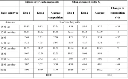
Without silver-exchanged zeolite Silver-exchanged zeolite X
Fatty acid type Exp 1 Exp 2 Average
composition
Exp 1 Exp 2 Average
Changes in
composition
(%)
Saturated % of total fatty acids
15:0 iso 10.85 9.83 10.34 8.39 8.05 8.22 + 20
15:0 anteiso 46.64 43.12 44.88 42.73 44.05 43.39 +3
16:0 2.69 2.71 2.70 3.21 3.95 3.58 + 32
17:0 iso 5.32 4.75 5.04 4.53 4.15 4.34 - 14
17:0 anteiso 11.55 11.66 11.61 12.74 12.71 12.73 -9
18:0 9.67 10.74 10.21 10.12 9.60 9.86
19:0 iso 2.26 2.42 2.34 3.07 3.04 3.06 + 30
19:0 anteiso 3.02 3.57 3.30 4.98 4.64 4.81 + 46
20:0 8.00 9.40 8.70 10.56 9.47 10.02 + 15
100:0 100:0
ISSN: 2319-8753
I
nternational
J
ournal of
I
nnovative
R
esearch in
S
cience,
E
ngineering and
T
echnology
(An ISO 3297: 2007 Certified Organization)
Vol. 2, Issue 11, November 2013
6239
The composition of fatty acids extracted from strains with no zeolite and with zeolite X (zeolite X with no silver ions) were fairly equal but differed with that obtained composition with the profiles obtained with silver-loaded zeolite X. This suggests that changes in fatty acids composition were primarily due to the action of silver ions that were released from the zeolite framework. Silver ions released from the zeolites have been reported to be effective against a wide range of microorganisms and appear to have multiple target sites of microorganisms [3, 26, 32]. The saturated/unsaturated ratio in E. coli K12 W-T increased on exposure to silver-exchanged zeolite X. Similar results were obtained by Mrozik et al [16] when they detected an increase in cyclopropane fatty acid composition in response to naphthalene). Exposure to silver-exchanged zeolite X also changed the composition of branched and hydroxyl fatty acids in E. coli K12 W-T and P. aeruginosa NCIMB 8295. In S. aureus NCIMB 6571 however, all the fatty acids detected were saturated with an increase in the composition of branched fatty acids. Tsitko et al [35] obtained similar results when he studied the impact of different aromatic compounds on whole cell fatty acid composition of Rhodococcus opacus strains GM-14, GM-24 and 1CP. Our results showed a decrease in the composition of 14:0, 10:0 3OH and 17:0 cy fatty acids in P. aeruginosa NCIMB 8295 when their cells were challenged with silver-loaded zeolite X. Gram-positive bacteria cell wall contains three to twenty times more peptidoglycan than their gram-negative counterpart [3, 11]. Since peptidoglycans are negatively charged, it binds some portion of silver. Consequently more silver ions reach the plasma membrane of gram-negative than gram positive bacteria. In addition, gram-negative bacteria have been reported to contain lipids in the cell wall as well as in the cytoplasmic membrane [13]. Fatty acids ofWithout silver-exchanged zeolite Silver exchanged zeolite X
Fatty acid Exp 1 Exp 2 Average Exp 1 Exp 2 Average Changes (%)
Saturated % of total fatty acids
10:0 3OH 2.79 3.24 3.02 0.00 0.00 0
12:0 4.72 5.15 4.94 4.43 4.44 4.44 0
12:0 2OH 2.71 3.27 2.99 2.93 2.95 2.94 0
12:0 3OH 4.26 3.88 4.07 4.44 4.11 4.27 0
14:0 0.91 n.d 1.05 0.92 0.98 0
16:0 28.26 28.82 28.54 32.16 31.37 31.77 + 11
17:0 cy 0.92 n.d 1.08 0.97 1.03 0
18:0 0.90 1.18 1.04 0.88 1.10 0.99 0
19:0 cy ω8c 1.49 n.d 1.53 1.49 1.51 0
Unsaturated % of total fatty acids
16:1 ω7c 14.71 15.35 15.03 12.70 12.82 12.76 - 15 18:1ω9c
(new)
0.00 0.00 0.00 0.93 0.75 0.84 0
ISSN: 2319-8753
I
nternational
J
ournal of
I
nnovative
R
esearch in
S
cience,
E
ngineering and
T
echnology
(An ISO 3297: 2007 Certified Organization)
Vol. 2, Issue 11, November 2013
6240
cytoplasmic origin are mostly cyclopropane fatty acids. These were detected in both E. coli K12 W-T and P. aeruginosa NCIMB 8295. The absence of cyclopropane fatty acid in S. aureus NCIMB 6571 suggests that all fatty acids were in the peptidoglycan. Hence, factors that influence their susceptibility to antimicrobial agents are likely to be more complex than those of gram-positive bacteria. The saturated/unsaturated ratio in P. aeruginosa NCIMB 8295 decreased from 4.6 to 3.7. Mrozik et al [15] obtained similar results of fatty acid profiles for P. aeruginosa when they studied the influence of naphthalene on fatty acid composition of Pseudomonas sp. JS 150. Cells of E. coli contained significant proportion of 17:0 cy and 19:0 cy
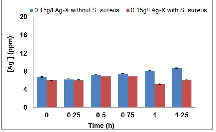
8c. Another observation in the fatty acids composition was the appearance of new fatty acid in S. aureus NCIMB 6571 (18:1 9c, Table 2).Fig. 1 Silver ions released from silver-exchanged zeolite X into TSB with or without E. coli with time at concentration of 0.15 g l-1 silver-exchanged zeolite X.
Fig. 2 Silver ions released from silver-exchanged zeolite X into TSB with or without E. coli with time at concentration of 0.15 g l-1 silver-exchanged zeolite X.
[Ag
+
]
(ppm
)
Time (h)
[A
g
+
]
(ppm
)
ISSN: 2319-8753
I
nternational
J
ournal of
I
nnovative
R
esearch in
S
cience,
E
ngineering and
T
echnology
(An ISO 3297: 2007 Certified Organization)
Vol. 2, Issue 11, November 2013
6241
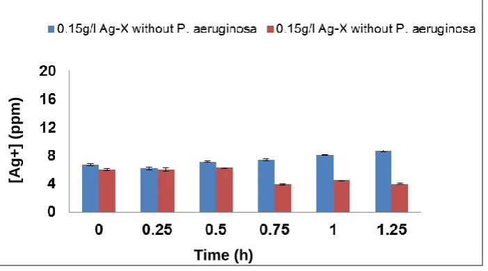
The mechanism of formation of new fatty acids has not been documented (Gutierrez et al., 1999; Mrozik et al. 2004a). However, they may be involved in the protection of strains tested against disruptions of the membrane-cell wall structure [7, 8, 16]. Interaction of silver ions with the fatty acid composition of E. coli K12 W-T S. aureus NCIMB 6571 and P. aeruginosa NCIMB 8295 resulted in significant changes in the composition. These changes are likely to affect the charge, hydration and conformity of their membrane lipids with a consequent changes in the phase behaviour of the lipids [13, 18]. In Figure 1 (a – c), the release profiles of silver again varied from strain to strain on their exposure to 1.0 g l-1 in TSB. In the presence of E. coli (Figure 1a) the release profile of silver ions increased between 0 and 0.15 hours followed by a progressive decrease in the release of silver ions until the end of the exposure period.In the presence of S. aureus (Figure 1b), however the release profile of silver ions increased in a similar manner between 0 and 0.15 hours followed by a decrease in release between 0.75 and 1.25 hours. For P. aeruginosa (Figure 1c), the release in silver ions was progressive from 0 to 0.75 hours and decreased progressively until the end of the exposure period. The results of this study have shown that the amount of silver ions needed to effect antimicrobial activity is dependent of the cell structure of the bacteria. As samples were withdrawn from the flask silver ions in the culture were reduced. This implies that more silver needed to be released from the zeolite framework to replace what was removed. According to Le Chatelier’s principle the position of the equilibrium will shift to the right hence more silver ions were likely to be released from the zeolite framework. However, since the release of silver ions is controlled by the zeolite framework, the amount of silver ions released at any time depends on the location of ions in the framework. Silver ions at exchangeable sites are normally more difficult to displace than those at the surface or in the pores[38]. It is likely that this observation might account for the anomalous trend of the silver release from the zeolite framework into the TSB. The uptake of silver ions in bacteria depends on the type of strain being inhibited or killed [5]. Currently, information available on the release of silver ions from silver compounds is very little since results reported in literature are conflicting [2]. While results showed that the amount of silver ions released to effect antimicrobial activity has been found to be directly proportional to the rate of kill in vitro [20, 21, 31, 34], Ovington, [22] Parsons et al [23] showed the contrary. The results obtained from this study showed that silver ions were released from the zeolite in an anomalous trend and inhibition of growth rate. The discrepancies between results have been attributed to the lack of detail on how some tests were performed and the established chemical principles of solubility [2].As an example it has been shown that a hydrofiber dressing containing silver and carboxymethylcellulose (Ag/CMC) released 0.8 ppm of silver in water and in saline but released 85 ppm of silver in thiosulfate [23, 36]. Nanocrystalline silver released 50 ppm or 70 ppm in water (depending on experimental conditions), 0.8 ppm in saline for the same analysis and 640,000 ppm
Fig. 3 Silver ions released from silver-exchanged zeolite X into TSB with or without E. coli with time at concentration of 0.15 g l-1 silver-exchanged zeolite X.
[A
g+]
(
pp
m)
ISSN: 2319-8753
I
nternational
J
ournal of
I
nnovative
R
esearch in
S
cience,
E
ngineering and
T
echnology
(An ISO 3297: 2007 Certified Organization)
Vol. 2, Issue 11, November 2013
6242
in thiosulfate [23, 36]. Hence, extent of silver elution depends on the nature of the type of media and the experimental conditions.IV CONCLUSION
The first objective of this work was to determine whether a relationship exists between the fatty acid composition and susceptibility of bacteria to silver ions released in a controlled manner from a zeolite framework. The saturated/unsaturated of ratio of fatty acids detected in cells grown in the presence of silver-loaded zeolite X decreased for P. aeruginosa NCIMB 8295 but increased for E. coli K12 W-T compared with zeolite X and without any zeolite. This indicates that silver ions released from the zeolite framework exerted substantial stress on the lipid of the bacterial cells. However, there was no strict relationship between the individual fatty acids and susceptibility. The saturated/unsaturated of ratio of fatty acids detected in cells grown in the presence of silver-loaded zeolite X decreased for P. aeruginosaNCIMB 8295 but increased for E. coli K12 W-T compared with zeolite X without silver.
REFERENCES
[1] Bellantone, M., Williams, H. D., and Hench, L. L., ‘‘Broad-spectrum bactericidal activity of Ag2O-doped bioactive glass’’, Antimicrobial Agents
and Chemotherapy, Vol 46(6), 1940 – 1945, 2002.
[2] Brett, D. W., ‘‘A discussion of silver as an antimicrobial agent: Alleviating the confusion’’, Ostomy/Wound Management, Vol. 52(1), pp. 34 – 41, 2006.
[3] Feng, Q. L., Wu, J., Chen, G. O., Cui, F. Z., Kim, T. N., and Kim, J. O., ‘‘A mechanistic study of the antibacterial effect of silver ions on Escherichia coli and Staphylococcus aureus’’, Journal of Biomedical Material Research, Vol. 52(4), pp. 662 – 668, 2000.
[4] Gutierrez, J. A., Nichols, P., and Couperwhite, I., ‘‘Changes in whole cell-derived fatty acids induced by benzene and occurrence of the unusual 16:1
6c in Rhodococcus sp. 3’’. FEMS Microbiology Letters, Vol. 176, pp. 213 – 218, 1999.[5] Haefeli, C., Franklin, C., and Hardy, K., ‘‘Plasmid-mediated resistance in Pseudomonas stutzeri isolated from a silver mine’’, Journal of Bacteriology, Vol. 158(1), pp. 389 – 392, 1984.
[6] Heipieper, H. J., and de Bont, J. A. M., ‘‘Adaptation of Pseudomonas Putida S12 to ethanol and toluene at the level of fatty acid composition membranes’’, Applied and Environmental Microbiology, Vol. 60, pp. 4440 – 4444, 1994.
[7] H. J. Heipieper, R. Diefenbach, and H. Keweloh, ‘‘Conversion of cis unsaturated fatty acids to trans, a possible mechanism for the protection of phenol-degrading Pseudomonas Putida P8 from substrate toxicity’’, Applied and Environmental Microbiology, Vol. 58, pp. 1847 – 1852, 1992.
[8] Heipieper, H. J., Meinhardt, F., and Segura, A., ‘‘The cis-trans isomerase of unsaturated fatty acids in Pseudomonas and Vibrio: biochemistry, molecular biology and physiological function of a unique stress adaptive mechanism’’, FEMS Microbiology Letters, Vol. 229(1), pp. 1 – 7, 2003.
[9] Inoue, Y., Hoshino, M., Takahashi, H., Noguchi, T., Murata, T., Kanzaki, Y. Hamashima, H., and Sasatsu, M., ‘‘Bactericidal activity of Ag-zeolite mediated by reactive oxygen species under aerated conditions’’ Journal of Inorganic Biochemistry, Vol. 92, pp. 37 – 42, 2002.
[10] Inoue, Y., Kogure, M., Matsumoto, K., Hamashima, H., Tsukada, M., Kazutoyo, E. K., and Tanaka, T., ‘‘Light Irradiation Is a Factor in the Bactericidal Activity of Silver-Loaded Zeolite’’, Chemical and Pharmaceutical Bulletin, Vol. 56(5), p 592, 2008.
[11] Kawahara, K., Tsuruda, K., Morishita, M., and Uchida, M., ‘‘Antibacterial effect of silver-zeolite on oral bacteria under anaerobic conditions.’’Dental Materials, Vol. 16, pp. 452 – 455, 2000.
[12] Kwakye-Awuah, B., Radecka, I., Kenward, M. A., and Williams, C., ‘‘Antimicrobial action and efficiency of silver-loaded zeolite X’’, Journal of Applied Microbiology, Vol. 104(5), pp. 1516 – 1524, 2008a.
ISSN: 2319-8753
I
nternational
J
ournal of
I
nnovative
R
esearch in
S
cience,
E
ngineering and
T
echnology
(An ISO 3297: 2007 Certified Organization)
Vol. 2, Issue 11, November 2013
6243
[14] Matsumura, Y., Yoshikata, K., Kunisaki, S., Tsuchido, T., ‘‘Mode of bacterial action of silver zeolites and its comparison with that of silver nitrate.’’, Applied Environmental Microbiology, Vol. 69(7), pp. 4278 – 4281, 2003.[15] Mrozik, A., Łabużek, S., and Piotrowska-Seget, Z., ‘‘Changes in cellular fatty acid composition induced by phenol, catechol in Pseudomonas vesicularis and Pseudomonas stutzeri’’, Biotechnologia, Vol. 1, pp. 89 – 97, 2004a.
[16] Mrozik, A., Łabużek, S., and Piotrowska-Seget, Z., ‘‘Changes in fatty acid composition in Pseudomonas putida and Pseudomonas stutzeri during naphthalene degradation’’, Microbiological Research, Vol. 160(2), pp. 149 – 157, 2005.
[17] Mrozik, A., Piotrowska-Seget, Z., and Łabużek, S., ‘‘Changes in whole cell-derived fatty acids induced by naphthalene in bacteria from genus Pseudomonas’’, Microbiological Research, Vol. 159(1), pp. 87 – 95, 2004b.
[18] Mykytczuk, N. C. S., Trevors, J. T., Leduc, G. D. Ferroni, G. D., ‘‘Fluorescence polarization in studies of bacterial cytoplasmic membrane fluidity under environmental stress’’, Progress in Biophysics and Molecular Biology, Vol. 95(1–3), pp. 60–82, 2007.
[19] Occelli, M. I., and. Kessler, H. Synthesis of Porous Materials: Zeolites, Clays and Nanostructures, New York, USA, CRC Press, 1997.
[20] Ovington, L. G., ‘‘The value of silver in wound management’’, Podiatry Today, Vol. 12, pp. 59–62, 1999.
[21] Ovington, L. G., ‘‘The role of silver technology in wound healing Part 2: why silver is superior’’, WOUNDS, Vol. 13(B2), pp. 5 – 10, 2001.
[22] Ovington, L. G., ‘‘The truth about silver’’, Ostomy Wound Management, Vol. 50(9A), pp. 1S–10S, 2004.
[23] Parsons, D., Bowler, P. G., Myles, V., and Jones, S., ‘‘Silver antimicrobial dressings in wound management: a comparison of antibacterial, physical and chemical characteristics, WOUNDS, Vol. 17(8), pp. 222–232, 2005.
[24] Poon, K. M., and Burd, A., ‘‘In vitro toxicity of silver: implication for clinical wound care’’, Burns, Vol. 30, pp. 140 –147, 2004.
[25] Russell, A. D., and Day, M. J., ‘‘Antibiotic and biocide resistance in bacteria’’ Microbios, Vol. 85, pp. 45–65, 1996.
[26] A. D. Russell, A. D., and Hugo, W. B., ‘‘Antibacterial activity and action of silver’’, Progress in Medicinal Chemistry, Vol. 33, pp. 351 – 370, 1994.
[27] Russell, A. D., Dancer, B. N., Power, E. G. M., ‘‘Effects of chemical agents on bacterial sporulation, germination and outgrowth’’, Society for Applied Bacteriology Technical Series, Vol. 27, pp. 23–44, 1991.
[28]. Russell, A. D. Furr, R. J., and. Maillard, J. Y., ‘‘Microbial susceptibility and resistance to biocide’’, ASM News, Vol. 63, pp. 481 – 487, 1997.
[29] Sajbidor, J., ‘‘Effect of some environmental factors on the content and composition of microbial membrane lipids’’, Critical Reviews in Biotechnology, Vol. 17, pp.87 – 103, 1997.
[30] Sasser, M., Identification of Bacteria by Gas Chromatography of Cellular Fatty Acids, MIDI Technical Note 101, Microbial ID, Inc., Newark, DE, USA 1990.
[31] Schierholz, J. M., Fleck, C., Beuth, J., and Pulverer, G.,‘‘The antimicrobial efficacy of a new central venous catheter with long-term broad-spectrum activity’’, Journal of Antimicrobial Chemotherapy, Vol. 46, pp. 45 – 50, 2000.
[32] Silver, S., ‘‘Bacterial silver resistance: molecular biology and uses and misuses of silver compounds misuse’’, FEM Microbiology Reviews, Vol. 27, pp. 341 – 353, 2003.
[33] Sondi, I., and Salopek-Sondi, B., ‘‘Silver nanoparticles as antimicrobial agent: a case study on E. coli as a model for gram-negative bacteria.’’, Journal of Colloid and Interface Science, Vol. 275(1), pp. 177 – 182, 2004.
[34] Thibon,P.,. Le Coutour, X., Leroyer, R., and J. Fabry, J., ‘‘Randomized multi-centre trial of the effects of a catheter coated with hydrogel and silver salts on the incidence of hospital-acquired urinary tract infections’’, Journal of Hospital Infection, Vol. 45(2), pp. 117–124, 2000.
[35] Tsitko, I. V., Zaitsev, G. M., Lobanok,. A. G., and Salkinoja-Salonen, M. S.,‘‘Effect of aromatic compounds on cellular fatty acid composition of Rhodococcus opacus’’, Applied and Environmental Microbiology, Vol. 65, pp. 853 – 855, 1999.
[36] Wright, J. B., Hansen, L., and Burrell, R. E., ‘‘The comparative efficacy of two antimicrobial barrier dressings: in-vitro examination of two controlled-release silver dressings’’, WOUNDS, Vol. 10(6), pp. 179–188, 1998.
ISSN: 2319-8753
I
nternational
J
ournal of
I
nnovative
R
esearch in
S
cience,
E
ngineering and
T
echnology
(An ISO 3297: 2007 Certified Organization)
Vol. 2, Issue 11, November 2013