in Eye Development and Disease
Department o f Molecular Genetics
Institute o f Ophthalmology
University College London
Ordan J. Lehmann MA (Cantab.) BM Bch (Oxon) FRCOphth
Thesis submitted to the University of London for the degree of Doctor o f
Philosophy
All rights reserved
INFORMATION TO ALL USERS
The quality of this reproduction is dependent upon the quality of the copy submitted.
In the unlikely event that the author did not send a complete manuscript and there are missing pages, these will be noted. Also, if material had to be removed,
a note will indicate the deletion.
uest.
ProQuest U643320
Published by ProQuest LLC(2016). Copyright of the Dissertation is held by the Author.
All rights reserved.
This work is protected against unauthorized copying under Title 17, United States Code. Microform Edition © ProQuest LLC.
ProQuest LLC
789 East Eisenhower Parkway P.O. Box 1346
Page
Acknowledgements viii
Publications and presentations arising from research ix
Statement o f Originality xi
Introduction 1
Clinical overview of glaucoma 1
Classification o f glaucoma 2
Genetic basis o f glaucoma 3
Axenfeld-Rieger Syndrome 6
Forkhead genes 8
FOXCl 12
Foxcl 12
genes 16
Haploinsufficiency and altered gene dosage 17
Position effects 17
Molecular mechanisms of chromosomal rearrangements 18
Human Genome Project 19
Strategies for gene identification 20
Linkage analysis 21
Fluorescent in-situ hybridisation 23
Transgenesis 24
Aims of Thesis 25
Materials and Methods 28
Patient ascertainment 28
Patient identification 28
Patient examination 28
Photography and examination o f hair samples 29
DNA preparation 30
DNA extraction from lymphocytes 30
DNA extraction from buccal swabs 31
PCR amplification 34
Restriction enzyme digests 35
DNA fractionation 35
Agarose gel electrophoresis 35
Pulse field gel electrophoresis 36
Polyacrylamide gel electrophoresis 37
Heteroduplex analysis 37
DNA sequencing 38
Fluorescent in-situ hybridisation 39
Murine transgenesis 40
Assessment of cross-species conservation o f forkhead function 41 Computational analysis o f DNA sequence 42
Results 44
Clinical details of patients 44
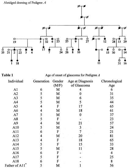
Pedigree A 44
Pedigree B 46
Pedigree C 46
PedigreesD - K 48
Pedigree L 49
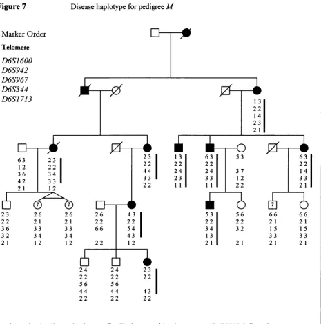
Pedigree M 50
Genetic analysis of pedigrees 53
Linkage analysis (pedigrees A and M) 53
Sequencing 57
Fluorescent in-situ hybridisation 59
Pedigree B 64
Segmental deletion (pedigree L) 66
Extent o f cytogenetic abnormalities 70
Analysis of ELF-2b 75
NIX analysis 75
Sequence analysis 77
Restriction enzyme digest 79
Murine model of segmental duplication 80
Foxcl/FOXCl and comeal phenotype 88
FoxeS comeal phenotype 91
FOXC2 ocular phenotype 97
Segmental duplication CNS phenotype 98
Discussion 100
Ocular features of pedigrees 100
Genotyping and FISH 101
Pedigree 101
Pedigree B ,C ,L 103
Evidence for a second glaucoma-causing gene on 6p25 105 Murine model of 6p25 segmental duplication 106
Hair phenotype 107
FOXCl and FoxeS comeal phenotype 108
FOXC2 ocular phenotype 110
Segmental duplication CNS phenotype 111
Conclusions 113
References 115
Appendix 131
1 Ocular anterior segment photographs 51 2 Ocular anterior segment photographs (Pedigree L) 52
3 Disease haplotype, pedigree A 55
4 Ethidium bromide stained gel, pedigree A {D6S967) 56
5 sequencing results, pedigreed 57
6 Fluorescent in-situ hybridisation, pedigree A 60
7 Disease haplotypes, pedigree M ' 62
8 Chromosome 6p25 contig 63
9 Ethidium bromide stained gels, pedigree B (D6S967, D6S344 and D6S942) 64
10 Haplotype analysis pedigrees A and B 65
11 Ethidium bromide stained gel -segmental deletion pedigree 66 12 Genotypic evidence for 6p25 segmental deletion 66
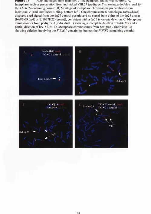
13 Fluorescent hybridisation results 68
14 Physical map of 6p25 69
15 Extent of 6p25 segmental duplications and deletion 71 16 Ethidium bromide stained gels illustrating extent o f segmental duplication 73 17 Haplotype analysis o f 6p25 telomeric deletion 74
18 NIX analysis o f clone M 75 7/24 75
19 E7,7^-26 sequence 77
20 ELF-2b electropherograms 78
21 Restriction enzyme sites for sequence changes identified in ELF-2b 78
22 S ad restriction enzyme digest 79
23 Screening of murine DNA for FOXC7-containing transgene 83 24 Digital photomicrographs o f hair samples 87 25 Graph displaying human and murine comeal thickness data 93 26 Photographs and histology of FOXC2 patients and dyl mice 94
27 Specular microscopy 96
28 FOXC2 mutations and phenotypes 97
Introduction
I Glaucoma-causing genes and loci 5
II Chromosomal distribution of human forkhead genes 26 III Phenotypes attributable to forkhead gene mutations 27
Results
1 Age of onset o f pedigree A 45
2 Age of onset o f pedigrees B and C 47
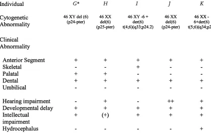
3 Clinical features of pedigrees D - F 48
4 Clinical features of pedigrees G - K 48
5 Two point lod scores between iris hypoplasia and 4q25/l 3ql4 markers 54 6 Two point lod scores between iris hypoplasia and 6p25 markers 54 7 Densitometry analysis of D6S967 alleles 57 8 Sequences of FOXCl and FOXF2 primers 58 9 FISH results pedigree /I (individual 13 and control) 61 10 Analysis of subset o f cells with a symmetric signal pattern 61
11 Summary o f FISH results 67
12 Summary o f 6p25 marker genotyping results 72
13 ELF-26 primer sequences 76
14 ELFparalogues 76
15 Sequences o f Foxcl and FOZC7-specific primers 80
16 Murine transgenesis results 81
17 Qualitative analysis of hair samples (observer data) 84
18 Statistical analysis o f observer data 85
19 Quantitative analysis of hair data (imaging data) 86 20 Statistical analysis o f quantitative data 86 21 Central comeal thickness data (pedigrees A - D ) 90
22 Central comeal thickness (controls) 91
My thanks go to my supervisors, Professors Shomi Bhattacharya and Roger Hitchings, for their kind support, encouragement and advice over the years. I am equally grateful to Professor Peng Khaw for letting me study such a fascinating collection o f patients, assisting with many aspects o f this research and for his longstanding support.
I am indebted to many friends and colleagues in the Department of Molecular Genetics, not least Neil Ebenezer for being so generous with his time, and to Louise Ocaka. In addition the advice and help o f Alison, Steph, Andrew and David has been invaluable. Tim’s support has been most welcome and, amongst other things, made ascertaining families in South Wales so much more fun. I acknowledge the
considerable help I have received from Professor Fred Fitzke and Dr Brian Clark (Institute of Ophthalmology), Dr Jane Sowden (Institute of Child Health), Professor Sue Povey and Dr Rosemary Ekong (Galton Laboratory), Professor Michael Walter (University of Alberta, Canada), Dr Simon John (Jackson Laboratory, USA) and Professor Peter Carlsson (University of Gothenburg, Sweden). Last but not least I thank all the families and Dr Beverly Searle from Unique for participating in the study.
Chromosomal duplication involving the Forkhead transcription factor gene FOXCl causes iris hypoplasia and glaucoma
Lehmann OJ, Ebenezer ND, Jordan T, Fox M, Ocaka L, Payne A, Leroy BP, Clark BJ, Hitchings RA, Povey S, Khaw PT, Bhattacharya SS.
American Journal o f Human Genetics (2000) 67:1129-1135.
A novel keratocan mutation causing autosomal recessive cornea plana
Lehmann OJ, El-ashry MF, Ebenezer ND, Ocaka L, Francis PJ, Wilkie SE, Patel RJ, Picker L, Jordan T, Khaw PT, Bhattacharya SS.
Investigative Ophthalmology and Visual Science (2001) 42:3118-22.
Interstitial 6p25 duplications and deletions cause ocular developmental abnormalities and glaucoma
Lehmann OJ, Ebenezer ND, Ekong R, Ocaka L, Mungall A, Fraser S, McGill JI, Hitchings RA, Khaw PT, Sowden J, Povey S, Walter M, Bhattacharya SS, Jordan T. Investigative Ophthalmology and Visual Science (2002) 43: 1843-1849
Novel anterior segment phenotypes resulting from forkhead gene alterations: evidence for cross-species conservation o f function
Lehmann OJ, Tuft S, Brice G, Smith R, Blixt Â, Bell R, Jordan T, Hitchings RA, Khaw PT, John S, Carlsson P, Bhattacharya SS.
Submitted to Investigative Ophthalmology and Visual Science (June 2002)
Investigating the association between OP A I polymorphisms and glaucoma:
comparison between normal tension and high tension primary open angle glaucoma Aung T, Ocaka L, Ebenezer ND, Morris AG, Brice G, Child AH, Hitchings RA, Lehmann OJ. Bhattacharya SS.
Human Genetics (2002) 110:513-514.
A major marker for normal tension glaucoma: association with polymorphisms in the OPAl gene. Aung T, Ocaka L, Ebenezer ND, Morris AG, Krawczak M, Thisleton DL, Alexander C, Vortruba M, Brice G, Child AH, Francis PJ, Hitchings R, Lehmann OJ. Bhattacharya SS.
Human Genetics (2002) 110: 52-56.
The phenotype o f normal tension glaucoma patients with and without OP A I polymorphisms.
Glaucoma Genetics. Lehmann OJ. European Glaucoma Society Meeting (2000) Iris hypoplasia associated with a 6p25 chromosmal duplication
Lehmann OJ, Jordan TL, Ebenezer ND, Ocaka L, Khaw PT, Hitchings RA, Child A, Brice G, Plant C, Bhattacharya SS.
[ARVO Abstract]. Invest Ophthalmol Vis Sci (2000) 41:4 S822. Abstract # 4634 Progress with FOXCI gene dosage.
Lehmann OJ, Jordan TL, Ebenezer ND, Ocaka L, Hitchings RA, Khaw PT, Walter MA, Povey S, Sowden J, Bhattacharya SS.
[ARVO Abstract]. Invest Ophthalmol Vis Sci (2001) 42:4 S530. Abstract # 2846 Paired interstitial duplications and deletions: a novel cause o f ocular developmental abnormalities and glaucoma
Lehmann OJ, Ebenezer N, Jordan TL, Ekong R, Hitchings RA, Khaw PT, Sowden J, Povey S, Walter MA, Bhattacharya SS.
American Society of Human Genetics (2001) S342. Abstract # 928
Duplication as well as haploinsufficiency of the forkhead/winged helix transcription factor FOXCI cause human anterior segment dysgenesis.
Mrirzayans F, Saleem R, Gould DB, Marshall J. Lehmann O. Jordan T, Raymond V, Mears AJ, Walter MA.
American Society o f Human Genetics (2001) S653. Abstract # 2780 Linkage analysis of a large Amish pedigree with glaucoma
Gallagher SP, Lehmann OJ. Leonardo D, Ebenezer N, Ocaka L, Child A, Hitchings RA, Sarfarazi M, Bhattacharya SS.
[ARVO Abstract]. Invest Ophthalmol Vis Sci (2001) 42:4 S563. Abstract # 3020 A gene for autosomal dominant Microphthalmia maps to 6p25
Leroy BP, Aragon-Martin JA, Martin KR, Webster AR, Trump D, Lehmann OJ. Ebenezer ND, Moore AT, Payne AM, Bhattacharya SS.
[ARVO Abstract]. Invest Ophthalmol Vis Sci (2001) 42:4 S651. Abstract #3053 Genetic exclusion o f 2 pedigrees with primary angle closure glaucoma from loci for nanophthalmos and microphthalmia
Aung T, Chew PT, Seah SK, Ang LP, Ocaka L, Ebenezer ND, Yap E. Lehmann OJ. Hitchings RA, Bhattacharya SS
The work presented in this thesis submitted for the degree o f doctor of philosophy is my own composition and save as otherwise stated the data presented herein is my own original work.
INTRODUCTION
The research described in this thesis involves molecular biology approaches to determine the cause of certain developmental forms o f glaucoma and the varied phenotypes associated with these genetic abnormalities. The following sections review the classification and genetic basis o f glaucoma, the role o f forkhead genes as well as PAX6 particularly in ocular development, as well as introducing concepts in molecular biology relevant to this thesis.
Clinical overview o f glaucom a
The glaucomas are a heterogeneous group of disorders characterised by an optic neuropathy in which retinal ganglion cell death leads to excavation of the optic nerve head (glaucomatous cupping) and visual field loss. They are responsible for more than six million cases o f blindness and represent the commonest cause o f irreversible visual loss world-wide (Quigley 1996). Glaucoma is frequently diagnosed late by which time the prognosis has worsened and even in Western countries, the
proportion o f undiagnosed cases is estimated to be ~50% (Quigley et al. 1997). This high figure has important public health implications for a condition that is treatable but in which visual loss, at present, cannot be reversed.
Classification o f glaucom a
Glaucoma can be classified in a variety o f ways, including: anatomically (open angle versus closed angle), aetiologically (primary versus secondary), chronologically (congenital, juvenile or adult) or on the basis of phenotypic features such as lOP. The diversity o f classifications highlights the paucity o f our understanding o f the molecular mechanisms responsible for this common disease.
Primary open-angle glaucoma (POAG) represents the commonest form o f glaucoma in Caucasians. It is characterized by elevated lOP believed to be primarily caused by resistance to aqueous outflow at the level of the juxta-canalicular portion of the trabecular meshwork. Most patients with POAG present fi"om the sixth decade of life onwards, although this type of glaucoma can affect all ages. An important subtype of POAG is normal tension glaucoma (NTG) in which the intraocular pressures lie within the statistically normal population range. NTG is under-diagnosed and typically presents with more advanced disease. It is estimated to account for approximately one third of POAG cases (range 20 - 50%) (Kamal and Hitchings 1998).
Primary angle-closure glaucoma (PACG) primarily results fi*om pupil block, iris or peripheral anterior synechial obstruction of the normal aqueous outflow, although the size o f the globe and position of the iris-lens diaphragm are other factors believed to be involved. PACG is the main type o f glaucoma in populations o f Chinese and Mongoloid descent and is also highly prevalent in India. As a result PACG is not just the commonest form o f glaucoma in Asia but represents the commonest type of glaucoma worldwide (Quigley 1996).
Risk fa cto rs f o r p rim a ry open angle glaucoma
The evidence that elevated lOP is the major risk factor for developing glaucoma comes from a series o f studies, including the Batimore survey. This demonstrated that 1.2% of the population with an lOP < 21mmHg had glaucoma in comparison to
10.3% population with TOP > 22mmHg (Somner et al. 1991). The lOP level has also been shown to correlate with the relative risk of developing glaucoma and the
severity of the field damage at presentation (Somner et al. 1991 ; Jay and Murdoch 1993). Even in NTG patients, the eye with the higher lOP exhibits the more severe degree o f field loss (Cartwright and Anderson 1988). Other major risk factors include age - the prevalence o f glaucoma in white subjects rises from ~1% at age 50 to ~4% at 80 years (Quigley 1997) and ethnicity (Leske et al. 1995). Important though these factors are, the significance o f heritable factors is increasingly being recognised.
Genetic basis o f glaucoma
Glaucoma has long been thought to have a genetic component due to familial aggregation o f cases, although suitable large-scale twin studies have not been performed. For many decades glaucoma was estimated to have a heritable component o f 5-15% but recent data suggests that this may represent a very
conservative figure. Data from the Rotterdam study, a prospective population-based survey, show that the lifetime risk of developing glaucoma is approximately ten times higher amongst relatives of glaucoma patients than relatives of controls (Wolfs et al. 1998). This thorough study included in the analysis offspring o f glaucoma patients, who would be too young to have developed the condition, biasing the estimate obtained towards a lower value. In addition, due to the small number of glaucoma patients studied (n=48), the confidence intervals associated with the relative risk observed are very large (95% Cl = 1.2 - 73.9), suggesting that the estimate is imprecise.
Data from two teaching centres (Tasmania and Iowa) indicate that the familial component of glaucoma may be higher still with 50-60% o f patients reporting a family history o f the condition (Alward et al. 2000; Mackey et al. 2000). It is
also showed that individuals from large POAG pedigrees were frequently unaware of their positive family history (McNaught et al. 2000). As one large pedigree with severe early-onset disease in which all individuals were aware of their family
history, was included in the analysis, the level o f under-reporting of a family history is higher than the study stated (27%). This data taken together with glaucoma's generally late age o f onset, known high rate of under-diagnosis (which presumably was greater in previous decades), together with the possibility o f incomplete
penetrance and evidence for modifier genes (Bejjani et al. 1999, Vincent et al. 2002) highlight why even autosomal dominantly inherited types o f glaucoma may not be readily appreciated as having a clear genetic basis.
Further evidence for a significant genetic basis to glaucoma comes from the condition's known genetic heterogeneity. To date seven genes have been identified that cause glaucoma as part of their phenotype [PAX6 (MIM 106210), PITX2 (MIM 601542), CYPIBI (MIM 601771), MYOC (MIM 601652), FOXCI (MIM 601090), LM XIB (MIM 602575) and OPTN (MIM 602432] and the mapping of eight further genes has been reported {GLCIB 2cen-ql3, GLCIC 3q21-24, GLCID 8q23, GLCIF 7q35-36, PD Sl 7q35-36, PDS2 1 8 q l l - 2 1 , ^ G 2 13ql4, GLC3B lp36) (reviewed by Craig et al. 1999; WuDunn 2002). This does not include the four loci (Xp, 15ql2-
15, 14q32 and 6p25) and one gene {CHXIO) so far identified as causing
Table I List of glaucoma-causing genes/loci.
Genes/loci associated with glaucoma, shown below.
Phenotype
JOAG/POAG
POAG
POAG
POAG
POAG/NTG
POAG
Pigment Dispersion
Pigment Dispersion
Axenfeld-Rieger
Axenfeld-Rieger
Axenfeld-Rieger
Axenfeld-Rieger
Congenital Glaucoma
Congenital Glaucoma
Nail-Patella Syndrome
Aniridia
Nanophthalmos
Micropthalmia
Micropthalmia
Micropthalmia Micropthalmia
Locus Position Inheritance Gene
GLCIA Iq24.3-q25.2 AD MYOC
GLCIB 2cen-ql3 AD
GLCIC 3q21-24 AD
GLCID 8q23 AD
GLCIE 10pl5-14 AD OPTN
GLCIF 7q35q36 AD
GPDSl 7q35q36 AD
GPDS2 18qll-21 AD
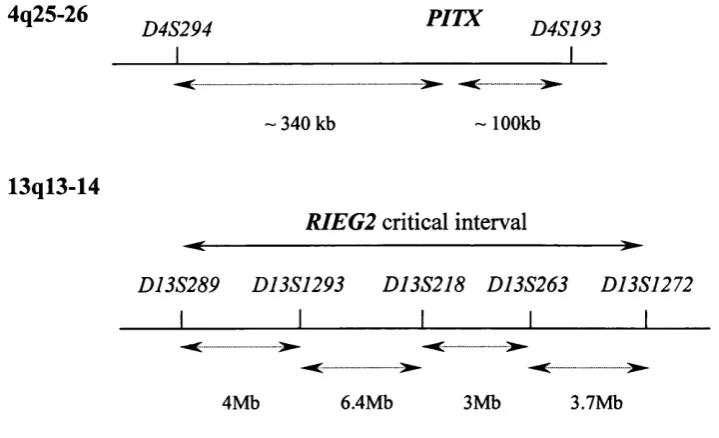
RIEGl 4q25 AD PITX2
RIEG2 13ql4 AD
IRIDl 6p25 AD FOXCI
16q24 AD
GLC3A 2p21 AR CYPBl
GLC3B lp36.2-36.1 AR
NPSl 9q34 AD LM XIB
PAX6 l l p l3 AD PAX6
NNOl l i p AD
Xp X
arMi 14q32 AR
CHXIO 14q24.3 AR CHXIO
adCMIC 15ql2-15 AD
M onogenic or Polygenic Disease
At present it is unclear whether (genetically-determined) cases o f glaucoma are a heterogeneous collection o f monogenic disorders or a complex genetic disorder with multiple genes acting (either alone or in conjunction with environmental factors) to determine an individual's susceptibilty to developing glaucoma. Answering this question would require complex segregation analysis and the necessary studies have not been performed. The simplest means of identifying susceptibiltiy loci for
glaucoma would require nonparametric linkage studies. This approach, with
relatively large numbers o f small glaucoma families (minimum size o f two affected siblings), allows investigation o f possible association between a locus and
development of glaucoma without the problem o f defining the nature o f inheritance or the possibility o f genetic heterogeneity. Although a number of possible loci have been tentatively identified through sib-pair analysis (Wiggs et al. 2000), these have yet to be confirmed.
In the last two years, evidence for the presence of modifier genes that modulate the penetrance and or severity o f certain forms of glaucoma has emerged. The first study, an analysis of Saudi pedigrees with primary congenital glaucoma identified 40 apparently unaffected individuals in 22 pedigrees with CYPIBI mutations and haplotypes identical to their affected siblings. This suggested the presence o f a dominant modifier locus capable o f modulating the severity o f the disease (Bejjani et al. 2000). More recently a single pedigree with autosomal dominant glaucoma was reported in which CYPIBI and MYOC mutations segregated. The mean age at diagnosis of glaucoma in the MYOC mutation carriers was 51 years compared to 27 years in individuals with both mutations, indicating that MYOC and CYPIBI may interact through a common pathway (Vincent et al. 2002).
Axenfeld-Rieger Syndrome
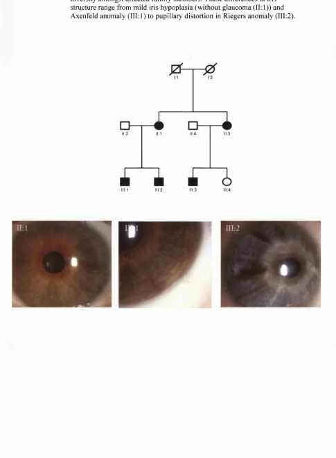
This group o f highly penetrant autosomal dominantly inherited disorders is
anomaly, whilst in Rieger anomaly, additional changes to the iris with
distortion/displacement of the pupil (correctopia) and holes in the iris (polycoria) are seen. Patients with the ocular features o f Rieger anomaly plus certain systemic abnormalities (including midfacial and dental abnormalities and/or umbilical hernia) are considered to have Rieger syndrome. In reality these disorders form an
overlapping clinical spectrum with mutations in at least four different genes resulting in the same phenotypes. To date two o f these genes, PITX2 and FOXCI, have been identified and a further two loci (13ql4 and 16q24) mapped through the study o f single large American and Brazilian pedigrees respectively (Phillips et al. 1996; Nishimura et al. 2001).
Mutations in PITX2, a paired homeobox gene identified from study o f Rieger
syndrome pedigrees, were shown to cause a wide clinical spectrum that encompasses the Axenfeld-Rieger subtypes (Semina et al. 1996). PITX2 is expressed in the
periocular mesenchyme and Pitx2^‘^ mice (homeobox deleted allele) have displaced irregular pupils, and lack anterior chambers, differentiated corneas and extraocular muscles. This suggests that haplo-insufficiency underlies the Axenfeld-Rieger phenotypes and demonstrates the role of Pitx2 in periocular mesenchyme
differentiation. Pitx2"s function in determining left-right patterning is of particular interest as pitx2^^' and pitx2'^' mice exhibit a range of defects including abnormal cardiac positioning and lung asymmetry (Lu et al. 1999, Lin et al. 1999) and some patients with Axenfeld-Rieger phenotypes and ocular asymmetry due to PITX2 mutations have been reported (Perveen et al. 1999).
FOXCI mutations also cause the full range of Axenfeld and Rieger phenotypes (as well as iris hypoplasia and possibly Peter’s anomaly) (Nishimura et al. 1998; Mears et al. 1998; Swiderski et al. 1999; Mirzayans et al. 2000). That these phenotypes result from different PITX2 and FOXCI mutations, coupled with phenotypic
Forkhead genes
More than ten years have elapsed since the identification o f the Drosophila transcription factor fork head (Wiegel et al. 1989) subsequently led to that o f the forkhead DNA binding domain (Wiegel and Jackie 1990). At the time it was already apparent that ancestral chordate genes could give rise to whole families o f vertebrate genes and, as illustrated by the homology between eyeless and PAX6 (Quiring et al. 1994), their structure and function were remarkably conserved. Since then more than 100 Forkhead genes have been identified in species ranging from yeast to humans. These genes code for a subgroup o f the helix-tum-helix family of proteins and the arrangement o f loops (or wings) connecting the P strands that flank one o f the three a helices, gives rise to a butterfly-like appearance, hence their alternative name o f "winged-helix" transcription factors (Clark et al. 1993).
Forkhead genes, share a conserved 100 amino acid DNA binding domain, which has a central role in normal gene function. Extensive evidence has emerged for the important role of these forkhead/winged helix transcription factors in development and cell differentiation through the description of such mutations in model organisms and humans. This has offered insight into the diverse biological processes that these genes influence, including tumorigenesis, cell cycle regulation, and differentiation.
Forkhead genes (Classification)
Forkhead genes (Tumorigenesis and cell-cycle control)
At the commencement o f this thesis the phenotypes o f only 2 forkhead genes {FOXEl and FOXNl) had been defined in humans. In the subsequent three years there has been extremely rapid progress identifying and defining the functions of members of this family. It is now apparent that they possess diverse roles in the development o f a wide range o f tissues as well as in tumorigenesis and cell cycle regulation. Each o f these topics will be reviewed briefly as they have relevance to different aspects o f this thesis.
The first indication of Fox gene involvement in tumorigenesis came from the
identification o f FOXKla, located at a site (17q25) o f translocations in some cases of acute myelogenous leukemia (Li et al. 1992). Subsequently, a translocation leading to fusion between PAXS and FOXOla (FKHR) was identified in some alveolar rhabdomyosarcomas (Galili et al. 1993). The resultant protein, possessing an intact PAX binding domain plus the C terminal half o f the forkhead binding domain, is a more potent transcriptional activator than PAX3 alone and may act by over
expressing PAX3 target genes (Fredericks et al. 1995). Introduction o f this chimaeric transcript into NIH3T3 cells induces expression o f transcription factors involved in myogenesis, including Myod, Myogenin, Sixl, and Slug (Khan et al. 1999). Since PAX3 normally inhibits myogenic differentiation it is possible that upregulation may cause tumour formation by suppressing terminal differentiation (Epstein et al. 1995).
expression has been shown to block cell-cycle progression at phase G l, by
transcriptionally activating the cell-cycle inhibitor CDKNIB (p27 or KIPl), whereas FOXOSa activity, which can be regulated by the oncogene AK Tl (protein kinase B), could play a role in apoptosis by inducing the expression of genes such as TNFSF6 (tumour necrosis factor superfamily 6) (Brunet et al. 1999).
Therefore a reasonable amount of evidence suggests that the Fox ‘O ’ subclass, which are orthologues of daf-16, a forkhead factor that regulates longevity in
Caenorhabditis elegans (Lin et al. 1997, 2001), are involved in cell-cycle regulation and that inactivation could be an important step in oncogenic transformation. The cloning of the murine orthologues of these genes (Biggs et al. 2001) and evidence for the wider role o f forkhead genes in cell cycle control (Alvarez et al. 2001) will permit further advances o f our understanding o f these genes. In addition indications exist that other Fox genes may play a role in tumorigenesis including; FOXGlb (Brain Factor 1, BFl), a transcriptional repressor and the homologue of the cell derived oncogene from the retrovirus avian sarcoma virus 31 and FOXN2 (Human T cell Leukaemia Virus Enhancer Factor, HTLF).
Forkhead genes (Extraocular Phenotypes)
Fox genes have fundamental roles in the formation o f a wide range o f organs including cardiac, meningeal, skeletal, renal, immunological, pulmonary and
mesenteric development. Just three examples will be considered, that are relevant to their roles in regulating tissue proliferation, hair development and neural
development.
unusual. It raises the possibility that heterozygous mutation may result in an, as yet unrecognised, milder phenotype(s) in a manner similar to heterozygous Foxcl^'' mice. Foxel is co-expressed with Titfl and Pax8 in the developing thyroid where they are believed to regulate cell differentiation. Thyroid hormones are synthesised by follicular cells derived from an endodermal bud which originates from the posterior pharyngeal floor. This bud migrates to between the fourth pharyngeal pouches, with which it frises, and functional differentiation of the thyroid follicular cells, as shown by the expression of thyroglobulin, follows migration. Since Foxel is expressed in the migrating primordium but down-regulated prior to differentiation it may be involved either in promoting the migration process or in repressing
differentiation of the TFCs until migration has occurred (Zannini et al. 1997). In any event homozygous mutations result in thyroid agenesis. Foxel is also expressed in the craniopharyngeal ectoderm involved in palate formation. In cleft palate there is absence o f normal tissue whereas in choanal atresia there is retention o f abnormal tissue. Either could result from enhanced or reduced proliferation or apoptosis suggesting a role for FOXEl in both processes.
The second example concerns hair development, a complex process involving formation of clumps of epithelial cells in the lower layers o f the epidermis. This induces the condensation o f specialized mesenchymal cells that in turn provide the inductive signals required for formation o f the epithelial portion o f the hair follicle. Participation of forkhead genes in this process has been revealed by the finding o f a Foxql mutation in satin mice (Hong et al. 2001), so named after the striking silky appearance of their coat, which has a high degree o f sheen. This phenotype has been attributed to aberrant differentiation o f the hair shaft and interestingly, the human homologue FOXQl is one of a triplet of Fox genes on 6p25. A second forkhead gene (Foxnl, formerly winged-helix nude) is responsible for the murine nude phenotype, characterized by the congenital absence of hair and a severe immunodeficiency. Foxnl is expressed in the skin and thymus where it maintains the balance between growth and differentiation (Nehls et al. 1996, Brisette et al. 1996). The human nude phenotype, also characterised by severe T cell immunodeficiency as well as nail dystrophy and alopecia, is caused by a homozygous mutation in the human
The last example concerns intellectual development and the recent identification of a novel forkhead gene involved in the development o f speech and language. FOXP2 was shown to be disrupted in an individual with a chromosomal translocation and a severe speech and language disorder. A mutation in a highly conserved (FOXP2) forkhead domain residue was then found to segregate with a similar phenotype in a large pedigree. This suggests that haplo-insufficiency o f FOXP2 at a key stage of embryogenesis leads to abnormal development of structures important for speech and language, representing the first gene to have been implicated in such neural pathways (Lai et al. 2001).
F O X C I
FOXCI (formerly FKHL7) is one of seven genes recognised to cause glaucoma. It was independently identified by two groups using different methodologies: cloning of a translocation breakpoint in an individual with primary congenital glaucoma and a balanced translocation (Nishimura et al. 1998), and a positional cloning approach (Mears et al. 1998). FOXCI mutations were shown to cause Axenfeld anomaly, Rieger anomaly, Rieger Syndrome and iris hypoplasia both in isolated cases and in pedigrees with an autosomal dominant pattern of inheritance. However, four such pedigrees that map to 6p25 do not contain a mutation in the coding region of the FOXCI gene (Mears et al. 1998; Jordan et al. 1997; Morissette et al. 1997). In two of these families FOXCI was excluded fi*om the disease-causing interval by mapping data on the basis o f recombination events. These findings suggested the existence of a second glaucoma-causing gene on 6p25 (Mears et al. 1998).
F o x c l
At the start o f this thesis, data was also available on the naturally-occurring Foxcl mutant which in the homozygous state results in the congenital hydrocephalus (ch) phenotype (Kume et al. 1998). The first description o f the ch phenotype (Griineberg
laryngeal cartilages were all severely abnormal with a range o f other defects
affecting the skeleton, cerebellum, cistema magna and tissue surrounding the fourth ventricle. Subsequently ch heterozygotes were shown to exhibit milder skeletal anomalies than ch homzygotes (Hong et al. 1999).
Kume and colleagues demonstrated that the ch allele was caused by a mutation in Foxcl (a C—►T transition in the forkhead box) resulting in a truncated protein of 122 amino acids (compared to the normal 553aa). The authors extended the range o f skeletal defects described in ch mice, highlighted the wider range o f organs affected by the ch mutation (including cardiac and renal development) and described novel changes in both meningeal and ocular development. Using a null allele {Foxcl^°^^), Foxcl^^^^ was shown to be highly expressed in the developing meninges with disruption of the normal meningeal architecture in mutant embryos; an observation potentially relevant to the causation of glaucoma in individuals with FOXCl
mutations. Foxcl^^'^^ homozygotes were also found to have a range o f ocular abnormalities including open eyelids at birth, disorganised comeal architecture and iris hypoplasia. In addition four individuals with telomeric deletions encompassing FOXCl and ocular anterior segment developmental anomalies were reported (Kume et al. 1998). This observation, taken together with several previous reports of ocular anomalies in patients with 6p25 cytogenetic abnormalities (such as Law et al. 1998), provided the first indication that altered dosage of a gene or genes in the
duplicated/deleted region was responsible for the ocular phenotype(s) observed.
FoxcVs expression pattern is very similar to that o f Foxc2, with which it shares close homology and the co-ordinated function of these two genes in ocular development has now been demonstrated (Smith et al. 2000). Co-ordinated expression is a feature o f other Fox paralogues, including Foxdl and Foxd2 in renal development. The similar ocular anomalies that result from haplo-insufficiency o f Foxc2 and FOXCl and the absence of direct interaction between Foxcl and Foxc2, provide evidence that these genes share a common downstream pathway. Recently this has been confirmed by data that Foxcl and Foxc2 have similar dose-dependent functions in cardiovascular, renal and somite development (Kume et al. 2000; Kume et al. 2001; Topczewska et al. 2001).
Forkhead genes (other ocular phenotypes)
The prevalence o f ocular disease amongst the ten human Fox genes whose
phenotypes have been defined, is noteworthy. Mutations in FOXCl, FOXC2, FOXES and F0XL2 affect a range of tissues including the eyelashes, eyelids, comea, iris and lens, and a fifth gene {Foxn4\ expressed in developing retina, has recently been identified (Gouge et al. 2001).
Mutations in FOXC2 cause lymphedema-distichiasis, the third heritable human disorder attributed to a forkhead gene (Fang et al. 2000). FOXC2 was identified through cloning o f a translocation breakpoint in an individual with neonatal lymphedema, the same approach used by one laboratory to identify FOXCl (Nishimura et al., 1998). FOXC2 neatly illustrates the principle that forkhead proteins, like other transcription factors, may have one function during embryonic development and organogenesis, and a completely distinct function in adult,
most sensitive to expression levels (and therefore gene dosage). In adults on the other hand, FOXC2 appears to be a master regulator o f energy
expenditure/storage/glucose metabolism. There is a similar situation with the FoxA (HNF3) genes, which control essential steps in embryogenesis such as notochord, floorplate, and endoderm differentiation, but control liver metabolism in adults (Kaestner et al. 2000).
Mutations in FOXL2 cause a complex developmental eyelid disorder,
blepharophismosis-epicanthus inversus syndrome (BPES), in which individuals display narrowed palpebral apertures (blepharophismosis), drooping o f the upper eyelid (ptosis) and inversion o f the arrangement of one o f the lid folds (epicanthus inversus) (Crisponi et al. 2001). Patients are divisible into two categories according to whether their BPES phenotype includes ovarian failure. Mutations causing type I BPES (with ovarian failure) generate a truncated allele whilst those associated with type II (BPES alone) generally occur downstream of the forkhead domain. This genotype-phenotype correlation is consistent with the interpretation that type I mutations disturb the DNA-binding properties of the forkhead domain, generating a null allele, whilst type II mutations form a hypomorphic allele with reduced
transactivating properties (De Baere et al. 2001).
The importance o f Fox genes in eye development is further highlighted by the role of FOXES, in the development o f the anterior segment. FoxeS mutations, which cause the naturally occurring dysgenetic lens {dyl) mutant, result in a similar phenotype to FoxcF^^^^ or FoxcF^', in that the lens vesicle fails to separate from the overlying ectoderm. This closely resembles Peters anomaly a genetically heterogeneous disorder, so far attributable to mutations in PAX6, FOXCl, PITX2, EYAl and CYPIBI (Hanson et al. 1994; Nishimura et al. 2000; Perveen et al. 2000; Azuma et al. 2000; Vincent et al. 2002). The recent identification o f FOXES mutations in individuals with anterior segment developmental anomalies and cataract (Semina et al. 2001), and in Peters anomaly (Ormestad et al. 2002) highlights the close
P aired box genes
Pax (paired box) genes are a family o f transcription factors isolated as a result of their homology with the Drosophila segmentation gene paired. Pax proteins share a
128 amino acid DNA binding domain and of the nine PAX genes identified, the role o f PAX6 is perhaps best understood. Mutations in PAX6/Pax6 are responsible for the human aniridia and murine Small eye phenotypes respectively, both of which are associated with comeal opacification and cataracts. The recognition that the Drosophila orthologue of PAX6 was responsible for the classic mutant eyeless (ey), had broad evolutionary implications. It demonstrated that the same orthologue controls development of the compound insect and vertebrate eye and that
comparative ocular developmental genetics is a powerful tool for elucidating the genetic pathway controlling eye morphogenesis.
A number of Drosophila genes have been identified through the study ey paralogues and these are categorized into four gene families: PAX6 {eyeless, twin o f eyeless {toy), eyegone {eyg)); EYA {eyes absent), SIX {sine oculis {so), Optix) and DACH {dachshund). The functional relationships between these genes is presently better understood in Drosophila than in vertebrates. Through induction o f ectopic eye formation toy has been shown to directly activate ey (and induce other genes
including sine oculis {so) and eyes absent {eya)) and lie above them in the hierarchy of ocular developmental genes. In tum the Notch signaling pathway lies upstream of ey and toy in eye spéciation (Kumar et al. 2001). Whilst a full review o f PAX6 and vertebrate eye development is beyond the scope o f this thesis (see, Hanson 2001), mammals have homologues o f these gene families {PAX6, EYAl-4, SIXl-6 and DACHl and DACH2) and the effects o f mutations in these genes are slowly being reported. The close functional similarity o f these fruit fly/murine/human orthologues makes naturally-occurring/transgenic mutants excellent models for the study of human disease.
PAX6 also illustrates both the developmental consequences and the exquisite
developmental anomalies with other organs, in which PAX6 is expressed, unaffected (Schedl et al. 1996). This indicates that both increased and decreased levels of Pax6/PAX6 expression result in ocular abnormalities and that the eye is especially
sensitive to altered gene dosage.
M olecular aspects o f haploinsufficiency and altered gene dosage
The mechanisms underlying haploinsufficiency are incompletely understood but one model for transcription factors is that they activate different spectrums o f target genes. In the case o f the C.elegans orthologue o f FOXA,pha-4 activates all known pharyngeal genes raising the possibility that direct transcriptional regulation of entire gene networks may be a common feature of all organ identity (and Fox) genes (Gaudet and Mango, 2002). Target genes may have different binding affinities for each transcription factor, and according to the level o f functioning transcription factor in a cell, a different pattern of gene expression would result. Combining this model with stochastic effects could explain some o f the phenotypes caused by gene mutation or altered dosage. Pax5 has been shown to exhibit allele-specific
expression in which Pax5 is transcribed from one of its two alleles at different stages of development (Nutt et al. 1999). The observation of similar mono-allelic
expression m IL 4 and Ly49 receptors (Riviere et al. 1998; Held et al. 1998) suggests this may be a widespread phenomenon. It is tempting to speculate that such a
mechanism may apply to other transcription factors and thus could explain why single mutations in PAX2, PAX6 and PAX8 cause renal-coloboma syndrome, aniridia and congenital hypothyroidism respectively.
Position effects
Numerous instances exist o f pathogenic chromosomal rearrangements whose breakpoints lie many kilobases fi*om the coding elements o f the gene responsible. Such position effects are believed to result from deleterious changes to gene
expression, due to change in the gene’s position relative to its normal chromosomal environment. Possible mechanisms include transposition to sites where the
position effects and this may reflect either the number or wide distribution o f their regulatory elements. Position effects have been reported with PAX6, PITX2 and POU3F4, and can be induced by translocations with breakpoints up to 900kb away (reviewed in Kleinjan and van Heyningen 1998). Fox genes are also susceptible with reports o f balanced translocations causing positional effect inactivation o f FOXCl, FOXC2, and FOXL2 in glaucoma, lymphedema-distichiasis and BPES respectively (Nishimura et al. 1998; Fang et al. 2000; Crisponi et al. 2001). These mechanisms contrast with other forms of gene inactivation such as the CTG repeat expansion in DMPK which causes cataracts in dystrophia myotonica by directly disrupting SIX5 expression (Sarkar et al. 2000; Klesert et al. 2000).
M olecular M echanism s fo r Chromosomal Rearrangements
Chromosomal rearrangements are associated with a wide variety o f genetic disorders and are divisible into two groups depending on whether the rearrangements are inter- chromosomal (translocations) or intra-chromosomal. The latter category comprises a wide range o f anomalies including telomeric deletions, segmental duplications and deletions, inversions and marker chromosomes.
Telomeric (or terminal) deletions involve loss of the distal portion of one
chromosomal arm. Although they can occur on any chromosome, certain deletions are observed more frequently than others. This may reflect the relative viability of monosomy in different chromosomal regions or the susceptibility of certain areas to rearrangements. There is evidence in some deletion syndromes (e.g. Jacobsen syndrome) of an association between certain repeat sequences and the position of breakpoints (Jones et al. 2000) although for most telomeric deletions, the breakpoints do not occur at a single site.
patients is consistent with these anomalies arising through a common mechanism, such as homologous recombination between low-copy number repetitive sequences (Emanuel et al. 2001). In two examples (CMT/HNPP and SMS) the sequences responsible for these contiguous gene duplication/deletion syndromes have been characterised enabling patient-specific junctional fragments to be amplified. In GMT (type 1 A) the duplication arises from unequal crossing over mediated by 24 kb flanking repeats whilst the reciprocal deletion is associated with a peripheral neuropathy (hereditary neuropathy with liability to pressure palsies (HNPP). Both phenotypes result from altered copy number of the dosage-sensitive myelin gene (PMP22) (Lupski 1991; Lupski 1992; Reiter 1996). In Smith Magenis Syndrome, the size of the duplicated/deleted region is much larger (~ 5 Mb) and the
rearrangements are mediated by repeats ~ 200kb in length (Chen et al. 1997; Potocki et al. 2000).
Only relatively short lengths o f sequence homology (134-232 bp) are required for homologous recombination and unequal crossing over to occur (Waldmann et al.
1998) and in some disorders, such as the rare X-linked condition Pelizaeus-
Merzbacher disease, large blocks of repetitive sequence have not been identified. In this disease, aberrant CNS myelination occurs secondary to altered dosage o f the proteolipid protein gene; and the segmental duplications and deletions are of differing size suggesting that a different mechanism is involved (Woodward et al. 1998; Hodes et al. 2000). Possibilities include the presence of multiple small blocks o f repetitive sequence or alternatively sequences that may predispose to the
chromosomal breaks.
Human Genome Project
Much of the research described in this thesis was facilitated by the increasing availability o f assembled sequence fi*om the human (and related) genome(s). Access to such genomic sequence data was especially helpful in defining the approximate extents o f the segmental cytogenetic abnormalities observed in some of the
pedigrees.
by programmes to create genetic and then physical maps of clones covering the yeast and C.elegans genomes. The development of shotgun sequencing methods finally made sequencing of larger genomes feasible. Between 1995 and 1999, bacterial artificial chromosomes (BACs) were successfully used to construct a clone tiling path and -15% of the human genome was sequenced. In the next 18 months, perhaps spurred on by the efforts o f the biotechnology firm Celera, a draft genome sequence was published (International Human Genome Sequencing Consortium 2001; Venter et al. 2001). Whilst both employed shotgun sequencing, the public consortia first arranged the genome into an overlapping collection o f (primarily) BAG clones, which were then individually sequenced. In contrast, Celera adopted a whole- genome shotgun assembly method in which random portions o f genomic sequence were reassembled using computational techniques. The relative merits of these different approaches remains controversial although the extent to which the whole- genome shotgun assembly method benefited from pre-existing physical map and sequencing data, has recently been reported (Waterston et al. 2002).
Systematic sequencing of chromosome 6 commenced in September 1996 and was largely performed at the Sanger Centre using bacterial clones o f genomic origin subcloned into M l3 and pUC vectors. Chromosome 6 is -ISO Mb in size and when the sequence for dJ118B18 (the clone containing FOXCl) was available, the position and sequence of adjacent clones was not known. In the subsequent two and a half years, the sequencing of this terminal portion of 6p is virtually complete.
Strategies fo r gene identification
sequence data enables candidate genes in the region to be readily identified. The considerable amount o f database information available on each sequence (which frequently includes tissue expression pattern data, motif structure and known
paralogues/orthologues) often highlights the best (glaucoma-causing) candidate(s) to screen, effectively replacing traditional physical mapping techniques.
One variant o f this approach, which has been especially successful for identifying human forkhead genes, is translocation breakpoint mapping. This technique assumes that the phenotype in individuals with a translocation is breakpoint-mediated (direct or positional effect) gene inactivation. Fluorescent in-situ hybridsation (FISH), using YACs from the region as probes, enables the breakpoint to be defined as at least one probe will span the breakpoint and yield hybridsation signals from both
chromosomes. The interval spanning the breakpoint can be defined to less than 40kb through use o f progressively smaller probes (BACs and then either cosmids or PCR- generated probes). Subsequent database analysis of the clone that spans the
breakpoint will identify adjacent genes. Determining which o f these candidates is responsible for the phenotype is most easily achieved by screening a panel of patients with the same phenotype, for mutations in each candidate gene. The gene closest to the breakpoint may not be the cause of the phenotype as illustrated by the breakpoint mapping of FOXCl and FOXC2. In both cases the genes disrupted by or lying closest to the breakpoint {GMDS and FOXLl respectively) were not
responsible for the disease phenotype.
Linkage analysis
Linkage analysis is a technique for mapping inherited disorders. It relies upon the likelihood of two loci being separated by a recombination event being proportional to the distance between them. These recombination events occur on average at least once on the arm o f each chromosome during the pachytene phase of meiosis
(International Genome Sequencing Consortium, 2001). By measuring the frequency of non-random segregation a statistical value can be calculated for the likelihood that two loci are linked.
compared with that o f a number of polymorphic markers. The markers available have changed considerably over the last two decades, progressing from blood group markers and restriction fragment length polymorphisms, via VNTRs to the
microsatellite markers used in this thesis, and most recently to single nucleotide polymorphisms. The most useful markers are those with a high degree of heterozygosity. A marker’s informativeness can be quantified by a value, its polymorphic information content (PIC) (Ott 1997), which is determined by the number of alleles and the frequency o f each in the population. Markers with values > 0.7 are prefered for linkage analysis and PIC can be calculated with the following formula:
n n-I n
PIC = 1 - E p / - S . E 2 p i p f i = 1 i= 1 j = i + 1
(where Pi and P, are the population frequencies o f the zth andyth alleles).
The mathematical measure of linkage is the lod score (Z) - the logarithm of the odds ratio that two loci are linked at a given recombination fraction (0) compared to the same segregation pattern if they are unlinked. This ratio is calculated for a range of recombination values between 0 = 0 and 0 = 0.5 (where loci are regarded as
independently assorted).
Z (0) = logio [L(0) / L(0.5)] where: L(0) = likelihood if the two loci are linked and have a recombination fraction o f 0
L(0.5) = likelihood if the two loci are not linked
By convention a lod score of > 3.0 is regarded as significant evidence that two loci are linked. This equates to the odds in favour of linkage being at least 1000:1 or a 1 in 20 probability (or P=0.05) that the observed linkage has occurred by chance. A smaller lod score is required for demonstrating linkage in X-linked disorders (> 2.0). The presence o f a negative lod score suggests that two loci are unliked and, by convention, a lod of < -2.0 is required to disprove linkage. Although large
their logarithmic nature, under certain circumstances lod scores from different pedigrees can be combined. Lod scores can be calculated with a variety o f computer packages not least the MLINK program (Lathrop and Lalouel 1984) and web based programs (Genetic Linkage User Environment (GLUE), Human Genome Mapping Project).
Confirmation that a sequence change represents a mutation
Once a sequence change is identified in an affected individual it is necessary to demonstrate that it is responsible for the disease phenotype. The minimum proof required is that the sequence change co-segregates in the pedigree with the disease phenotype, is not present in a panel o f ethnically matched control chromosomes, and generally lies within the gene’s open reading frame altering the amino acid sequence of the protein. Identification of a second mutation, segregating in an unrelated pedigree, excludes the possibility of the sequence change being in linkage
disequilibrium with the real disease-causing gene. It is now increasingly important to demonstrate the mutation’s effect on protein structure, in vitro function and ideally in a model organism.
Fluorescent in-situ hybridisation
separated in three dimensions, which can lead to signal masking. To compensate for this, large numbers of interphase nuclei are generally imaged from both affected and control individuals and the results analysed to determine whether the pattern in affected individuals differs significantly from that seen in controls.
Transgenesis
The introduction o f specific DNA sequences (or transgenes) into animal germ lines represents a valuable technique for investigating the role and regulation o f genes in a model organism. At least three approaches exist for generating transgenic models - retroviral injection o f preimplantation embryos, manipulation o f embryonal stem cells and finally direct microinjection o f mouse embryos. The latter approach, direct microinjection of the transgene, involves several steps including: design and
isolation of the construct, microinjection o f murine embryos, oviduct transfer and genomic analysis of transgenic animals.
In the design of a transgene, the inclusion o f the gene’s regulatory elements is
important. Where these are unknown, as in the case o f FOXCl, it is usual to insert as large a construct as possible such as a cosmid or PAC. Some evidence exists that the presence o f vector derived sequence may inhibit mammalian transcription and removal of such sequences prior to injection, which also serves to linearise the DNA, may be advantageous (Jessen et al. 1988). Many researchers however achieve
Aims o f thesis
The initial aim of the thesis was to identify novel glaucoma-causing genes by linkage analysis o f large pedigrees with iris anomalies and early onset glaucoma. As
evidence for altered gene dosage as the cause for these 6p25-linked phenotypes was obtained, the emphasis switched to: attempting to understand the causation of these duplication, determining whether related cytogenetic anomalies were present in 6p25, generating a murine model o f the 6p25 segmental duplication and determining some of the wider phenotypes attributable to altered FOXCl gene dosage or
Name Location
F0XD2 lp32-34
F0XD3 lp31-32
FOXES lp32
FOXN2 2pl6-22
FOXPl 3pl3
FOXL2 3q23
FOXDl 5ql2-ql3
FOXJl 5q34
FOXOlb 5q35.2-35
FOXF2 6p25
FOXCl 6p25
FOXQl 6p25
FOXOSa 6q21
FOX02 6q21
FOXP2 7q31
FOXHl 8q24
FOXD4 9q21
FOXEl 9q22
FOXJ2 12pl3
FOXMl 12pl3
FOXN4 12q24
Name Location
FOXOla 13ql4.1
FOXKlb 14
FOXAl 14ql2-13
FOXGla 14ql3
FOXGlb 14ql3
FOXGlc
FOXN3 14q24
FOXBl 15q21-26
FOXFl 16q24.3
FOXLl 16q24.3
F0XC2 16q24.3
FOXOSb 17pll
FOXNl 17qll-ql2
FOXJl 17q22-25
FOXKla 17q25
FOXKlc 17q25
FKHL18 2 0 q lL l-q lL 2
FOXE2 22ql3-qter
F 0 X 0 4 Xql3
FOXP3 XplL23
Table III FO XCl^ (6p25) FOXC2U (16q24.3) FOXEIX (9q22) FOXE3X (lp32) Foxil FOXL2X (3q23) FOXNIX (17qll-ql2) FOXOla (13ql4)
F 0 X 0 4
(Xql3)
FOXP2
(7q31)
FOXP3
(Xpll.23)
Phenotypes attributable to Forkhead Gene Mutations
Human
Axenfeld-Rieger Syndrome
Glaucoma, Iris Hypoplasia
Lymphedema-Distichiasis
Murine (naturally occurring murine mutant)
Ocular/memingeal/cardiac/skeletal & renal anomalies
{Congenital hydrocephalus, ch)
Ocular/cardiac/renal anomalies
Thyroid agenesis, cleft palate, choanal atresia, polyhydranmios spiky hair
Ocular anterior segment anomalies & cataract
Ocular anterior segment anomaly
{Dysgenetic lens, dyl)
Common cavity syndrome
Blepharophimosis/Ptosis/ Epicanthus Inversus Syndrome (BPES)
Premature ovarian failure
Immunodeficiency, alopecia, nail dystrophy
Rhabdomyosarcoma
Severe immunodeficiency/absence o f hair
{Winged-helix nude)
Myeloid-Lymphoid Leukaemia (MLL)
Speech & language disorder
Neonatal diabetes, enteropathy Lymphoproliferative disorder
& endocrinopathy fatal in hemizygous males
{Scurfy mouse)
FOXQIX
(6p25)
t ocular phenotype J hair phenotype
Satin hair
MATERIALS & METHODS
Ascertainment o f Patients
Patient Identification
The majority o f patients and families with anterior segment malformations and glaucoma were identified from the Moorfields Eye Hospital (MEH) Paediatric Glaucoma Clinic, a tertiary referral clinic treating patients from across the United Kingdom. Additional patients were identified from other sources including the MEH Genetic Database, referrals from other units and UNIQUE - a support group for individuals with rare chromosomal abnormalities.
After individuals were identified from families with definite evidence o f hereditary glaucoma, priority was given to the ascertainment o f large families (> 8 meioses) that were particularly suitable for linkage analysis. Contact details for each patient / relative were sought using the hospital information systems / directory enquiries and were invited to participate, either by letter and or telephone contact.
Informed consent was obtained from all participants - in the case o f children or individuals with developmental delay, consent was provided by their parents. The ethics committee of Moorfields Eye Hospital approved the study (protocol numbers 492, 633, HITR 1011).
P atient Exam ination
A variety of data were collected from each individual including demographic details, age at diagnosis, and medical or surgical treatment. All family members underwent a comprehensive ophthalmic assessment with slit-lamp examination and, where
indicated, anterior segment and optic disc photography, visual field examination and scanning laser ophthalmoscopy. Examinations in patient's homes were performed with a portable slit lamp (Clement Clarke, UK). Blood or buccal swab samples were collected from patients, primarily to obtain genomic DNA, but in certain instances additional samples were collected either for lymphocyte culture (for subsequent FISH) or to establish EBV transformed cell lines. The latter samples were deposited at ECACC (European Collection of Cell Cultures, Salisbury, UK).
Patient Photography
Ocular anterior segment photographs were recorded onto standard ASA400 slide film using a Cannon macro lens, extension tube and ring flash system. Photos were also taken using a Zeiss slit lamp camera and both sets of images were digitised using an HP5490C flat bed (slide) scanner before being saved as compressed JPEG files. More recently anterior segment photographs were obtained using a Topcon digital anterior segment slit lamp camera system.
Examination o f H air Samples
Hair samples were requested from members o f pedigrees A and B by post. Five (1-2 cm) portions o f hair from each family member were mounted with DPX on glass microscope slides, labelled with an individual reference number. Dried slides were photographed with a DP 10 digital camera mounted on a BX50 Olympus microscope, which uses a transmitted halogen light source. To ensure a consistent level of
microscope illumination, internal standards were used. Three of the five hair samples on each slide were selected for photography by a masked consultant ocular
histopathologist (Dr BJ Clark), using the following settings (magnification 40 times, neutral density filters 6 and 25 out, lamp preset at 9 volts and iris diaphragm
uncompressed JPEG files, were imported into Adobe Photoshop (Adobe Systems Inc, CA, USA), cropped to yield 2000 x 2000 pixel images o f each hair shaft and passed to Professor F. Fitzke (who was also masked) for quantitative colour analysis. Each image was split into its component red (R), green (G) and blue (B) values using a Matrix Inspector program (Prof. F. Fitzke). Analysis o f each component revealed considerable scatter between the three hair images. As analysis using the mean o f the RGB values and the mean o f the three images from each individual’s hair gave similar results, these were used in the subsequent statistical analysis (SigmaStat, SPSS Science, Chicago, USA).
The slides were qualitatively assessed by ten masked observers (6 male, all with normal colour vision) to determine the perceived darkness of each specimen. Each observer arranged the slides, on a photographic gel transilluminator, in order of decreasing darkness. The ranking of each sample by the ten was then tabulated and the data analysed (t-test).
Preparation of DNA
DNA extraction fro m peripheral blood lymphocytes
2Omis of venous blood were collected in EDTA tubes using the Vacuetted vacutainer blood collection system and stored at -20°C until required. Prior to DNA extraction (Nucleon II kit, Scotlab Bioscience) samples were thawed at room temperature, transferred to 50ml sterile falcon tubes (Greiner) and 40ml of reagent A (lOmM Tris- HCl pH8.0, 320mM sucrose, 5mM MgCb, 1% Triton X-100) added. After mixing by inversion to lyse the erytherocytes, the preparation was centrifuged (3500rpm, 6 min) and the supernatant discarded.
transferred to a fresh tube and the DNA precipitated by addition o f two volumes of 100% ethanol. The DNA was washed with 70% ethanol, air dried and dissolved in 400pl of water to yield a stock solution that was stored at -20° C (along with a separate 1 in 10 dilution working solution). On average 300-400pg DNA was obtained from 9ml of whole blood.
DNA extraction fro m buccal swabs
Three buccal swabs (Cytobrush Plus, Medscand, Malmo, Sweden), collected from children or adults with developmental delay, were stored in 2.0ml 0.9% saline at - 20°C. Prior to extraction samples were thawed and after resuspending the buccal cells in fresh saline, were transferred to a fresh tube. After centrifugation (1500 rpm,
1 min), the pellet of buccal mucosa cells (and other oral material) was re-suspended in fresh PBS (600pl) (PBS: 8g NaCl, 0.2g KCl, 1.44g Na2HP0 4 made up to 1 litre
with distilled water, pH 7.4 with HCl). 20pl protease solution and 600pl buffer AL (all reagents provided in the QIAamp DNA minikit, QIAGEN, UK) were added, vortexed for 15 seconds and incubated for 10 minutes at 56°C. After addition of 600pl 100% ethanol, the mixture was applied in 700pi aliquots to a QIAamp spin column and centrifuged (8000 rpm, 1 min). After discarding the flow through, the column was washed with 500pl buffer A W l, centrifuged (8000 rpm, 1 min) before 500pl buffer AW2 was added and centrifuged (14000 rpm, 1 min). After
centrifugation (13,000 rpm, 1 min) to remove residual buffer, DNA was eluted from the spin column colunrn by applying 100pi water (incubated at room temperature 1 minute, centrifuged 8000 rpm 1 minute). The typical yield from a set o f three buccal swabs was 1 pg DNA.
Extraction o f cloned DNA
and agar: lOg NaCl, lOg bacto-tiyptone, 5g bacto-yeast extract to 1 litre dH20, pH to 7.0 with 5M NaOH; for agar add 20g agar/litre o f LB broth; autoclave]. Glycerol stocks were made at this stage, by adding 0.5ml o f 50% sterile glycerol to 0.5ml of culture in a sterile screw-capped tube - these were stored at -20°C. The cultures were centrifuged at 3500g for 10 minutes and processed using the Qiagen Plasmid
Maxikit. The resulting pellet was re-suspended in 500pl buffer PI (50mM Tris HCl, pH 8.0, lOmM EDTA, lOOpg/ml RNAse A) mixed with 500pl o f buffer P2 (200mM NaOH, 1% SDS) and the tube gently inverted to avoid shearing genomic DNA. This cell lysis stage, o f a maximum o f 5 minutes, was followed by addition of neutralising buffer P3 (500pl 3M potassium acetate, pH 5.5), which forms a precipitate (of protein, bacterial chromosomal DNA and cell debris) that was removed by centrifugation (13,000 rpm for 10 minutes). Plasmid DNA in the supernatant was precipitated by addition of 0.8 volume of 100% isopropanol and centrifugation (13000 rpm, 1 minute). The DNA pellet was washed with 70% ethanol, air-dried, dissolved in 50-100pl distilled water and quantified by agarose gel electrophoresis of a 5 pi aliquot.
DNA extraction fro m low copy num ber plasm ids (d Jl 18B18)
For low copy number plasmids a modified protocol was followed using QIAGEN Giga-kits. The starter culture of 5ml LB-broth (plus kanamycin), inoculated with E.coli containing dJ118B18, after overnight culture was diluted into 2.5 litres of LB- broth (with kanamycin) and again cultured overnight (at 37°C with vigorous
incorporate an additional precipitation step to remove endotoxin contaminants. The DNA pellet was redisolved in embryo tested water, dialysed on Millipore dialysis discs for 2 hours and additional ultrapure water added to achieve a final DNA concentration o f l-2ng/pl.
Purification o f DNA
Phenol chloroform extraction
This procedure removes impurities such as proteins and salts from DNA solutions. An equal volume o f phenol chloroform isoamyl alcohol (25:24:1) was added and mixed by inversion prior to centrifugation (6,000 rpm, 3 minutes). The upper (aqueous) layer was then carefully removed to a clean 1.5ml eppendorf and an equal volume of chloroform was added, mixed and centrifuged as before. The phenol extraction was repeated 2-3 times prior to the chloroform stage if the protein content of the sample was high. After the chloroform extraction the DNA-containing aqueous layer was removed in to a clean 1.5ml eppendorf and subjected to ethanol precipitation.
Ethanol precipitation
To precipitate DNA, two volumes o f cold 100% ethanol and 1/10* volume of 3M sodium acetate were added and placed at -80°C for 30 minutes. After centrifugation (13,000 rpm, 30 minutes, at 4°C) the DNA pellet was washed with 70% ethanol, air- dried, and dissolved in the appropriate volume of distilled water for subsequent use.
Purification o f PC R products f o r cycle-sequencing
PCR Amplification of DNA
PCR was performed routinely in this study, typically in 20pl volumes with 50 ng genomic DNA, 10 pmol of each primer, 200mM dNTPs, 1.5mM MgCl and 1 unit of Taq DNA Polymerase (Promega, Madison, USA). The PCR conditions generally consisted of 35 cycles o f amplification with an initial denaturing step at 94°C for 5 minutes followed by 35 cycles of dénaturation at 94°C for 30 seconds, annealing for 30 seconds and extension at 72°C for 30 seconds. The reaction was completed with a final extension step at 72°C for 5 minutes and a slow cooling to 4°C if the PCR product was to be subjected to heteroduplex analysis. The annealing temperature of the PCR was determined by the melting temperature of the two primers and is generally 2°C less than the lower melting temperature of either of the two primers - determined by the nucleotide sequence o f the primer and the following equation: [Tm = 4(C+G)+ 2(A+T)]. If no product was generated a spectrum o f annealing
temperatures were tried, using a gradient PCR block, to determine the optimal annealing temperature.
PCR amplification o f GC-rich sequences poses a particular challenge and a modification of published methodology was used to amplify FOXCl, FOXF2 and FOXQl. Final concentrations of 10% glycerol and 5% formamide were included in the reaction mix together with hot-start PCR to alleviate the problems associated with amplifying sequences with a high melting temperature.