Discreteness of Brainwaves Estimating Based
on Functional Connectivity for Improving
EEG Based Biometric System
Bhagavatheeswari 1, Suja Priyadharsini 2
P.G. Student, Department of Electronics and Communication Engineering, Anna University Regional Campus,
Tirunelveli, Tamilnadu, India1
Assistant Professor, Department of Electronics and Communication Engineering, Anna University Regional Campus,
Tirunelveli, Tamilnadu, India2
ABSTRACT: An EEG system capable of measuring brain waves in analyzing individual for biometric purpose has received plenty of attention in recent years. These brain waves (EEG), measured as the electrical activity on the scalp, can reveal numerous information concerning a person. This paper discusses the efficacy of the EEG signal for human identification using fifty six channels of 2 totally different sorts of recording. It is one amongst the foremost fraud resistant biometrics. Most of the previous analysis relies on the extraction of features characterizing the activity of a single brain region. But in the functional connectivity features are extracted from the activity of different brain region. A recognition accuracy improves using functional connectivity between totally different brain regions compared to the activity of a single brain region and shows higher distinctiveness. The main aim of this proposed approach is to use channel supported functional connectivity between totally different brain regions as a viable biometric feature. Two totally different characterizations such as Eyes-Open and Eyes-Closed resting state conditions of brain activities are considered. In this proposed methodology, considering the functional connectivity concept and adding features using different wavelet feature extraction methods to improve the recognition accuracy results in both closed and eyes-open resting state condition compared to previous approaches when integrating between totally different regions in the brain zones such as frontal, central and parieto-occipital regions.
KEYWORDS: Electroencephalogram (EEG), Functional Connectivity, Correct Recognition Rate, Biometric, Resting State
I. INTRODUCTION
and feeling deep emotions. Delta waves are the slowest recorded brain waves in human beings and they are related to the deepest levels of relaxation state. Resting state is a beneficial condition for biometric application since it will not need the active involvement of the topic throughout the electroencephalogram recording, thus reducing fatigue, inconvenience and artifact occur. From a neuro-physiological perspective, ongoing electroencephalogram activity throughout resting state elicit patterns of synchronous oscillations in the exact frequency ranges from 1 to 40 Hz that share and support basic psychological feature functions.
II. RELATEDWORK
The research cluster at University of Kent is led by Ramaswamy Palaniappan [3], [5] and [6] has been shown that folks have a sure distinct brain and heart patterns that are specific to individual. The benefit of innovative technology is that it is additional fraud resistant compared to conventional biometrics like fingerprints.
Tien Pham et al [7] proposed the feasibility of using Electroencephalography (EEG) brain signals for authentication purpose. In general, there are three sorts of authentications including token based, password based and biometric based. An EEG based authentication system has the combined benefits of both password based and biometric based authentication systems without drawbacks. Therefore, it makes an EEG signal based mostly authentication appropriate for the particularly high security system.
And Hong Ji Lee et al [8] proposed the performance of using EEG signal for reproducibility for personal authentication. The documentation shows that finding repeatable and stable brainwave patterns in EEG data is feasible. In this study, we found the characteristics to identify people using features extracted from EEG data with resting state. And the method was verified using data measured from the same subjects on different day. LDA had 100% correct classification with 50 seconds data length on 4 subjects. The advantages of this research were that using only one bipolar channel, and the simple and easy method related to power analysis was used for identity of subjects.
Swati S Bobde et al [9] proposed a multi-biometric model that combining voice, fingerprint and facial scanning can be installed on a mobile phone and also making e-transactions for additional security. It says that a multi-biometric system is obtained by the unification of multiple individual biometrics models. A number of models integrating face, hand geometry, keystroke dynamics and iris recognition system have flooded the markets in recent years.
La Rocca et al [10] proposed the distinctiveness of the human brain based on EEG spectral coherence connectivity. In this paper, they stated that using features extracted from different brain region, providing better results compared to using features extracted from single brain region, but in this proposed method, considering the functional connectivity concept that is using features extracted from different brain regions and adding more features using different connectivity methods that do not require stationarity that is wavelet based feature extraction methods are used to obtain the 100% of recognition accuracy in more number of channel pairs in both eyes-closed and eyes-open resting state condition.
III. METHODOLOGY
Fig 1: Block Diagram of the Proposed Methodology
The Block Diagram of the proposed methodology Fig 1 describes the operation of pre-processing, feature extraction methods and SVM classifier. In the per-processing, it performs operations like down sampling and filtering. In feature extraction, it deals with spectral coherence connectivity and wavelet based methods. In classification, svm classifier is used for identification of persons.
i. Pre-processing
In pre-processing stage, a filter is used to confine the available frequency range up to 50 Hz. In this a finite impulse response (FIR) filter could be a filter response to any finite length input is of finite period, as a result of it settles to zero in finite time. Then the input data are subsequently downsampled to 100 Hz after applying low pass filter. For each subject eyes-closed and eyes-open condition the obtained EEG signals are segmented into consecutive non-overlapping six epochs. These epochs are used to extract specific features for the assessment of person recognition.
ii. Feature Extraction
Feature extraction starts from a primary set of measured information and builds derived values or features intended to be non redundant, informative, facilitating the subsequent learning and generalization steps. In signal processing, a pattern recognition in which pattern within a signal are measured and using that input signal feature values are estimated and then classification process are carried out depending on those measurements. In this proposed methods five features are extracted.
a) Spectral Coherence Connectivity
In this proposed technique, functional connectivity [10] is estimated by calculating the spectral coherence (COH). This method is frequently used due to its intuitive and practical interpretation. Spectral coherence quantifies the level of synchronous between two stationary signals at a specific frequency f.
COH(f) = | ( )|
Gxy(f) the cross-spectral density between x and y the two input signal channel, and Gxx(f) and Gyy(f) the auto-spectral densities, respectively for the first and second channel of the pair. For each electrode pair, consider a feature vector consisting of COH values ranging from 1 to 50 Hz. The total number of electrodes are equal to 56, we have x=1,...,Nch-1 and y=x+1,...,Nch and each segmented epoch consists of 21 feature vectors for a channel pair.
b) Wavelet Cross Spectrum
The cross wavelet transform of two channels and is defined as WXY=WXWY*, where * denotes complexconjugation [11] at scales a and position b and S represents the smoothing parameter.
W (a, b) = S(W (a, b)W∗(a, b)) (2.2)
Using this method each segmented epoch consists of 1000 features for each channel pair x and y.
c) Wavelet Coherence
Cross wavelet power confesses areas with high common power. Another beneficial measure is how coherent the cross wavelet transform is in frequency, time space [11]. Coherence between two channels is a standard method of measuring the synchronicity between two signals. This represents the strength of connectivity between regions in the brain.
WC (a, b) = ( , )
( ( , ) ) ( ( , ) )
(2.3)
W the cross-spectral density between the two input channels, and W and W represents the auto-spectral densities
for the first and second channels of the pair at scales and position respectively and represents the smoothing parameter. Using this method each segmented epoch consists of 1000 features for each channel pair.
d) Wavelet Packet Based Energy and Entropy Feature
Wavelet packet based methods was preferred as a result of its ability to produce data in frequency and time domain of a non-stationary signal. It separates the signal into its low frequency and high frequency components and the frequency is downsampled at each level results in a complete wavelet packet tree for a complete signal analysis [12]. Then the signal is decomposed into completely different scales to obtain wavelet coefficients.
The discrete wavelet transform can be implemented by a symlet wavelet filter with least asymmetry and highest number of vanishing moments for a specified support width. The EEG signals were then applied a three-level wavelet packet decomposition for further processing. Energy and Entropy are applied to wavelet packet coefficients to decrease the dimension of feature vectors. The energy and entropy values of each coefficient vector were estimated or calculated according to the following equations to obtain the feature set of a channel pair.
EnergyE =∫ x (t)dt (2.4)
Entropy∈(x) =− x log(x ) (2.5)
iii. Classification
The classification process is used to identify the person using the Support Vector Machine (SVM) classifier. SVM is used for classifying both linear and non-linear data. It uses the non-linear mapping to transform the original training data into the higher dimension. The new dimension searches the linear optimum hyper plane that is decision boundary. It separates the one class from another one. The SVM determines the hyper plane using support vectors and margin [13]. Extracted features using different feature extraction methods are given as the input to the svm classifier to identify the person.
The misclassification or confusion matrix (M) [10] is used to evaluate the recognition performance. The result is calculated based on the correct recognition rate (CRR), is defined as the mean or average over the diagonal of the emerging misclassification matrix M
CRR = ∑ M[n, n] ∗100 (2.6)
Where Mrepresents the misclassification or confusion matrix and N represents the number of subjects.
IV. RESULT AND DISCUSSION
Scalp EEG signals were gathered from the on-line information PhysioNet [14]. The database consists of healthy subjects EEG signals recorded in 2 totally different baseline conditions, i.e. 1-minute EO resting state and 1-minute EC resting state. The EEG data were recorded with a 64-channel system with an original sampling rate of 160 Hz.
The result of the analysis is based on the correct recognition rate determined for each subject based on EEG signals. Two conditions such as Eyes-Closed and Eyes-Open resting state condition are investigated separately and the related output is here compared in the Table I, Table II, Table III, Table IV, Table V and Table VI.
In this totally six thousand samples are considered. It is segmented into six epochs and each epoch consists of thousand samples. From this five epochs are considered for training and one epoch for testing and average of 10 executions are carried out to see the performance and to see which combination of channels are providing better result.
TABLE I
CRR Obtained in the Frontal Region in Eyes-Closed Condition
S.No Channel Name Channel Number CRR
1 F3,F5 32,31 100
2 F1,F3 33,32 90
3 F5,F7 31,30 100
4 F6,F8 37,38 70
5 F4,F6 36,37 60
6 Fp2,Fpz 24,23 90
7 F2,F6 35,37 70
8 Fp1,Fp2 22,24 90
9 Af7,Af3 25,26 80
10 Afz,Af4 27,28 70
11 Af4,Af8 28,29 90
12 F5,F1 31,33 90
Table I compares the CRR of different combinations of channels for eyes-closed condition in the frontal region of the brain zones. It is observed that the channel pairs F3-F5 and F5-F7 achieved the maximum of 100% recognizing accuracy respectively.
TABLE II
CRR Obtained in the Central Region in Eyes-Closed Condition
S.No Channel Name Channel Number CRR
1 Tp7,T7 45,41 100
2 Fc2,Fcz 5,4 100
3 Fc2,Fc4 5,6 100
4 C6,T8 14,42 60
5 Fc1,Fc4 3,2 90
6 C2,Cz 12,11 90
7 Cp2,Cpz 19,18 90
8 Cp6,T8 21,42 90
Table II compares the CRR of different combinations of channels for eyes-closed condition in the central region. It is observed that the channel pairs like T9-T7, Fc2-Fcz and Fc2-Fc4 achieved the maximum of 100% recognizing accuracy respectively.
TABLE III
CRR Obtained in the Parieto-Occipital Region in Eyes-Closed Condition
S.No Channel Name Channel Number CRR
1 P2.P4 52,53 90
2 Po7,P5 56,48 90
3 P1,P3 50,49 80
4 P4,Pz 53,51 60
5 Po7,P4 56,47 90
6 P4,P6 53,54 100
TABLE IV
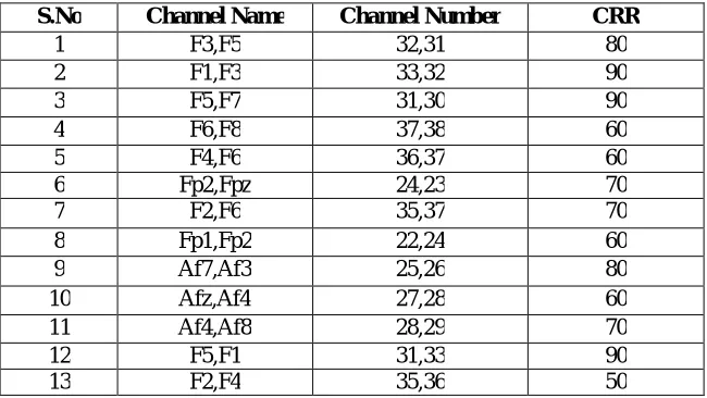
CRR Obtained in the Frontal Region in Eyes-Open Condition
S.No Channel Name Channel Number CRR
1 F3,F5 32,31 80
2 F1,F3 33,32 90
3 F5,F7 31,30 90
4 F6,F8 37,38 60
5 F4,F6 36,37 60
6 Fp2,Fpz 24,23 70
7 F2,F6 35,37 70
8 Fp1,Fp2 22,24 60
9 Af7,Af3 25,26 80
10 Afz,Af4 27,28 60
11 Af4,Af8 28,29 70
12 F5,F1 31,33 90
13 F2,F4 35,36 50
Table IV shows the CRR of different combinations of channels for eyes-open condition in the frontal region of the head. It is observed that the channel pairs like F1-F3, F5-F7 and F5-F1 achieved the maximum of 90% recognizing accuracy respectively.
TABLE V
CRR Obtained in the Central Region in Eyes-Open Condition
S.No Channel Name Channel Number CRR
1 Tp7,T7 45,41 60
2 Fc2,Fcz 5,4 90
3 Fc2,Fc4 5,6 100
4 C6,T8 14,42 70
5 Fc1,Fc4 3,2 90
6 C2,Cz 12,11 100
7 Cp2,Cpz 19,18 70
8 Cp6,T8 21,42 50
9 Cp5,Tp7 15,45 70
10 C5,T7 8,41 50
11 Tp8,Cp6 46,21 50
12 Cp5,T7 15,41 60
TABLE VI
CRR Obtained in the Parieto-Occipital Region in Eyes-Open Condition
S.No Channel Name Channel Number CRR
1 P2.P4 52,53 90
2 Po7,P5 56,48 100
3 P1,P3 50,49 60
4 P4,Pz 53,51 80
5 Po7,P4 56,47 90
6 P4,P6 53,54 90
Table VI shows the CRR of different combinations of channels for eyes-open condition in the parieto-occipital region of the brain zone. It is observed that the channel pair Po7-P5 achieved the maximum of 100% recognizing accuracy.
The results are obtained stating that in eyes-closed resting state condition maximum of channel pairs in the central region shows the maximum of 100% recognizing accuracy compared to the frontal and parieto-occipital region of the brain zones. In eyes-open resting state condition the maximum of 100% recognizing accuracy is achieved in the central and parieto-occipital region and maximum of 90% recognizing accuracy is attained in the frontal region of the brain zones.
V. CONCLUSION
In the recent years there has been an increasing interest in EEG-based biometric systems [15]. Compared to other characteristic that are usually considered in biometrics (e.g. fingerprints, iris, voice, signatures, etc.) EEG activity presents main advantages, among others: i) It is a most fraud resistant technique ii) The technical advantages are for future implementation of smart EEG-based biometric system for criminal identification and it can be used as a biometric for highly confidential purposes like bank lockers etc.
The majority of the methods only consider the activity of a single brain region without taking into account its essential relationship with different regions. In this proposed technique, features estimate among specific zones (i.e. frontal, central, parieto-occipital) and correct recognition rate are determined. The proposed approach test on a number of subjects have been recorded with a high-density EEG system with 56 electrodes available.
In a future implementation, the embedded based hardware system can be developed to use EEG as a biometric for highly confidential purposes like security applications. For example, it can be used for enrollment to enter into the bank locker room and for entering in a military campus etc.
The obtained results indicate that the combined use of five different features significantly enhances the overall performance compared to existing approved techniques. Notably a perfect recognition of 100% accuracy is achieved in more number of channel pairs in eyes-closed and eyes-open resting states by considering EEG channel pairs in the three regions of the head for ten subjects.
REFERENCES
[1] Somayaji, A., Van Oorschot, P.C., and Thorpe, J., “Pass-thoughts: Authenticating with our minds”, In Proc. 2005 Workshop on New Security Paradigms (NSPW), pages 45–56, 2005.
[2] Campisi, P., La Rocca, D., and Scarano, G., “Eeg biometrics for individual recognition in resting state with closed eyes”, In Proc. Int’l Conf. Biometrics Special Interest Group (BIOSIG), pages 1–12, 2012.
[4] Amit Kumar Ray., Sanjay Kumar Singh., and Yogendra Narain Singh., “Bioelectrical Signals as Emerging Biometrics: Issues and Challenges”, Vol.No.2012, pp. 1-13, 2012.
[5] Mandic, D.P., and Palaniappan, R., “Biometrics from brain electrical activity: A machine learning approach”, IEEE Trans. Pattern Anal. Mach. Intell., pp.738–742, 2007.
[6] Bassett, D.S., and Gazzaniga, M.S., “Understanding complexity in the human brain. Trends in Cognitive Sciences”, pp.200 – 209, 2011. [7] Dat Tran., Dinh Phung., Wanli Ma., Phuoc Nguyen., Tien Pham., and Wanli Ma., “A Study on the Feasibility of Using EEG Signals for
Authentication Purpose” , LNCS 8227, pp. 562–569, 2013.
[8] Hong Ji Lee., Hyun Seok Kim., and Kwang Suk Park., “A Study on the Reproducibility of Biometric Authentication based on Electroencephalogram (EEG)”, Int’l Conf . Neural Engg., pp.13-16, 2013.
[9] Ms. Swati S Bobde ., and Prof. D. N. Satange., “Biometrics in Secure e-Transaction”, Vol. 2, pp. 243-248, 2013.
[10] Babiloni, F., Campisi, P., Cserti, P., De Vico Fallani, F.,Kozmann, G., La Rocca, D., and Vegso, B., “Human Brain Distinctiveness Based on EEG Spectral Coherence Connectivity”, IEEE Trans. Biomed. Engg., Vol. 61, No.9, pp.2406-2412, 2014.
[11] Grinsted, A., Jevrejeva, S., and Moore, J.C., “Application of the cross wavelet transform and wavelet coherence to geophysical time series”, Vol. 11, pp. 561-566, 2014.
[12] Justin Leo Cheang Loong., Khazaimatol S Subari., Muhammad Kamil Abdullah., and Nurul Naida Ahmad., “Analysis of the EEG Signal for a Practical Biometric System”, Vol. 4, No.8, 2010.
[13] Dhilipan, A., Preethi, J., and Sreeshakthy, M., “A Survey on EEG Based Emotion Analysis using various Feature Extraction Techniques”, IJSETR, Vol No.3, pp.3113-3120, 2014.
[14] Database physionet bci. [Online]. Available:http://www.physionet.org/ pn4/eegmmidb/