Exploring potential super infection in SARS-CoV2 by genome-wide analysis and receptor–ligand docking
Chuanjun Shu*1,2, Xuan Huang*3,Juergen Brosius4, Cheng Deng2,#
1Department of Bioinformatics, School of Biomedical Engineering and Informatics, Nanjing Medical University,
Nanjing, 211166, China.
2Jiangsu Key Laboratory for Biodiversity and Biotechnology, College of Life Sciences, Nanjing Normal University,
Nanjing, 210023, China.
3Reproductive Medical Center, Jinling Hospital Affiliated to The Medical School of Nanjing University, Nanjing,
210002, China.
4Institutes for Systems Genetics, West China Hospital, Sichuan University, Chengdu, 610041, China.
*Contributed equally.
#corresponding information: Cheng Deng, [email protected].
Abstract
SARS-CoV2 (corona virus) has spread globally at an unprecedented rate; so far, increasing SARS-CoV2-infected individuals have been identified. Although the situation in China is improving and is currently under control, the outbreak in other countries and its pandemic management is only beginning to develop. Based on 154 SARS-CoV2 genome sequence analyses, we used receptor–ligand docking to identify one potential point mutation (V354F) on the spike structure which enhances spike binding to ACE2 receptors underlying potential super infection. Importantly, the V354F site on spike S1 had been identified in 5/10 infected French patients living in Paris, who sharing 100% identical SARS-CoV2 genomes. With Covid-19 cases increasing rapidly in France that could lead to a new explosion, we suggest that the French government should identify all potential super spreaders and treat them accordingly. In summary, our study provides on of the measures to avoid the potential second worldwide explosion of SARS-CoV2.
Main text
In December 2019, a pneumonia outbreak associated with the 2019 novel coronavirus (2019-nCoV, also named as SARS-CoV2) occurred in Wuhan, Hubei Province, China (Pan et al., 2020; D. Wang et al., 2020; Zhu et al., 2020a, 2020b). Subsequently, the new coronavirus pneumonia (NCP) -termed COVID-19 has rapidly spread around the world(Guan et al., 2020; Huang et al., 2020; Wu, Leung, & Leung, 2020). Up to now- March 18, 2020, 198,032 (81,151-China and 116,881-other counties) have been identified(Guan et al., 2020; D. Wang et al., 2020). Through the coordinated efforts, the situation tends to improve and be under control in China. Thus far, more than half of the patients in China have been recovered and the total number of patients continues to decrease. However, the potential danger of a second worldwide spread is still imminent.
Currently, there are no effective therapeutics for treatment of the SARS-CoV2 available. SARS-CoV2 is a single-stranded RNA beta-coronavirus(Woo et al., 2005; A. Wu et al., 2020). The SARS-CoV2 genome encodes non-structural proteins (such as papain-like protease, 3-chymotrypsin-like protease (also known as main protease Mpro), RNA-dependent RNA polymerase, and helicase), structural proteins (such as spike glycoprotein, membrane protein, envelope protein, and nucleocapsid protein) and accessory proteins (such as ORF3a, ORF8)(A. Wu et al., 2020). The four non-structural proteins are critical enzymes for the viral life cycle(Shimamoto et al., 2016). Spike binds to the human cellular receptor angiotensin-converting enzyme 2 (ACE2) and mediates fusion between the viral and cellular membranes(Wong & K.,
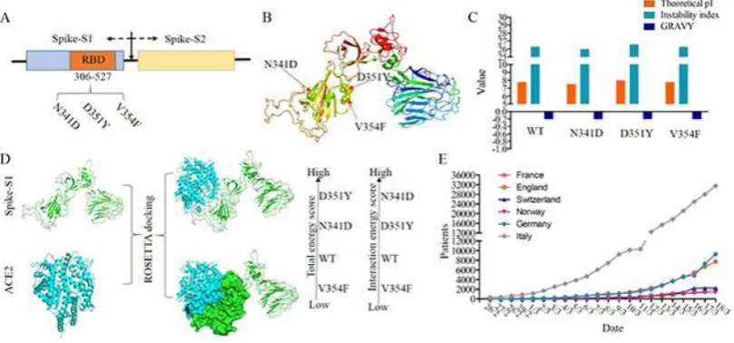
2003). The receptor binding domain (RBD) for ACE2 is distributed in the S1 region
objectives were to investigate potential super infection based on evolution analysis of SARS-CoV2 and structural pharmacological analysis of receptor-ligand (ACE2-Spike) binding ability.
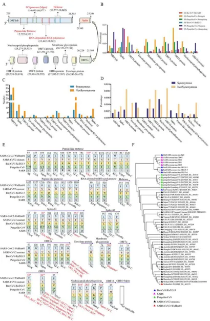
as single nucleotide polymorphism. The substitution sites were mainly distributed in the ORF1ab and spike regions (Fig 1C). But ORF8a, ORF3a, ORF10, and spike-S1 had a higher substitution speed in SARS-CoV2 (Fig 1D). With the ratio of non-synonymous sites/synonymous sites >1, our results indicated that SARS-CoV2 probably underwent adaptive evolution after infection of human hosts.
These non-synonymous substitutions could result in a SARS-CoV2 with super infectivity. To address potential super infection in SARS-CoV2, amino acid mutations caused by non-synonymous substitutions of SARS-CoV2-Wuhan01 against SARS-CoV2 mutants were analyzed and the corresponding amino acids in bat-CoV-RaTG13, pangolin-CoV, and SARS viruses were then used as controls. As shown in Fig 1E, most amino acid substitutions between SARS-CoV2 viruses always are conserved residues in bat-CoV-RaTG13, pangolin-CoV, and SARS viruses, except amino acid substitutions in spike-S1. This suggested that most amino acid substitutions probably are random events. However, 10 amino acid substitutions recurred in SARS-CoV2, i.e. I789V of papain-like protease, H36Y and V354F of spike-S1, G251V of ORF3a, D209H of membrane glycoprotein, V62L, L84S, and P85S of ORF8, and S194L, S202N, and P344S of nucleocapsid phosphoprotein (Fig 1E). Spike protein, binds to the human cellular receptor-ACE2 and responsible for virus infection, and according to WHO data, the basic reproduction number of the
infection (R0) for SARS-CoV2 is about 1.4 to 2.5. Hence, we should pay more
closer phylogenetic relationship between this mutant and its ancestors than that of SARS-CoV2-Wuhan01 (Fig 1F). According to these phylogenetic results, we proposed a hypothesis that Wuhan, China is an early substitutions site of the SARS-CoV2 outbreak but it is probably not the original site for the human infection. Hence, this SARS-CoV2 mutant-L84S could be a more ancestral virus rather than a virus with super infectivity.
Another SARS-CoV2 mutant with high frequency of occurrence, i.e. G251V of ORF3a, showed similar result (Fig 1E). The amino acid substitution of ORF3a, i.e., G251V, had the second highest substitution times (value=17) in the 10 mutant residues (Fig 1E). These SARS-CoV mutants were mainly isolated from patients of France and Singapore. An interesting observation is that five patients from France with 100% identical protein sequences (Supplementary materials Table 1) shared the same mutation - V354F and this amino acid substitution was in the RBD region of spike-S1 (V354F). Based on the recorded epidemiology data, only one patient came from Wuhan, China. Furthermore, these five patients with V354F lived in the same area (Location: Europe / France / Ile-de-France / Paris). The Ro (basic reproduction number) of the SARS-CoV2 mutant that was isolated from France probably reached 4. This Ro was far beyond the WHO predicted value of SARS-CoV2. Taken together, these results suggested that the SARS-CoV2 mutant which has two mutations (G251V of ORF3a and V354F of spike-S1) probably exhibits a super infectivity.
and chemical parameters of spike protein, and the mutant V354F had the smallest impact among these three mutants. We further tested whether these three mutations can result in different interaction models between ACE2 and spike. With the three-dimensional structure of ACE2 downloaded from the PDB database (PDB ID: 6acj, structures of wild type spike-S1 and its mutants were obtained by utilizing I-TASSER (Iterative Threading Assembly Refinement) algorithm. Complexes of ACE2-spike were obtained by a combination of using Discovery studio and Rosetta package. As shown in Fig2D, V354F had the lowest energy and interaction score in these ACE2-spike-S1 complexes, based on structural comparison and protein-protein interaction analysis (Fig 2D, supplement materials Table S1). These results further suggested that spike with the V354F amino acid substitution had a higher affinity for ACE2 than wild type spike and potentially leads to super infectivity. Meanwhile, in France SARS-CoV2 cases accumulate faster than in many other affected countries and could be one of the centers of a new explosion (Fig 2 E). Therefore, SARS-CoV2 mutant-V354F with a potential super infectivity should deserve more attention by government health institutions in France by epidemic surveillance and thus reducing the worldwide threat of the potential second explosion of SARS-CoV2.
Supplementary Material
Supplementary Materials are available online.
Methods and Materials
Data preparation
Whole genome sequences of SARS-CoV2, SARS, bat-CoV, and pangolin-CoV were downloaded from CNCB/BIG (https://bigd.big.ac.cn/ncov), the GISAID EpiFluTM
database and NCBI. According to gene locations, the nucleotide sequences of RBD regions of coronavirus and their corresponding amino acids were acquired by BioEdit software (Helge-Friedrich, 2004). Three-dimensional spatial structure of ACE2 was downloaded from PDB database (Sussman et al., 2010).
Genetic and phylogenetic analysis
Synonymous/non-synonymous substitution sites between coronavirus were calculated by DnaSP and BioEdit package(Rozas et al., 2017). The nucleotide/amino acid sequences of proteins and the similarities between sequences were aligned by BioEdit software. Unrooted tree topology based on multiple alignments of amino acids was established with the neighbor-joining method in MEGA 6.06 (Lewis, Kumar, Tamura, & Nei, 1995). Consistency of branching was tested using a bootstrap analysis with 500 resamplings of the data in MEGA 6.06.
Structure analysis
mutants) were predicted by Discovery Studio 3.0 software(Gao & Huang, 2011). These predicted pockets were utilized to construct an initial coarse model of the spike-S1-ACE2 complexes. The complex structures were refined by Rosetta software (RosettaDock and FelxPepDock module)(Rohl, Strauss, Misura, & Baker, 2003). The final structure was obtained based on energy scores. The interactions scores were calculated by Rosetta. High-quality 3-D images of the proteins were drawn by PyMOL(Ordog, 2008).
Acknowledgement
We thank Dr. Jürgen Brosius immeasurable help for this project. This work was supported by the National Key Research and Development Program of China (2018YFD0900602) and National Natural Science Foundation of China (31970388, 31701234), the Priority Academic Program Development of Jiangsu Higher Education Institutions (PAPD), the Natural Science Foundation of the Jiangsu Higher Education Institutions (17KJB180006), the Natural Science Foundation from Jiangsu Province BK20160043, BK20151546, 15KJA180004 and BK20171035, Jiangsu Distinguished Professor Funding.
References
Gao, Y. D., & Huang, J. F. (2011). An extension strategy of Discovery Studio 2.0 for non-bonded interaction energy automatic calculation at the residue level. Zoological Research.
Garg, V. K., Avashthi, H., & Tiwari, A. (2016). MFPPI – Multi FASTA ProtParam Interface. 12(2), 74-77. Guan, W.-j., Ni, Z.-y., Hu, Y., Liang, W.-h., Ou, C.-q., He, J.-x., . . . Zhong, N.-s. (2020). Clinical
characteristics of 2019 novel coronavirus infection in China. medRxiv,
2020.2002.2006.20020974. doi:10.1101/2020.02.06.20020974
Helge-Friedrich, T. (2004). Analysis for free: Comparing programs for sequence analysis. Briefings in
Bioinformatics(1), 1.
Huang, C., Wang, Y., Li, X., Ren, L., Zhao, J., Hu, Y., . . . Cao, B. (2020). Clinical features of patients
infected with 2019 novel coronavirus in Wuhan, China. Lancet.
doi:10.1016/S0140-6736(20)30183-5
Kirchdoerfer, R. N., Cottrell, C. A., Wang, N., Pallesen, J., Yassine, H. M., Turner, H. L., . . . Ward, A. B. (2016). Pre-fusion structure of a human coronavirus spike protein. Nature, 531(7592), 118-121.
Lewis, P. O., Kumar, S., Tamura, K., & Nei, M. (1995). MEGA: Molecular Evolutionary Genetics Analysis, Version 1.02. Systematic Biology, 44(4).
Ordog, R. (2008). PyDeT, a PyMOL plug-in for visualizing geometric concepts around proteins.
Bioinformation, 2(8), 346-347.
patients in Wuhan, China. Eur Radiol. doi:10.1007/s00330-020-06731-x
Rohl, C. A., Strauss, C. E., Misura, K. M., & Baker, D. (2003). Protein structure prediction using Rosetta.
383(383), 66.
Roy, A., Kucukural, A., & Zhang, Y. I-TASSER: a unified platform for automated protein structure and function prediction. Nature Protocols, 5(4), 725-738.
Rozas, J., Ferrer-Mata, A., Sánchez-DelBarrio, J. C., Guirao-Rico, S., Librado, P., Ramos-Onsins, S. E., & Sánchez-Gracia, A. (2017). DnaSP 6: DNA Sequence Polymorphism Analysis of Large Data Sets.
Mol Biol Evol, 34(12), 3299-3302. doi:10.1093/molbev/msx248
Shimamoto, Y., Hattori, Y., Kobayashi, K., Teruya, K., Sanjoh, A., Nakagawa, A., . . . Akaji, K. (2016). Fused-ring structure of decahydroisoquinolin as a novel scaffold for SARS 3CL protease inhibitors. Bioorg Med Chem, 23(4), 876-890.
Sussman, J. L., Lin, D., Jiang, J., Manning, N. O., Prilusky, J., Ritter, O., & Abola, E. E. (2010). Protein Data Bank (PDB): Database of Three-Dimensional Structural Information of Biological Macromolecules . Acta Crystallographica, 54(6-1), 1078-1084.
Wang, D., Hu, B., Hu, C., Zhu, F., Liu, X., Zhang, J., . . . Peng, Z. (2020). Clinical Characteristics of 138 Hospitalized Patients With 2019 Novel Coronavirus-Infected Pneumonia in Wuhan, China.
JAMA. doi:10.1001/jama.2020.1585
Wang, S., Guo, F., Liu, K., Wang, H., Rao, S., Yang, P., & Jiang, C. (2008). Endocytosis of the receptor-binding domain of SARS-CoV spike protein together with virus receptor ACE2. Virus
Research, 136(1-2), 0-15.
Wong, & K., S. (2003). A 193-Amino Acid Fragment of the SARS Coronavirus S Protein Efficiently Binds Angiotensin-converting Enzyme 2. Journal of Biological Chemistry, 279(5), 3197-3201. Woo, P. C., Lau, S. K., Chu, C. M., Chan, K. H., Tsoi, H. W., Huang, Y., . . . Yuen, K. Y. (2005).
Characterization and complete genome sequence of a novel coronavirus, coronavirus HKU1, from patients with pneumonia. J Virol, 79(2), 884-895. doi:10.1128/JVI.79.2.884-895.2005 Wu, A., Peng, Y., Huang, B., Ding, X., Wang, X., Niu, P., . . . Jiang, T. (2020). Genome Composition and
Divergence of the Novel Coronavirus (2019-nCoV) Originating in China. Cell Host Microbe. doi:10.1016/j.chom.2020.02.001
Wu, J. T., Leung, K., & Leung, G. M. (2020). Nowcasting and forecasting the potential domestic and international spread of the 2019-nCoV outbreak originating in Wuhan, China: a modelling study. Lancet, 395(10225), 689-697. doi:10.1016/S0140-6736(20)30260-9
Zhu, N., Zhang, D., Wang, W., Li, X., Yang, B., Song, J., . . . Research, T. (2020a). A Novel Coronavirus from Patients with Pneumonia in China, 2019. N Engl J Med. doi:10.1056/NEJMoa2001017 Zhu, N., Zhang, D., Wang, W., Li, X., Yang, B., Song, J., . . . Research, T. (2020b). A Novel Coronavirus
from Patients with Pneumonia in China, 2019. N Engl J Med, 382(8), 727-733. doi:10.1056/NEJMoa2001017
SARS-CoV2-Wuhan01, percentages of synonymous/non-synonymous substitution sites in ten CDS regions for bat-CoV-RaTG13, pangolin-CoV-Guangxi, and pangolin-CoV-Guangdong. (C) and (D) represent numbers and percentages of synonymous/non-synonymous substitution sites in ten CDS regions and five potential drug targets for SARS-CoV2, respectively. (E) Amino acid substitutions in SARS-CoV2 mutants, the corresponding residues in bat-CoV-RaTG13, pangolin-CoV, and SARS viruses. Red indicates that amino acid substitution times are greater than 2. (F) The phylogenetic tree of SARS-CoV2 mutants (L84S in ORF8).