1071-412X/05/$08.00
⫹
0
doi:10.1128/CDLI.12.4.513–519.2005
Copyright © 2005, American Society for Microbiology. All Rights Reserved.
Simultaneous Serodetection of 10 Highly Prevalent Mouse Infectious
Pathogens in a Single Reaction by Multiplex Analysis
Imran H. Khan,
1,4* Lon V. Kendall,
2Melanie Ziman,
1Scott Wong,
1Sara Mendoza,
1James Fahey,
3Stephen M. Griffey,
2Stephen W. Barthold,
1and Paul A. Luciw
1,4Center for Comparative Medicine,
1Comparative Pathology Laboratory,
2and Department of Pathology and Laboratory
Medicine,
4University of California, Davis, CA 95616, and The Jackson Laboratory, Bar Harbor, ME 04609
3Received 18 October 2004/Returned for modification 12 December 2004/Accepted 11 February 2005
Under current practices of mouse colony maintenance, sera from mice are analyzed for antibodies against
several widespread infectious pathogens by conventional immunoassays, generally enzyme-linked
immunosor-bent assay (ELISA). To test for multiple agents, these methods consume large volumes of mouse serum and are
laborious and time-consuming. More efficient immunoassays, using small amounts of sample, are therefore
needed. Accordingly, we have developed a novel multiplex diagnostic system that employs fluorescent
mi-crobeads, coated with purified antigens, for simultaneous serodetection of 10 mouse infectious agents.
Indi-vidually identifiable, fluorescent microbeads were coated with antigens from Sendai virus, mouse hepatitis
virus, Theiler’s mouse encephalomyelitis virus/GDVII strain, mouse minute virus, mouse cytomegalovirus,
respiratory enteric orphan virus (Reo-3 virus), mouse parvovirus, calf rotavirus for epizootic diarrhea virus of
infant mice, vaccinia virus for ectromelia virus, and
Mycoplasma pulmonis
. Standard sera, singly positive for
antibodies to individual infectious agents, were generated by inoculation of BALB/cj and C57BL/6j mice. Sera
from these experimentally infected mice, as well as sera from naturally infected mice, were analyzed using a
mixture of microbeads coated with antigens of the 10 infectious agents listed above. Results demonstrated that
the multiplex assay was at least as sensitive and specific as ELISA for serodetection. Importantly, the multiplex
assay required only 1 microliter of serum for simultaneous serodetection of the 10 mouse infectious agents in
one reaction vessel. Thus, this multiplex microbead assay is a reliable, efficient, and cost-effective diagnostic
modality that will impact serosurveillance of mice used in research.
The mouse is the most widely used animal in biomedical
research. Availability of specific-pathogen-free (SPF) mice for
use in research is essential to obtain consistently accurate data.
Experimental animals exposed to, or infected with, various
infectious agents may yield questionable data, thereby
con-founding the findings of a given study. Mice may be screened
for several important infectious pathogens (1, 3, 5, 6, 9, 10, 16,
17). Routine screening of a large number of animals, with
respect to a large number of infectious agents, is a
time-con-suming and tedious task under current practices.
Serosurveil-lance of mouse colonies is usually performed indirectly by
introducing sentinel mice to animal rooms. After allowing for
exposure, these sentinel mice are sacrificed, and their sera are
tested by conventional immunoassays, such as enzyme-linked
immunosorbent assay (ELISA) and/or indirect
fluorescent-an-tibody assay (IFA). Because conventional immunoassays allow
detection of only one infectious agent in a serum sample, large
amounts of sample are consumed for the detection of multiple
agents. Additionally, a composite of multiple individual tests
requires much time, materials, and labor. This in turn
encour-ages the use of sentinel mice instead of direct testing of
indi-vidual mice in colonies. Direct monitoring of animals, for
ac-curate knowledge of prevalence of common pathogens, is not
only desirable for colony maintenance but critical for health
care of valuable specialized mouse strains, such as genetically
engineered mice. Furthermore, the increasing demand for
re-search mice requires more efficient serodiagnostic assays that
are readily amenable to a high-throughput format.
We have selected a relatively new technology, designated
“multiple analyte profiling,” from Luminex Corp. (Austin,
TX), which allows simultaneous detection of multiple analytes
in a small amount of sample (2). Up to 100 analytes can be
measured in a single reaction. In addition, the multiple analyte
profiling technology has been designed for high-throughput
analysis. This method has been used to determine amounts of
15 different cytokines in small amounts of individual samples
(2) as well as for detection of serum antibodies to multiple
peptide epitopes (8), autoantigens (4), bacterial antigens (13,
14), and viral antigens (12).
The multiplex microbead assay is performed by the use of
unique, fluorescently coded sets of polystyrene microbeads
(5.6-
m diameter) (7, 15). A specific ratio of an orange and a
red fluorophore is embedded within the matrix of a specific
microbead set that enables identification of each bead set (7,
15). Microbead sets conjugated to known biomolecules are
mixed and added to the test sample. Analytes in the sample
react with biomolecules applied as a coating on the
mi-crobeads. Specific interactions are detected by a common
re-porter fluorochrome (e.g., phycoerythrin) conjugated to a
sec-ondary detection reagent. Thus, the multiplex microbead assay
has a substantial advantage over the conventional
immunoas-say techniques with its ability to perform simultaneous
detec-tion of antibodies to several infectious agents in one reacdetec-tion
container.
This study focused on the development of a multiplex
mi-* Corresponding author. Mailing address: Center for Comparative
Medicine, University of California at Davis, Hutchison Rd. and
County Rd. 98, Davis, CA 95616. Phone: (530) 752-1245. Fax: (530)
752-7914. E-mail: [email protected].
513
on August 17, 2020 by guest
http://cvi.asm.org/
crobead assay for detection of antibodies to 10 infectious
agents in mice. Two strains of mice (BALB/cj and C57BL/6j)
were inoculated with several viruses and
Mycoplasma pulmonis
to develop a panel of single-positive test sera. Microbeads
coated with antigens from these agents were highly specific and
sensitive for multiplex serodetection. Comparisons with
ELISA and IFA demonstrated that the multiplex microbead
assay is as sensitive and specific as these conventional assays in
mouse serodetection. Furthermore, a volume as small as 1
microliter of serum is sufficient to perform detection of
anti-bodies to multiple infectious agents.
MATERIALS AND METHODS
Purified viruses for conjugation to microbeads.Viruses purified by sucrose density centrifugation were purchased from Advanced Biotechnologies Inc. (Co-lumbia, MD). These preparations were supplied at a total protein concentration of 1 mg/ml in phosphate-buffered saline (PBS) (pH 7.2). The purified viruses included mouse hepatitis virus (MHV), Theiler’s mouse encephalomyelitis virus/ GDVII strain (GD7), mouse minute virus (MMV), mouse cytomegalovirus (MCMV), Sendai virus, vaccinia virus, and Nebraska calf diarrhea virus (NCDV). Vaccinia virus and NCDV are cross-reactive to, and allow detection of, antibodies against ectromelia virus and epizootic diarrhea virus of infant mice (EDIM), respectively.
Cultures of viruses andM. pulmonis.GD7, MMV, and respiratory enteric orphan virus (Reo-3) were cultured in baby hamster kidney 21 (BHK-21) cells grown to confluency in Dulbecco’s modified Eagle’s medium containing 2 mM glutamine, penicillin/streptomycin, and 10% fetal bovine serum. Virus prepara-tions were added to the confluent BHK-21 cells and allowed to adsorb to the cells
at 37°C in the presence of 5% CO2, for 1 h on a rocking shaker. Cells were
cultivated in Dulbecco’s modified Eagle’s medium, without antibiotics, and con-taining 2% heat-inactivated fetal bovine serum and 2 mM glutamine. Infected cell cultures were maintained until cytopathic effects were evident (48 to 120 h). Tissue culture medium containing virus was collected, clarified by centrifugation
at 1,000⫻gfor 10 min, and passed through a 0.45-m-pore-size filter. Virus was
stored at⫺80°C. For isolation of MMV, infected cells were collected by
centrif-ugation at 1,000⫻gfor 10 min. Cells were harvested by treatment with 0.1%
trypsin in 2 mM EDTA and resuspended in 5 ml of medium per T75 flask. To release the virus, infected cells were subjected to three sequential
freeze-and-thaw cycles. Cell debris was removed by centrifugation at 1,000⫻gfor 10 min.
The supernatant was clarified and stored frozen at⫺80°C. Virus titers were
determined by endpoint dilutions. Titers of GD7 and Reo-3 were measured on BHK-21 cells, and 324K cells were used to establish the titer of MMV.
M. pulmonis, obtained from the American Type Culture Collection (Manassas, VA), was cultured and isolated per supplier’s instructions. Cells were collected
by centrifugation of culture media at 3,700⫻gand washed three times with PBS.
Antigen was extracted by adding lysis buffer (1% Triton X-100 in PBS, containing protease inhibitor cocktail [Roche Diagnostics GmbH, Indianapolis, IN]) to the cell pellet. The contents were mixed by vortexing and incubated at 4°C for 20
min, and lysate was filtered through an 0.45-m-pore-size filter. Total protein
concentration was measured by the Bradford method (Bio-Rad, Hercules, CA).
The lysate was aliquoted and stored at⫺80°C until used for coating microbeads.
Production of positive sera. Mice of two different strains (BALB/cj and C57BL/6j) were inoculated with different infectious agents (24 mice per agent, per mouse strain). All mice were SPF and were a gift from Jax West Division of the Jackson Laboratory (Bar Harbor, ME). Two sources of virus preparation were used for inoculations. One was a known amount (measured as total protein) of sucrose-gradient-purified virus from Advanced Biotechnologies Inc. These included Sendai virus, vaccinia virus, and NCDV. Alternatively, infectious agents were grown and titer was determined as described above. These included GD7,
MMV, Reo-3, andM. pulmonis.
A few animals (4 to 12) from each mouse strain were initially test inoculated with various dilutions of different infectious agents prepared in Hanks’ minimal essential medium to estimate the minimal infectious dose. After the minimum dose was estimated, viruses were diluted to inoculate mice for the production of standard sera. The inoculation schedule is summarized in Table 1 for BALB/cj and in Table 2 for C57BL/6j mice. The natural route of infection was tested first. GD7, Reo-3, MMV, and Sendai virus were inoculated by intranasal route. For MHV, a cage of SPF mice was exposed to dirty bedding from MHV-infected mice. The MHV-infected mice that were used as a source of exposure were confirmed by ELISA to be seronegative for the other infectious agents including
GD7, EDIM, ectromelia virus, MMV, mouse parvovirus (MPV), Sendai virus,M.
pulmonis, and Reo-3. NCDV was inoculated by gastric lavage.
Mice were bled every 2 weeks from the retro-orbital sinus under anesthesia. Sera were collected and tested by ELISA kits from Charles River Laboratories (Wilmington, MA) for serodetection of individual infectious agents. If in 2 weeks the natural route of infection failed to yield an antibody response, mice were reinoculated by the natural route. If there was still no seroconversion after another 2 weeks, the mice were inoculated by intraperitoneal (i.p.) injection. For MMV, mice were initially inoculated twice by the intranasal route. Because these mice did not seroconvert, they were inoculated twice by the i.p. route. Two other
infectious agents (e.g.,M. pulmonisand vaccinia virus) were inoculated by the i.p.
route alone. Mouse sera positive for antibodies to MPV, generated in C3H/HeN and BALB/c mice, inoculated with MPV-1b virus by the oronasal route, were a gift from David Besselsen at the University of Arizona. These sera were positive by ELISA for antibodies to MPV by the donor laboratory. Standard serum positive for MCMV was purchased from Charles River Laboratories (Wilming-ton, MA).
After all mice seroconverted, as determined by commercial ELISA, they were euthanized with carbon dioxide and exsanguinated by cardiocentesis. Serum
from all animals was stored at⫺80°C.
Sera from naturally infected mice positive for antibodies to multiple infectious agents.Mouse sera from various sources are routinely submitted to the Com-parative Pathology Laboratory, University of California, Davis, for antibody analysis. As tested by commercial ELISA kits, serum samples from seven mice in a naturally infected mouse population were found to be positive for antibodies to 6 of the 10 prevalent infectious agents.
Antigen preparation for coating microbeads and ELISA plates.Viral andM. pulmonisantigens, for use in immunoassays, were prepared by 1:1 mixing of the infectious agent preparation with 0.5% Triton X-100 (Fisher Scientific, Fairlawn, NJ) and protease inhibitor cocktail (Roche) in PBS (pH 7.2). NCDV was further treated with 1 mM EDTA and sonication at low setting (Virtis Virsonic 60; Gardiner, NY). Vaccinia virus preparations were subjected to inactivation by psoralen treatment as previously described (18). Briefly, psoralen (Sigma
Chem-icals, St. Louis, MO) was added at a final concentration of 10g/ml to the
vaccinia virus antigen prepared above (0.5 mg/ml) and subjected to UV irradi-ation for 10 min at 254 nm (Chromato Vue Transilluminator, model T5-15; UVP, Inc., San Gabriel, CA). All antigen preparations were stored frozen at
⫺80°C. Prior to use, preparations were thawed and centrifuged in a
microcen-trifuge at 10,000⫻gfor 10 min to remove debris.
Coupling antigens to microbeads.Microbeads were purchased from Luminex Corp. (Austin, TX). Various antigen preparations were chemically cross-linked to the microbeads according to the manufacturer’s instructions. Bead stock was resuspended by vortexing and treatment in a sonicator bath (15 to 30 s) (Branson
1510; Danbury, CT). An aliquot of 2.5⫻106
beads was removed and centrifuged
at 21,000⫻gfor 2 min. Beads were resuspended in 80l of activation buffer (100
mM monobasic sodium phosphate; pH 6.3) by vortexing and sonication (15 to 30 s).
To activate the beads for cross-linking to proteins, 10l of 50-mg/ml
sulfo-N-hydroxysulfosuccinamide (Pierce, Rockford, IL) was added, and beads were
mixed by vortexing. Then 10l of 50-mg/ml 1-ethyl-3-[3-dimethylaminopropyl]
carbodiimide (EDC; Pierce, Rockford, IL) was added, and beads were mixed again by vortexing. All incubations of beads were performed in the dark. The bead mixture was shaken on a rotary shaker at room temperature for 20 min and
centrifuged at 21,000⫻gfor 2 min. Beads were washed twice with 250l of 50
mM morpholineethanesulfonic acid (MES; pH 6.0) buffer. To coat them with antigens, pelleted beads were resuspended in the relevant antigen preparation diluted in 50 mM MES (pH 6.0) buffer. Optimization of antigen concentration was performed by coating different microbead sets with a range of proteins
between 10 and 200g/ml for each antigen. These microbead sets were then used
to test against standard mouse sera, which are positive for antibodies to the relevant infectious agent. Microbead sets that provided the strongest specific signal for each antigen against the positive mouse sera were selected. The
optimized protein concentration for each antigen was as follows: 25g/ml of
vaccinia virus, 100g/ml of NCDV, 25g/ml ofM. pulmonis, 100g/ml of MHV,
25g/ml of Sendai virus, 100g/ml of GD7, 50g/ml of CMV, and 100g/ml
of MMV.
Recombinant protein rVP2, a specific antigen for MPV serodetection (9), was obtained from the Research Animal Diagnostic Laboratory (University of Mis-souri, Columbia, MO). Optimal concentration of rVP2 for coating on the beads
was determined to be 3.3g/ml. For Reo-3, infected cell lysate was used at 330
g/ml, and a separate bead set was coated with uninfected cell lysate (Reo-3
sham) at the same concentration.
Bead sets were also coated with biotin-conjugated goat immunoglobulin G
on August 17, 2020 by guest
http://cvi.asm.org/
(IgG; 100g/ml), a positive control protein for reaction with streptavidin con-jugated to R-phycoerythrin (streptavidin-phycoerythrin), and bovine serum
al-bumin (BSA;100g/ml) as a negative control protein (Pierce, Rockford, IL). For
coupling, mixtures of activated beads and proteins were incubated by shaking on a rocker for 2 h at room temperature. After coating with proteins, beads were
washed twice with 250l of wash buffer (0.1% Tween 20 in PBS, pH 7.4),
resuspended in 250l of blocking buffer (1% BSA; 0.1% Tween 20 in PBS, pH
7.4; 0.05% sodium azide), and shaken on a rocker at room temperature for 30 min. After blocking, beads were resuspended in 1 ml of blocking buffer and stored at 4°C.
Multiplex detection of antibodies in mouse serum.Immunoreactions were done in 96-well, filter-bottomed plates designed for high-throughput separations
(1.2-m-pore-size MultiScreen; Millipore Corporation, Bedford, MA).
Typi-cally, 2,000 beads for each individual bead set, coated with a specific antigen, were added per well. For example, for a 13-plex assay, 2,000 beads from each set coated with an antigen were mixed to provide a total of 26,000 beads per well. Mouse serum was diluted 1:250 in 5% BLOTTO (Pierce, Rockford, IL), and 50
l of this diluted serum was mixed with the 13-plex bead mixture per well. The
mixture of beads and serum was then incubated on a shaker for 1 h at room temperature. After incubation, liquid was drained from the bottom of the plate in a vacuum manifold designed to hold 96-well plates (Millipore Corporation,
Bedford, MA). The beads were washed two times by adding 100l of wash buffer
per well and draining under vacuum. For detection of mouse IgG, biotinylated horse anti-mouse IgG was used (Vector Laboratories, Burlingame, CA) at a
1:1,000 dilution in BLOTTO, and 100l was added per well. Beads were mixed
as before and incubated at room temperature for 30 min. Following incubation with the secondary antibody, beads were washed three times. To detect
biotin-ylated IgG, 100l of streptavidin-phycoerythrin (CalTag, Burlingame, CA) was
added at a dilution of 1:500 in wash buffer. The content of each well was mixed
and incubated at room temperature for 15 min. Beads were washed once with
wash buffer, resuspended in 100l of wash buffer per well, and analyzed in the
Luminex-100 instrument equipped with XY-Platform for automatic reading of a 96-well plate.
Luminex-100 instrument operation and data analysis.The Luminex-100 in-strument was manufactured by Luminex Corp. and purchased from Upstate USA Inc. (Lake Placid, NY). The instrument was used at default settings, set by the manufacturer for routine applications, as directed by the user’s manual. Data were acquired by Luminex Data Collection software (version 1.7). This software package allowed routine operation of the instrument and data acquisition. Cal-ibration beads, supplied by the manufacturer, were used to adjust instrument settings for bead set identification and for the detection of reporter (phyco-erythrin). Events were gated to exclude doublets and other aggregates. A min-imum of 100 independent, gated events were acquired for each bead set. A ratio of median fluorescence intensity (MFI) of antigen-coated bead sets to MFI of BSA-coated beads in each reaction was calculated. This ratio, designated as “signal,” was used as a measure of antibody detection. After acquisition by Luminex software, the data were further processed by Microsoft Excel software. Background or normal serum reactivity for the antigen-coated bead set was determined. For each bead set, an average of signals from quadruplicate wells of pooled normal mouse sera was calculated, and three times the standard deviation value was added to the average. For routine analysis, each experiment also contained a positive-control serum for all the infectious agents. Samples were analyzed in duplicate, and a minimum of two separate multiplex immunoassay experiments was performed. Sera were designated positive for antibodies to an infectious agent if the signal from the relevant antigen-coated bead set was greater than the background.
IFA.IFA analysis of serum samples was performed by Charles River
Labora-tories (Wilmington, MA).
TABLE 1. Production of standard sera in BALB/cj mice
Pathogen n
Inoculation route-dosea Seroconversionb
(% positive)
Primary Secondary Final ELISA Multiplex
Reo-3
24
i.n.
⫺
1.0 TCID50
i.n.
⫺
1.0 TCID50
None
100
100
Sendal virus
4
i.n.
⫺
1
g/10
l
None
None
100
100
18
i.n.
⫺
1
g/10
l
i.n.
⫺
1
g/10
l
None
100
100
GD7
3
i.n.
⫺
2
⫻
10
6PFU/10
l
None
None
100
100
4
i.n.
⫺
6
⫻
10
6PFU/10
l
None
None
100
100
4
i.n.
⫺
3
⫻
10
6PFU/10
l
i.n.
⫺
6
⫻
10
6PFU/10
l
None
100
100
4
i.n.
⫺
6
⫻
10
6PFU/10
l
i.n.
⫺
6
⫻
10
6PFU/10
l
None
100
100
4
i.n.
⫺
2
⫻
10
6PFU/10
l
i.n.
⫺
2
⫻
10
6PFU/10
l
i.n.
⫺
6
⫻
10
6PFU/10
l
100
100
MHV
10
d.b.
None
None
60
50
12
d.b.
d.b.
None
100
100
4
5 d.b. inoculations at
2-wk intervals
100
100
15
6 d.b. inoculations at
2-wk intervals
100
100
EDIM
11
g.l.
⫺
1
g/100
l
i.p.
⫺
1
g/100
l
i.p.
⫺
1
g/100
l
100
100
4
g.l.
⫺
3
g/100
l
g.l.
⫺
3
g/100
l
i.p.
⫺
3
g/100
l
100
100
4
g.l.
⫺
6
g/100
l
g.l.
⫺
6
g/100
l
i.p.
⫺
6
g/100
l
100
100
3
g.l.
⫺
9
g/100
l
g.l.
⫺
9
g/100
l
i.p.
⫺
9
g/100
l
100
100
M. pulmonis
4
i.n.
⫺
40
g/10
l
i.n.
⫺
40
g/10
l
i.p.
⫺
40
g/10
l
100
100
4
i.n.
⫺
77
g/10
l
i.n.
⫺
77
g/10
l
i.p.
⫺
77
g/10
l
100
100
4
i.n.
⫺
150
g/10
l
i.n.
⫺
150
g/10
l
i.p.
⫺
150
g/10
l
100
100
12
i.p.
⫺
150
g/10
l
i.p.
⫺
150
g/10
l
None
92
100
Vaccinia virus
24
i.p.
⫺
1
g/100
l
i.p.
⫺
1
g/100
l
i.p.
⫺
1
g/100
l
91
91
MMV
12
i.n.
⫺
1.0 TCID50
i.n.
⫺
2.0 TCID50
i.p.
⫺
2.0 TCID50
41
41
12
i.n.
⫺
1.0 TCID
50i.p.
⫺
2.0 TCID
50i.p.
⫺
2.0 TCID
5075
75
24
i.p.
⫺
3.0 TCID50
i.p.
⫺
3.0 TCID50
i.p.
⫺
3.0 TCID50
100
100
ai.n., intranasal; d.b., dirty bedding; g.l., gastric lavage; TCID
50, 50% tissue culture infectious dose.
bFor simplicity, only the final seroconversion results are presented.
on August 17, 2020 by guest
http://cvi.asm.org/
ELISA.Two types of ELISA systems were used. One system consisted of preconfigured commercial kits for the serodetection of different mouse infec-tious agents. These ELISA kits, purchased from Charles River Laboratories (Wilmington, MA), were used to test mouse sera according to the manufacturer’s instructions. The other ELISA system was developed with the purified antigens that were also used in the development of the multiplex microbead assay. This ELISA was used for routine testing of the antigens prior to coating on the microbeads. Immulon 4 HBX, flat-bottomed microtiter plates (ThermoLab-systems, Franklin, MA) were coated with viral preparations in the following
optimized protein concentrations: Sendai virus was used at 0.1g/ml, MHV at
0.01g/ml, GD7 at 1.0g/ml, MMV at 3.0g/ml, MCMV at 1.0g/ml, NCDV
at 0.01g/ml, and vaccinia virus at 0.3g/ml. MPV recombinant antigen was
used at 0.1g/ml. Reo-3-infected and uninfected cell lysates were used at 50.0
g/ml each, andM. pulmonis lysate was used at a 10.0-g/ml total protein
concentration. Antigens were diluted to a final optimized concentration in coat-ing buffer: Hanks balanced salt solution containcoat-ing 0.375% sodium bicarbonate.
Antigens were added to plates (100l/well) and incubated overnight at 4°C.
Plates were washed with wash buffer (0.1% Tween 20 in PBS, pH 7.4) and blocked with BLOTTO (5% nonfat powdered milk in wash buffer) for 2 h at
room temperature. To each well, 100 l of mouse serum, diluted 1:250 in
BLOTTO, was added. Plates were allowed to incubate at room temperature for 1 h and washed again with wash buffer. Detection antibody, biotinylated horse anti-mouse IgG (Vector Laboratories, Burlingame, CA), was diluted 1:10,000 in
wash buffer and added to the ELISA plate at 100l/well. Plates were incubated
with the detection antibody for 30 min at room temperature. After washing, 100
l of Vectastain ABC detection reagent, containing horseradish peroxidase H
(Vector Laboratories), was added to each well. Plates were washed and
devel-oped by adding 100l/well of TMB color development reagent (Sigma
Chemi-cals, St. Louis, MO). Plates were developed at room temperature for 10 to 12
min, and the reaction was stopped by adding 50l/well of 1 M sulfuric acid.
Color development was measured at 450 nm in the ELISA plate reader (Bio-Rad, Hercules, CA).
RESULTS
Production of standard, positive sera in BALB/cj and
C57BL/6j mice.
To generate standard sera, 24 BALB/cj and 24
C57BL/6j mice were inoculated with individual infectious
agents. Inoculations were initially attempted by various routes
of exposure to mimic transmission under colony conditions. If
this route failed to produce a serological immune response, as
detected by commercially purchased ELISA, then the i.p. route
of inoculation was used. Inoculation routes, viral doses, and
the final seroconversion results are presented for both mouse
strains in Tables 1 and 2.
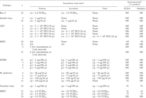
Standard serum production in BALB/cj mice (Table 1).
Five
viruses, Sendai virus, GD7, Reo-3, MHV and NCDV,
pro-duced infections in BALB/cj mice inoculated by the natural
route. However, more than one dose was usually required to
induce seroconversion in most of the animals. For NCDV, a
majority of the mice were inoculated by the i.p. route, which
resulted in 100% seroconversion (Table 1). For MVM,
M.
pulmonis
, and vaccinia virus, mice were inoculated by the i.p.
route alone. These three agents failed to generate antibodies
by other routes of exposure (Table 1).
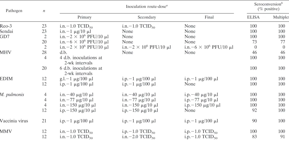
Standard serum production in C57BL/6j mice (Table 2).
The course of infection and antibody generation in the
C57BL/6j mice was similar to that described above for
BALB/cj mice. However, it appears that inoculations in
C57BL/6j mice by Sendai virus or GD7 virus were more
suc-cessful in generating antibody responses than in BALB/cj mice.
For example, 23 mice inoculated with Sendai virus and 22 mice
inoculated with GD7 by the intranasal route became
seropos-itive after only one inoculation. In contrast, most BALB/cj
exposed to Sendai virus or GD7 virus by routes mimicking
transmission under colony conditions required reinoculation
for the induction of antibody responses.
Analysis of single-positive mouse sera by multiplex
mi-crobead assay.
Serum samples, representative of individual
groups of mice inoculated with specific infectious agents, were
analyzed by multiplex microbead assay. Individual,
antigen-coated and control microbeads were mixed to produce the
TABLE 2. Production of standard sera in C57BL/6j mice
Pathogen n
Inoculation route-dosea Seroconversionb
(% positive)
Primary Secondary Final ELISA Multiplex
Reo-3
23
i.n.
⫺
1.0 TCID50
i.n.
⫺
1.0 TCID50
None
100
100
Sendai
23
i.n.
⫺
1
g/10
l
None
None
100
100
GD7
2
i.n.
⫺
2
⫻
10
6PFU/10
l
None
None
100
100
20
i.n.
⫺
6
⫻
10
6PFU/10
l
None
None
73
77
2
i.n.
⫺
2
⫻
10
6PFU/10
l
i.n.
⫺
2
⫻
10
6PFU/10
l
i.n.
⫺
6
⫻
10
6PFU/10
l
0
0
MHV
28
d.b.
None
None
46
46
4
4 d.b. inoculations at
2-wk intervals
100
100
20
6 d.b. inoculations at
2-wk intervals
100
100
EDIM
12
g.l.
⫺
1
g/100
l
i.p.
⫺
1
g/100
l
i.p.
⫺
1
g/100
l
100
100
12
i.p.
⫺
1
g/100
l
i.p.
⫺
1
g/100
l
None
100
100
M. pulmonis
4
i.n.
⫺
40
g/10
l
i.n.
⫺
40
g/10
l
i.p.
⫺
40
g/10
l
100
100
4
i.n.
⫺
77
g/10
l
i.n.
⫺
77
g/10
l
i.p.
⫺
77
g/10
l
100
100
4
i.n.
⫺
150
g/10
l
i.n.
⫺
150
g/10
l
i.p.
⫺
150
g/10
l
100
100
12
i.p.
⫺
150
g/10
l
i.p.
⫺
150
g/10
l
None
92
100
Vaccinia virus
21
i.p.
⫺
1
g/100
l
i.p.
⫺
1
g/100
l
i.p.
⫺
1
g/100
l
90
100
MMV
12
i.n.
⫺
1.0 TCID50
i.p.
⫺
1.0 TCID50
i.p.
⫺
1.0 TCID50
100
100
12
i.n.
⫺
1.0 TCID
50i.n.
⫺
2.0 TCID
50i.p.
⫺
1.0 TCID
5083
91
a
i.n., intranasal; d.b., dirty bedding; g.l., gastric lavage; TCID50, 50% tissue culture infectious dose.
b
For simplicity, only the final seroconversion results are presented.
on August 17, 2020 by guest
http://cvi.asm.org/
13-plex bead mixture (see Materials and Methods). Sera were
diluted 1:250, unless stated otherwise, and incubated with the
13-plex microbead mixture. The results of the multiplex assay,
presented in Table 3, show that specific antibodies to different
infectious agents were detected with high sensitivity by the
antigen-coated bead sets. Importantly, these beads did not
display a significant nonspecific reaction to mouse sera,
com-pared to background signal from normal mouse sera. Thus,
these findings demonstrate that the multiplex microbead assay
is highly specific and sensitive for the detection of antibodies to
the panel of 10 mouse infectious agents.
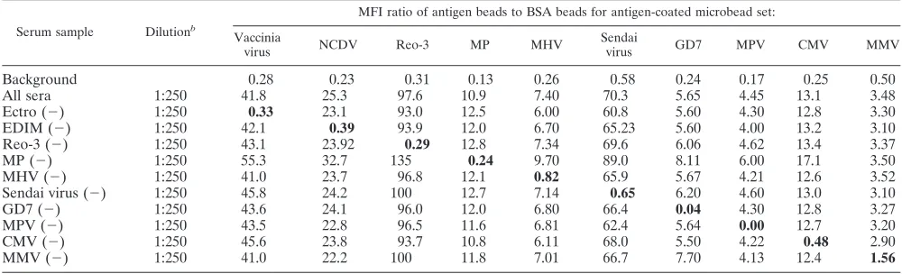
Analysis of mixed, single-positive mouse sera by multiplex
microbead assay.
The ability of the multiplex microbead assay
to detect antibodies against several infectious agents in single
serum samples was tested. Because mouse sera positive for
antibodies to all nine viruses and
M. pulmonis
are rare, the sera
in Table 3 were mixed in a 1:1 ratio to simulate multiple
infections. In addition, several combinations of the mixed
pos-itive sera, where one pospos-itive serum was omitted at a time,
were also prepared. The final dilution of the mixed sera was
the same as in Table 3. Various mixtures of positive sera were
incubated with the mixture of antigen-coated microbeads. The
results demonstrate that in both the mixture containing all of
the positive sera and the mixtures where one positive serum
sample was omitted, the multiplex microbead assay detected
specific antibodies (Table 4). Two observations are
notewor-thy. First, for 7 of 10 antigen-coated microbead sets (vaccinia
virus, NCDV,
M. pulmonis
, MHV, Sendai virus, CMV, and
MMV), the nonspecific reactivity was slightly higher than the
background. This increase may be the result of interserum
interactions of sera from different animals. Second, the values
of positive serum reactivity to each microbead set were about
half of those observed in analysis of the single-positive sera in
Table 3. The simplest explanation for this reduction in specific
signal is that there may be negative interference for antibody
detection when sera from multiple animals are mixed.
Inter-estingly, in the absence of
M. pulmonis
-positive serum, a higher
reactivity in the serum mixture towards all the microbead sets
was observed.
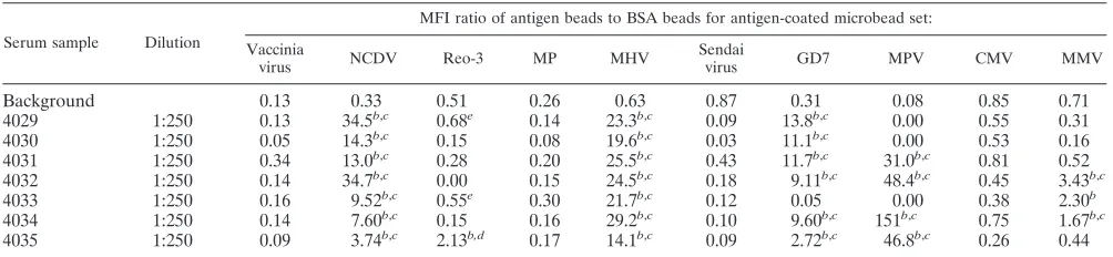
Multiplex microbead assay for serodetection in mice
in-fected with multiple infectious agents.
To evaluate the
multi-plex microbead assay under field conditions, sera from mice
naturally infected with multiple infectious agents were tested.
Seven serum samples, positive for antibodies to multiple
infec-TABLE 3. Antibody detection in known singly positive sera by multiplex microbead assay
aSerum sample Dilution
MFI ratio of antigen beads to BSA beads for antigen-coated microbead set: Vaccinia
virus NCDV Reo-3 MP MHV
Sendai
virus GD7 MPV CMV MMV
Background
0.28
0.23
0.31
0.13
0.26
0.58
0.24
0.17
0.25
0.50
Ectro
1:2
⫻
10
4b167.0
0.03
0.00
0.03
0.00
0.05
0.00
0.00
0.00
0.00
EDIM
1:250
0.19
63.0
0.29
0.09
0.08
0.43
0.03
0.11
0.11
0.42
Reo-3
1:250
0.14
0.16
189.0
0.16
0.16
0.27
0.00
0.01
0.15
0.34
MP
1:250
0.13
0.13
0.29
19.0
0.23
0.10
0.00
0.00
0.34
0.73
MHV
1:250
0.08
0.04
0.30
0.11
12.0
0.10
0.00
0.10
0.27
0.33
Sendai virus
1:250
0.17
0.24
0.35
0.14
0.18
153.0
0.00
0.00
0.06
0.44
GD7
1:250
0.07
0.15
0.26
0.15
0.11
0.37
12.0
0.02
0.09
0.35
MPV
1:250
0.09
0.11
0.25
0.08
0.08
0.17
0.00
11.0
0.01
0.31
CMV
AD
0.05
0.05
0.18
0.08
0.64
0.08
0.00
0.04
27.0
0.06
MMV
1:250
0.22
0.20
0.62
0.14
0.30
0.17
0.35
0.20
0.21
5.00
aResults are averages of two independent experiments. Boldface indicates the most specific results. Abbreviations: MP,M. pulmonis; Ectro, ectromelia virus; AD,
as directed by the manufacturer (Charles River, MA).
bHigher dilution needed due to high antibody titer.
TABLE 4. Multiplex microbead assay on mixed, singly positive mouse sera from Table 3
aSerum sample Dilutionb
MFI ratio of antigen beads to BSA beads for antigen-coated microbead set: Vaccinia
virus NCDV Reo-3 MP MHV
Sendai
virus GD7 MPV CMV MMV
Background
0.28
0.23
0.31
0.13
0.26
0.58
0.24
0.17
0.25
0.50
All sera
1:250
41.8
25.3
97.6
10.9
7.40
70.3
5.65
4.45
13.1
3.48
Ectro (
⫺
)
1:250
0.33
23.1
93.0
12.5
6.00
60.8
5.60
4.30
12.8
3.30
EDIM (
⫺
)
1:250
42.1
0.39
93.9
12.0
6.70
65.23
5.60
4.00
13.2
3.10
Reo-3 (
⫺
)
1:250
43.1
23.92
0.29
12.8
7.34
69.6
6.06
4.62
13.4
3.37
MP (
⫺
)
1:250
55.3
32.7
135
0.24
9.70
89.0
8.11
6.00
17.1
3.50
MHV (
⫺
)
1:250
41.0
23.7
96.8
12.1
0.82
65.9
5.67
4.21
12.6
3.52
Sendai virus (
⫺
)
1:250
45.8
24.2
100
12.7
7.14
0.65
6.20
4.60
13.0
3.10
GD7 (
⫺
)
1:250
43.6
24.1
96.0
12.0
6.80
66.4
0.04
4.30
12.8
3.27
MPV (
⫺
)
1:250
43.5
22.8
96.5
11.6
6.81
62.4
5.64
0.00
12.7
3.20
CMV (
⫺
)
1:250
45.6
23.8
93.7
10.8
6.11
68.0
5.50
4.22
0.48
2.90
MMV (
⫺
)
1:250
41.0
22.2
100
11.8
7.01
66.7
7.70
4.13
12.4
1.56
aResults are averages of two independent experiments. MP,M. pulmonis;Ectro, ectromelia virus. Boldface indicates the least specific results.
bFinal dilution of each serum sample in the mixture.
on August 17, 2020 by guest
http://cvi.asm.org/
tious agents, as determined by commercial ELISA kits, were
obtained from mice in an enzootically infected colony.
Multi-plex microbead analysis was performed in a blinded
experi-ment. Diluted sera (1:250) were incubated with the 13-plex
microbead mixture. A strong correlation was observed
be-tween the results obtained by commercial ELISA kits and the
multiplex microbead assay (Table 5). All animals that were
positive by ELISA for antibodies to EDIM (
n
⫽
7), MHV (
n
⫽
7), GD7 (
n
⫽
6), MPV (
n
⫽
3), MMV (
n
⫽
2), and Reo-3
(
n
⫽
1) were also positive for the same agents by the multiplex
assay. Additionally, multiplex analysis detected antibodies to
Reo-3 in one animal (4035) that tested negative by ELISA; this
animal was subsequently confirmed to be positive for
antibod-ies to Reo-3 by IFA. Two other mice, 4029 and 4033, were
indeterminate for Reo-3 antibodies by the multiplex
mi-crobead assay; sera from these mice displayed values slightly
above the background level (Table 5). Both of these mice were
indeterminate for Reo-3 antibodies by IFA as well. One serum
sample that was positive for antibodies to MMV (4033) by the
multiplex microbead assay was negative for MMV not only by
ELISA but also by IFA. It is possible that the analysis of this
animal represents a false-positive result by the multiplex
mi-crobead assay. Alternatively, the multiplex assay may be more
sensitive than ELISA and IFA for the detection of antibodies
to MMV.
DISCUSSION
This report describes the development of a multiplex
mi-crobead assay to detect antibodies to 10 infectious agents,
using only 1 microliter of mouse serum. A major advantage of
the multiplex format is its ability to detect antibodies to several
antigens in a small amount of sample; this feature substantially
saves sample, time, and labor compared to conventional
im-munoassays. The multiplex microbead technology is very
flex-ible, because microbead sets conjugated with different antigens
can be mixed to include only those that are needed; this feature
avoids waste of important antigens. The flexibility of
mi-crobead multiplex system also allows incorporation of internal
controls directly into the test sample (11). This built-in system
of controls enables the direct and simultaneous measurement
of specificity and sensitivity in detection of immune responses
to multiple antigens. Additionally, in comparison to ELISA,
the multiplex microbead assay provides a much larger dynamic
range, which allows accurate analysis of antibody levels without
additional dilution of sera with high antibody titers.
Further-more, the multiplex microbead assay is readily adaptable to
high-throughput format. This adaptability is important for
han-dling a large number of mouse samples for serodetection.
To develop the multiplex microbead assay system, we
pro-duced mouse sera positive for antibodies to individual
infec-tious agents (Tables 1 and 2). Attempts were made to inoculate
these mice by routes mimicking natural transmission. Because
our goal was to develop multiplex serodetection with wide
applicability to mouse strains that range in magnitude of
anti-body responses, the BALB/cj and C57BL/6j strains were used
to produce monospecific sera. Sera from these experimentally
inoculated mice were critical for unambiguous evaluation of
sensitivity and specificity of the multiplex microbead assay.
Mouse sera obtained from commercial sources are generated
by inoculating mice with a mixture of several antigens. These
sera may be positive for antibodies to multiple agents but are
provided as positive for only one agent; antibodies to any other
agents, if present, are not specified. Such sera may be useful to
test the performance of the conventional immunoassays such
as ELISA, IFA, and Western blotting. However, these
com-mercial sera are unsuitable for use in the multiplex microbead
assay. This complication was further exacerbated by lot-to-lot
variation of the sera, resulting in high titers against a different
set of antigens in each lot. Accordingly, the monospecific sera
produced in this project will be valuable reference material for
future assay development and validation, particularly as the
multiplex microbead assay is expanded for serodiagnosis of
additional infectious agents.
The multiplex serodiagnostic assay was tested by using sera
from animals containing antibodies to single infectious agents.
As shown in Table 3, microbeads coated with individual
anti-gens reacted to mouse sera in a specific manner. The data also
demonstrated that the microbeads did not display nonspecific
reactivity to sera containing irrelevant antibodies. The
speci-ficity of the multiplex microbead assay was further tested in
simultaneous serodetection of multiple agents in a single
re-action. Two experiments were performed: (i) standard positive
sera were mixed and reacted against the antigen-coated bead
TABLE 5. Multiplex assay for serodetection in field samples of mice
aSerum sample Dilution
MFI ratio of antigen beads to BSA beads for antigen-coated microbead set: Vaccinia
virus NCDV Reo-3 MP MHV
Sendai
virus GD7 MPV CMV MMV
Background
0.13
0.33
0.51
0.26
0.63
0.87
0.31
0.08
0.85
0.71
4029
1:250
0.13
34.5
b,c0.68
e0.14
23.3
b,c0.09
13.8
b,c0.00
0.55
0.31
4030
1:250
0.05
14.3
b,c0.15
0.08
19.6
b,c0.03
11.1
b,c0.00
0.53
0.16
4031
1:250
0.34
13.0
b,c0.28
0.20
25.5
b,c0.43
11.7
b,c31.0
b,c0.81
0.52
4032
1:250
0.14
34.7
b,c0.00
0.15
24.5
b,c0.18
9.11
b,c48.4
b,c0.45
3.43
b,c4033
1:250
0.16
9.52
b,c0.55
e0.30
21.7
b,c0.12
0.05
0.00
0.38
2.30
b4034
1:250
0.14
7.60
b,c0.15
0.16
29.2
b,c0.10
9.60
b,c151
b,c0.75
1.67
b,c4035
1:250
0.09
3.74
b,c2.13
b,d0.17
14.1
b,c0.09
2.72
b,c46.8
b,c0.26
0.44
a
Results are averages of two independent experiments. MP,M. pulmonis.Serum samples are from individual mice.
b
Positive by the multiplex microbead assay.
c
Confirmed as positive by ELISA.
d
Negative by ELISA but confirmed positive by IFA.
e
Negative by ELISA and indeterminate by multiplex assay and IFA.
on August 17, 2020 by guest
http://cvi.asm.org/
sets (Table 4) and (ii) field sera from mice infected with several
agents were obtained from a mouse facility and tested (Table
5). Both experiments clearly demonstrated a high degree of
sensitivity and specificity for the multiplex microbead assay. In
Table 4, where positive sera from several mice were mixed,
minor nonspecific reactivity of the antigen-coated beads was
observed, e.g., in the serum mixture lacking MMV-positive
serum, resulting in a decrease in the signal-to-noise ratio.
These results may reflect potential interactions and
interfer-ence among sera from different animals. However, any such
inconsistencies were resolved in sera from animals infected
with multiple agents, yielding results that were clearly
inter-pretable (Table 5). Similar types of minor interferences have
also been observed in multiplex immunoassay in a mixture of
individual sera from rhesus macaques positive for antibodies to
a single infectious agent. Again, such interferences were not
observed in individual rhesus macaque sera positive for
anti-bodies to multiple infectious agents (I. Khan and P. Luciw,
unpublished data).
Clinical validation of the multiplex microbead assay will
require analysis of a large number of field serum samples that
are also tested by conventional immunoassays, which detect
antibodies to one infectious agent at one time. Accordingly,
several thousand samples will be analyzed to establish
statisti-cally significant levels of sensitivity and specificity. In addition,
a high-throughput format of the multiplex microbead assay is
currently under evaluation to facilitate validation.
The increased efficiency of the multiplex microbead
immu-noassay and the requirement for only one microliter of serum
will promote direct testing of individual mice. In current
prac-tice for health maintenance of SPF colonies, indirect testing by
the use of sentinel mice may have certain disadvantages. For
example, a recent report showed that some sentinel mice used
in serosurveillance were infected with MPV at the time of
arrival from the rodent vendor and were the source of MPV
infection in the colony animals (16). Thus, direct testing of
mice may improve the accuracy of health monitoring in mouse
colonies. Because the multiplex microbead assay requires only
1 microliter of sample, this method is ideally suited for
sero-surveillance of mouse colonies in general, and in particular of
more valuable mice, such as genetically engineered lines. In
addition, the ability to detect and measure multiple analytes in
a small volume of serum (or plasma) allows for analysis of
samples collected in the live phase of a research protocol.
ACKNOWLEDGMENTS
We thank Leslie Jones and Tilhuan Yilma (International Laboratory
for the Molecular Biology of Tropical Disease Agents, University of
California, Davis) for inactivation of vaccinia virus. We also thank Paul
Rhyne and Christine Daly (Upstate USA Inc., Lake Placid, NY) for
suggestions on improving the manuscript. We thank Nicole Corley for
her invaluable assistance with the production of positive sera.
This work was supported by PHS grant RR14034 to S. W. Barthold
from the National Center for Research Resources, NIH.
REFERENCES
1.Andersen, C. A., J. C. Murphy, and J. G. Fox.1986. Surveillance of mice for
antibodies to murine cytomegalovirus. J. Clin. Microbiol.23:1152–1154.
2.Carson, R. T., and D. A. Vignali.1999. Simultaneous quantitation of 15 cytokines using a multiplexed flow cytometric assay. J. Immunol. Methods
30:41–52.
3.Compton, S. R., F. R. Homberger, F. X. Paturzo, and J. M. Clark.2004. Efficacy of three microbiological monitoring methods in a ventilated cage
rack. Comp. Med.54:382–392.
4.Gilburd, B., M. Abu-Shakra, Y. Shoenfeld, A. Giordano, E. B. Bocci, F. delle Monache, and R. Gerli.2004. Autoantibodies profile in the sera of patients with Sjogren’s syndrome: the ANA evaluation—a homogeneous,
multi-plexed system. Clin. Dev. Immunol.11:53–56.
5.Iwarsson, K.1990. Laboratory animal health monitoring: an introductory survey of university mouse breeding colonies in Stockholm. Acta Physiol.
Scand. Suppl.592:139–140.
6.Jacoby, R. O., and J. R. Lindsey.1997. Health care for research animals is
essential and affordable. FASEB J.8:609–614.
7.Kettman, J. R., T. Davies, D. Chandler, K. G. Oliver, and R. J. Fulton.1998. Classification and properties of 64 multiplexed microsphere sets. Cytometry
33:234–243.
8.Komatsu, N., S. Shichijo, M. Nakagawa, and K. Itoh.2004. New multiplexed flow cytometric assay to measure anti-peptide antibody: a novel tool for monitoring immune responses to peptides used for immunization. Scand.
J. Clin. Lab. Investig.64:535–546.
9.Livingston, R. S., D. G. Besselsen, E. K. Steffen, C. L. Besch-Williford, C. L. Franklin, and L. K. Riley.2002. Serodiagnosis of mice minute virus and mouse parvovirus infections in mice by enzyme-linked immunosorbent assay with baculovirus-expressed recombinant VP2 proteins. Clin. Diagn. Lab.
Immunol.9:1025–1031.
10.Lussier, G., and J. P. Descoteaux.1986. Prevalence of natural virus
infec-tions in laboratory mice and rats used in Canada. Lab. Anim. Sci.36:145–
148.
11.Martins, T. B.2002. Development of internal controls for the Luminex instrument as part of a multiplex seven-analyte viral respiratory antibody
profile. Clin. Diagn. Lab. Immunol.9:41–45.
12.Opalka, D., C. E. Lachman, S. A. MacMullen, K. U. Jansen, J. F. Smith, N. Chirmule, and M. T. Esser.2003. Simultaneous quantitation of antibodies to neutralizing epitopes on virus-like particles for human papillomavirus types 6, 11, 16, and 18 by a multiplexed Luminex assay. Clin. Diagn. Lab. Immunol.
10:108–115.
13.Pickering, J. W., T. B. Martins, R. W. Greer, M. C. Schroder, M. E. Astill, C. M. Litwin, S. W. Hildreth, and H. R. Hill.2002. A multiplexed fluorescent microsphere immunoassay for antibodies to pneumococcal capsular
polysac-charides. Am. J. Clin. Pathol.117:589–596.
14.Pickering, J. W., T. B. Martins, M. C. Schroder, and H. R. Hill.2002. Comparison of a multiplex flow cytometric assay with enzyme-linked immu-nosorbent assay for quantitation of antibodies to tetanus, diphtheria, and
Haemophilus influenzaetype b. Clin. Diagn. Lab. Immunol.9:872–876. 15.Prabhakar, U., E. Eirikis, and H. M. Davis.2002. Simultaneous
quantifica-tion of proinflammatory cytokines in human plasma using the LabMap assay.
J. Immunol. Methods1:201–218.
16.Pullium, J. K., K. A. Benjamin, and M. J. Huerkamp.2004. Rodent vendor apparent source of mouse parvovirus in sentinel mice. Contemp. Top. Lab.
Anim. Sci.43:8–11.
17.Suzuki, E., J. Matsubara, M. Saito, T. Muto, M. Nakagawa, and K. Imaizumi.1982. Serological survey of laboratory rodents for infection with Sendai virus, mouse hepatitis virus, reovirus type 3, and mouse adenovirus.
Jpn. J. Med. Sci. Biol.35:249–254.
18.Tsung, K., J. H. Yim, W. Marti, R. M. Buller, and J. A. Norton.1996. Gene expression and cytopathic effect of vaccinia virus inactivated by psoralen and
long-wave UV light. J. Virol.70:165–171.