Scholarship@Western
Scholarship@Western
Electronic Thesis and Dissertation Repository
August 2012
Telomere length dynamics in aging mice
Telomere length dynamics in aging mice
Paul J. Comartin Western University
Supervisor
Dr. Geoffrey Pickering
The University of Western Ontario Graduate Program in Biochemistry
A thesis submitted in partial fulfillment of the requirements for the degree in Master of Science © Paul J. Comartin 2012
Follow this and additional works at: https://ir.lib.uwo.ca/etd
Part of the Biochemistry Commons
Recommended Citation Recommended Citation
Comartin, Paul J., "Telomere length dynamics in aging mice" (2012). Electronic Thesis and Dissertation Repository. 658.
https://ir.lib.uwo.ca/etd/658
This Dissertation/Thesis is brought to you for free and open access by Scholarship@Western. It has been accepted for inclusion in Electronic Thesis and Dissertation Repository by an authorized administrator of
Thesis format: Integrated Article
by
Paul Comartin
Graduate Program in Biochemistry
A thesis submitted in partial fulfillment of the requirements for the degree of
Master of Science
The School of Graduate and Postdoctoral Studies The University of Western Ontario
London, Ontario, Canada
ii
THE UNIVERSITY OF WESTERN ONTARIO School of Graduate and Postdoctoral Studies
CERTIFICATE OF EXAMINATION
Supervisor
______________________________ Dr. J. Geoffrey Pickering
Supervisory Committee
______________________________ Dr. Dean Betts
______________________________ Dr. Robert Hegele
Examiners
______________________________ Dr. Murray Huff
______________________________ Dr. Caroline Schild-Poulter
______________________________ Dr. Sean Cregan
______________________________ Dr.
The thesis by
Paul Joseph Comartin
entitled:
Telomere length dynamics in aging mice
is accepted in partial fulfillment of the requirements for the degree of
Master of Science
______________________ _______________________________
iii
Leukocyte telomere length (TL) shortens with age and is associated with age-related
pathologies. However, inherited and acquired variation in telomere length in individuals
complicates clinical interpretations of TL as a biomarker of aging and age-related
pathologies. Therefore, it is critical to identify a post-mitotic tissue as a surrogate marker of
TL at birth. In my thesis project, I used quantitative PCR to trace TL dynamics of a variety of
tissue types of inbred mice during 1st year of life. I found that TL of smooth muscle of aortic
media did not shorten with age and represents birth TL. Notably, birth TL effectively offset
genetic variation of TL in a genetically diverse mouse population. In addition, I further
revealed that impaired collagen turnover in mice, which leads to premature aging symptoms,
accelerates TL shortening. In summary, I identified that aortic media provides a powerful
internal reference for birth TL with potentials to improve the accuracy of evaluating telomere
shortening in individuals.
Keywords
Telomere length dynamics, T/S ratio, birth telomere length, genetic variation, inbred mice,
iv
Acknowledgments
Completion of the work in this thesis would not have been possible without the collaborative
efforts of everyone in the Pickering lab and my advisory committee.
First and foremost, a special thank you goes to my supervisor, Dr. Geoffrey Pickering, for his
ongoing support and direction. His expertise as a cardiologist and a researcher has proved
immensely beneficial in developing my ability to think critically as a researcher while
understanding clinical relevance.
I would like to extend a thank you to my advisory committee, Dr. Robert Hegele and Dr.
Dean Betts, for their endless support. Their wisdom in the area of vascular research and
telomere biology, respectively, has been invaluable to gain in depth perspectives to advance
my research.
Finally, I would like to thank all members of the Pickering lab for making the journey of my
masters project a valuable experience. In particular, I would like to thank Oula Akawi for
getting me on my feet when I first came into the lab. Also, a special thanks to Zengxuan
Nong for helping me harvest tissue from all the mice used in this study. Lastly, I would also
like to thank our lab technician Caroline O’Neil for her help making sure all reagents and
mice are ordered on time and to our post-doctoral scholar Hao Yin for his insight and
v
CERTIFICATE OF EXAMINATION ...ii
Abstract ... iii
Acknowledgments ... iv
Table of Contents ... v
List of Tables...viii
List of Figures ... ix
List of Appendices ... xi
List of Abbreviations ... xii
Chapter 1 -Introduction ... 1
1.1 General introduction ... 1
1.2 Telomere structure ... 1
1.2.1 T-Loop ... 3
1.2.2 Shelterin ... 3
1.3 Telomere function ... 5
1.3.1 Genomic stability ... 5
1.3.2 End-replication problem ... 7
1.4 Telomeres and cell senescence ... 7
1.5 Cellular senescence and aging ... 9
1.6 Telomere length (TL) regulation ... 11
1.6.1 Factors that cause telomere shortening ... 11
1.6.2 Factors involved in telomere length maintenance ... 15
1.7 Telomere length measurement ... 21
1.7.1 TRF analysis ... 21
vi
1.7.3 Flow-FISH ... 23
1.7.4 STELA ... 24
1.7.5 qPCR ... 25
1.8 Telomere dynamics in the aging population ... 26
1.8.1 Telomere length with age ... 26
1.8.2 Telomere length and chronic illness ... 29
1.8.3 Telomere length (TL) variation in the population ... 32
1.8.4 Goals of this study ... 38
1.9 References ... 39
Chapter 2 -Aortic media defines telomere length dynamics in mice ... 63
2.1 Abstract ... 63
2.2 Introduction ... 64
2.3 Materials and methods ... 67
2.3.1 Animals and tissue ... 67
2.3.2 Tissue preparation ... 67
2.3.3 DNA isolation and quantitation ... 68
2.3.4 Telomere length measurement by qPCR ... 68
2.3.5 Statistical analysis ... 70
2.4 Results ... 70
2.4.1 Optimization of qPCR telomere length measurement for mice ... 70
2.4.2 Telomere length changes with age are strain specific ... 72
2.4.3 Age-related changes of telomere lengths in C57BL/6 mice differ among tissues ... 74
2.4.4 Aortic media telomeres are stable in early life development ... 74
vii
2.4.7 Aortic media referencing reveals age-dependent telomere attrition in
spleen and aortic intima of Diversity Outbred mice... 80
2.4.8 Telomere length shortening is induced by impaired collagen turnover .... 84
2.5 Discussion... 86
2.5.1 Strain-specific differences in telomere length dynamics ... 86
2.5.2 Tissue-specific differences in telomere length dynamics of C57BL/6 mice87 2.5.3 Aortic media has stable telomeres during early life development ... 91
2.5.4 Telo-mapping reveals telomere length heterogeneity in the aorta ... 92
2.5.5 Diversity Outbred mice as a murine model for telomere biology ... 93
2.5.6 Correcting for telomere length heterogeneity in Diversity Outbred mice . 94 2.5.7 Impaired collagen turnover accelerates telomere attrition ... 95
2.6 References ... 96
Chapter 3 -General Discussion ... 103
3.1 Limitations and immediate next steps ... 103
3.2 Future implications ... 104
3.2.1 Model for estimating birth telomere length ... 105
3.2.2 Diversity Outbred mouse model to study genetic diversity and telomere length dynamics... 106
3.3 References ... 108
viii
List of Tables
Table 1.1 - Telomere length variation across vertebrate species ... 1
Table 1.2 - Differences between mouse and human telomere length dynamics ... 26
Table 1.3 - Short leukocyte telomere length and associated cardiovascular disorders ... 29
ix
Figure 1.1 - Telomeres are located at the ends of linear DNA ... 2
Figure 1.2 - Secondary structure of telomeres. ... 4
Figure 1.3 - Anaphase breakage fusion bridge cycles ... 6
Figure 1.4 - Antagonistic pleiotropy of senescent cells in young and old individuals ... 10
Figure 1.5 - Factors affecting telomere length dynamics in vertebrate cells ... 12
Figure 1.6 - Elongation of telomeres by telomerase... 16
Figure 1.7 - Mechanisms of alternative lengthening of telomeres ... 20
Figure 1.8 - Detection of extra-telomeric DNA with different methods of telomere length measurement ... 22
Figure 2.1 - Reliability of T/S ratio measurements using different DNA quantities ... 71
Figure 2.2 - Inbred mouse strain differences in spleen telomere lengths ... 73
Figure 2.3 - Telomere length dynamics with age in different tissues of C57BL/6 mice... 75
Figure 2.4 - Telomere length changes in early life of C57BL/6 aorta and heart ... 76
Figure 2.5 - Telomere lengths of different layers in the aorta of C57BL/6 mice ... 78
Figure 2.6 - Telomere length in different regions of the aortic media in C57BL/6 mice ... 79
Figure 2.7 - Spleen telomere length referenced to aortic media telomere length as a function of age in DO mice... 82
Figure 2.8 - Intima telomere lengths normalized to aortic media telomere lengths with age. 83 Figure 2.9 - Telomere length in different tissues of aged collagenase-resistant mice ... 85
x
xi
Appendix A: Statement of permission for the use of animals for experimental research ...110
xii
List of Abbreviations
Abbreviation Meaning
53BP1 P53 binding protein 1
ABP ALT-associated PML body
ALT Alternative lengthening of telomeres
ATM Ataxia telangiectasia mutated
ATR Ataxia telangiectasia and rad3 related
BIR Break induced replication
BMI Body mass index
CAD Coronary artery disease
C-FOS FBJ murine osteosarcoma viral oncogene homolog
CHK2 Checkpoint kinase 2
CV Coefficient of variation
CVD Cardiovascular disease
DKC1 dyskerin gene
DDR DNA damage response
DNA Deoxyribonucleic acid
E2F Transcription factor family including E2F- and
DP-like subunits
ECM Extracellular matrix
FISH Fluorescence in situ hybridization
GWAS Genome wide association studies
HSC Hematopoietic stem cell
Kb kilobases
LFS Li-Fraumani syndrome
LTL Leukocyte telomere length
MDC1 Mediator of DNA-damage checkpoint 1
MOV10 Moloney leukemia virus 10
xiii
PBS Phosphate buffered saline
PCNA Proliferation cell nuclear antigen
PD population doubling
PML Promyelocytic leukaemia body
PNA peptide nucleic acid
POT1 Protection of telomeres 1
qPCR quantitative polymerase chain reaction
Rap1 Repressor/activator protein 1
RCR Rolling circle replication
ROS Reactive oxygen species
SD Standard deviation
SNP Single nucleotide polymorphism
STELA Single Telomere Length Analysis
TERC Telomerase RNA component
TERT Telomerase reverse transcriptase
TIF Telomere induced foci
TIN2 TRF1-interacting protein 2
TL Telomere length
TPP1 POT1-TIN2 organizing protein
T-SCE Telomere sister chromatid exchange
TRF Telomere restriction fragment
TRF1 Telomeric repeat binding factor 1
TRF2 Telomeric repeat binding factor 2
Chapter 1 -
Introduction
1.1
General introduction
The idea of modifying the course of aging and age-related diseases is a concept that
humans naturally desire to explore with great potential for clinical application. Telomeres
represent a potentially modifiable biomarker of aging that has been linked to age-related
diseases (Oeseburg et al., 2010). Tremendous research efforts, with over 13000 hits on
PubMed, growing continuously each day, are made to uncover the link between
telomeres with aging and various age-related diseases. Telomeres protect chromosomes,
but undergo attrition as DNA replicates or is insulted by environmental stresses
(Oeseburg et al., 2010). In particular, shortening of telomere length (TL) in leukocytes is
a well-established event in human aging (Aviv, 2012). However, an etiological link
between telomeres and aging is yet to be fully elucidated. Understanding the dynamics
of telomeres with normal aging in model organisms (e.g. mice) can help to uncover the
role telomeres play at the organismal level with age.
1.2
Telomere structure
Telomeres are DNA-protein complexes containing repetitive units of DNA that reside at
the end of each chromosome arm in eukaryotes (Figure 1.1) (Oeseburg et al., 2010). The
DNA sequence of telomeres is highly conserved among vertebrates and consists of
several kilobases of TTAGGG repeats (Meyne et al., 1989), ending in a 100-200 bp 3’
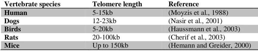
overhang (Makarov et al., 1997). However, TL is highly variable across different
species (Table 1.1).
Table 1.1 - Telomere length variation across vertebrate species
Vertebrate species Telomere length Reference
Human 5-15kb (Moyzis et al., 1988)
Dogs 12-23kb (Nasir et al., 2001)
Birds 5-20kb (Haussmann et al., 2003)
Rats 20-100kb (Cherif et al., 2003)
Figure 1.1 - Telomeres are located at the ends of linear DNA
Telomeres are located at the ends of linear chromosomal DNA consisting of repetitive
1.2.1
T-Loop
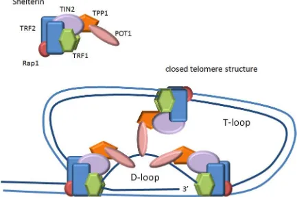
The 3’ overhang of telomere forms the secondary structure, which varies between taxa.
In vertebrates it forms a T-loop structure and internal D-loop (Figure 1.2) by looping
around to invade upstream telomeric DNA (de Lange, 2005). The 3’overhang is
maintained during each round of DNA replication by a nuclease called Apollo (Wu et al.,
2010). T-loop size is proportional to TL (Rahman et al., 2008). In fact, mice T-loop size
is 18 kb long, whereas in humans it is only 3 kb (Griffith et al., 1999), reflective of their
differing TLs (20-80 kb versus 10-15 kb, respectively) (Kipling and Cooke, 1990;
Zijlmans et al., 1997).
1.2.2
Shelterin
Telomeres are further stabilized by a protein complex called shelterin (de Lange, 2005).
Shelterin is composed of six proteins (Figure 1.2) – telomeric repeat binding factor 1 and
2 (TRF1 and TRF2), protection of telomeres 1 (POT1), repressor/activator protein 1
(Rap1), POT1-TIN2 organizing protein (TPP1, also referred to as TINT1, PIP1, and
PTOP), and TRF1-interacting protein 2 (TIN2). Three of these proteins (TRF1, TRF2,
and POT1) bind directly to telomeric repeats. TRF1 and TRF2 bind to double-stranded
repeats via their SANT/Myb domains with high specificity and affinity and each have a
flexible hinge that enables the formation of the T-loop (de Lange, 2005; Diotti and
Loayza, 2011). POT1 binds directly to the single-stranded 3’overhang via its OB folds,
which is important in the formation of the D-loop (Baumann and Cech, 2001; de Lange,
2005; Lei et al., 2004; Palm and de Lange, 2008). Unlike human cells that have only one
POT1 gene, mice have two variants that are highly homologous (POT1a and POT1b) but
have distinct roles at telomeres (He et al., 2006; Hockemeyer et al., 2006). The other
three proteins (Rap1, TPP1, and TIN2) are recruited by TRF1 and TRF2 to assist in
stabilizing telomeres (de Lange, 2005). TIN2 acts as the bridge between different
shelterin components by binding TRF1 and TRF2 in separate domains and recruiting the
TPP1-POT1 complex (Chen et al., 2008; Kim et al., 2004; Liu et al., 2004; Ye et al.,
2004). Furthermore, a novel isoform of TIN2 (TIN2L) tethers telomeres to the nuclear
Figure 1.2 - Secondary structure of telomeres.
Protein components of shelterin complex are involved in the formation and stability of
T-loop and internal D-T-loop secondary structures of telomeres. Image modified from
TPP1 binds TIN2 and POT1 in separate domains and is thought to be central in recruiting
POT1 to telomeres (Chen et al., 2007; Kibe et al., 2010). Lastly, Rap1 is recruited by
TRF2 and forms a tightly associated complex that is essential for Rap1’s binding to
telomeres (Celli and de Lange, 2005; Li and de Lange, 2003; Li et al., 2000). Overall
the shelterin complex is involved in telomere stability and telomere length regulation (de
Lange, 2005).
1.3
Telomere function
Telomeres are considered as a telomeric cap because of the important functions it serves
for linear DNA. Over the years scientists have uncovered that telomeres play a
fundamental role in the stability and mobility of the genome and prevent erosion of
coding DNA.
1.3.1
Genomic stability
The chromosome ends were first proposed to be important for chromosome stability by
the combined work of Barbara MClintock and Hermann Muller on their work with maize
and fruit flies, respectively (McClintock, 1939; Muller, 1938). Muller coined the term
telomere from the greek words telos (end) and meros (part). Both scientists observed that
without telomeres, chromosomes would fuse together and break off during mitosis
(Figure 1.3), which was catastrophic to cellular physiology.
Telomere dysfunction either by critically shortened telomeres or missing shelterin
components can activate the DNA damage response (DDR) and signal up-regulation of
cell cycle checkpoints such as p53 (Bodnar et al., 1998; Karlseder et al., 1999; Lee et al.,
1998). In some cells, cell cycle checkpoints can be by passed, propagating breakage
fusion bridge cycles and genomic rearrangements with risk of neoplastic transformation
(Figure 1.3). Thus cells require telomeres as a way to protect natural ends from being
recognized as double strand breaks, in order to suppress the DNA repair machinery and
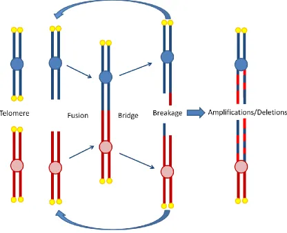
Figure 1.3 - Anaphase breakage fusion bridge cycles
Without telomeres (yellow circles) at the ends of chromosomes, a fusion event can take
place between bare ends. During anaphase when chromosomes are pulled to opposite
poles of the cell, DNA will break between bridged chromosomes. Anaphase
breakage-fusion-bridge cycles cause genomic shuffling (amplifications and deletion events) and
instability that can lead to further complications of cancer. Image modified from (Else,
Furthermore, during meiosis telomeres form bouquet structures that are integral to the
coordination of the spindle apparatus to ensure proper alignment and segregation of
genetic material (Maddar et al., 2001; Tomita and Cooper, 2007).
1.3.2
End-replication problem
One of the most important functions of telomeres was discovered by Olovnikov and
Watson who noticed that linear chromosomes cannot replicate their very ends, termed the
“end-replication problem” (Olovnikov, 1973; Watson, 1972). The end-replication
problem leads to inevitable loss of telomeres at every cell division. DNA polymerase can
only replicate in the 5’ to 3’ direction and requires a short RNA primer for initiation
(Shore and Bianchi, 2009). Because DNA is double stranded, one strand will be
synthesized in short “okazaki” fragments. However, at the 5’ end of the lagging strand,
there will be a gap due to removal of the last RNA primer (Shore and Bianchi, 2009).
Currently, it is not known whether or not the last RNA primer is placed at the very end of
telomeres or is variable (Sfeir et al., 2005b). Telomere ends undergo further processing
following DNA replication to ensure there is a 3’ overhang with a precise end sequence
(Sfeir et al., 2005a). Interestingly, the length of this overhang is highly variable between
different cell types and is closely associated with telomere length (Lee et al., 2008).
Initially, cells were thought to lose telomeres at a constant rate, keeping track of the
number of cell divisions like a “mitotic clock” (Von Zglinicki, 2003). However, there is
considerable heterogeneity in the rate of telomere shortening (Martin-Ruiz et al., 2004)
and in the number of divisions a cell can undergo (Hayflick, 2003; Smith and Hayflick,
1974). Notably, the end-replication problem directly limits the number of possible cell
divisions (Olovnikov, 1973), later known as replicative senescence.
1.4
Telomeres and cell senescence
There are several ways in which cells senesce but one of the main mechanisms is
telomere shortening and dysfunction, specifically referred to as replicative senescence or
mortality 1 (M1) senescence (Harley et al., 1990). During each round of cell division,
telomeres shorten by ~50-200 bp due to oxidative stress and the end replication problem
telomeres to reach a critical length (“Hayflick limit”) that signals a DNA-damage
response (DDR), which involves protein kinases (ATM and CHK2), adaptor proteins
(53BP1 and MDC1) and chromatin modifiers (γ-H2AX) (Campisi and d'Adda di
Fagagna, 2007). Telomere dysfunction induced foci (TIF), defined as cells with more
than 50% of 53BP1 foci colocalised with telomere repeats, are often used to identify
telomere-dependent senescent cells (Jeyapalan et al., 2007). Telomere-dependent
activation of DDR further leads to the activation of p53 via its phosphorylation and
upregulation of its downstream target p21 (Blackburn, 2005; Campisi and d'Adda di
Fagagna, 2007; Hornsby, 2002). Expression of p21 maintains pRB in a
hypophosphorylated active state, which inactivates the transcriptional factor E2F. E2F is
responsible for inducing the expression of genes that encode proteins responsible for
cell-cycle progression (i.e. replication-dependent histones, c-FOS, cyclin A and B, and
proliferation cell nuclear antigen (PCNA)). Therefore, repressing E2F activity causes
cells to enter a state of permanent cell cycle arrest (senescence) (Blackburn, 2005;
Campisi and d'Adda di Fagagna, 2007; Hornsby, 2002). If any of these cell cycle
checkpoints are by-passed (most commonly p53), perhaps due to mutations, the cell will
continue to divide and telomeres will continue to shorten until a crisis phase (M2 phase)
is reached. In M2 phase, telomeres are virtually non-existent and chromosome fusion
events occur due to activation of the non-homologous end joining (NHEJ) pathway,
which can result in genomic shuffling as shown in Figure 1.3 (Shay and Wright, 2005).
One of the key factors in triggering senescence or apoptosis in aging cells is shortened
telomeres that can no longer form a closed t-loop. In fact, inhibition of TRF2 from
shelterin in vitro causes the activation of DDR and senescence (Celli and de Lange, 2005;
Takai et al., 2003). In a similar way, human cells transfected with short telomere
oligonucleotides that mimic the effect of an open t-loop also trigger a p53 dependent cell
cycle arrest (Saretzki et al., 1999).
It is noteworthy that cellular senescence can also be induced by telomere-independent
mechanisms such as damage induced by reactive oxygen species (ROS), which follows
Holbrook, 2000). Murine fibroblasts in culture will senesce after 15-20 population
doublings despite having exceptionally long telomeres (>20 kb) when exposed to
atmospheric oxygen concentrations of 20%, which is above biological levels of 3%
(Hornsby, 2003; Parrinello et al., 2003). In contrast, human fibroblasts with shorter
telomeres (10-15 kb) senesce between 50-60 population doublings, suggesting better
protective mechanisms against ROS compared to mouse cells (Hornsby, 2003; Parrinello
et al., 2003). To some degree this argues against the relationship between telomere
shortening and aging, particularly in mice, however, in vitro studies do not fully translate
in vivo (Varela et al., 2011). Furthermore, the shortest mice telomeres are just as short to
those in human cells (Zijlmans et al., 1997), and a single short telomere is sufficient to set
off the cascade of events leading to senescence (Abdallah et al., 2009; Bendix et al.,
2010). Finally, it is not known whether the threshold telomere length that induces
senescence in humans and in mice is different. Perhaps in mice that have an average
T-loop size 6-7 times greater than in humans (Rahman et al., 2008), require longer
telomeres to maintain a closed T-loop structure.
1.5
Cellular senescence and aging
Cellular senescence describes a cell that has undergone permanent cell cycle arrest and
ceased to divide. This phenomenon was first observed in 1961 by Leonard Hayflick, who
observed that primary human fibroblasts in culture can only divide a limited number of
times (40-60 population doublings (PD)), later referred to as the “Hayflick limit”
(Hayflick, 1965; Hayflick and Moorhead, 1961). The exact number of divisions a cell
can undergo is dependent on cell type and organism (Campisi and d'Adda di Fagagna,
2007). For instance, stem cells and cancer cells can divide indefinitely in culture,
whereas human somatic cells cannot (Itahana et al., 2004). However, murine somatic
cells senesce between 15 and 20 PDs and can immortalize spontaneously in culture
(Itahana et al., 2004). A senescent cell is characterized by a flattened morphology in 2D
culture, altered gene expression, and secretion of various matrix-degrading enzymes
(Campisi and d'Adda di Fagagna, 2007).
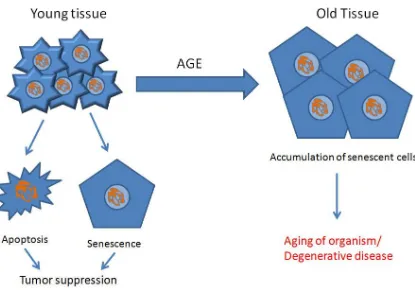
Cellular senescence is considered both beneficial to young organisms and harmful to old
Figure 1.4 - Antagonistic pleiotropy of senescent cells in young and old individuals
Young tissue is predominantly composed of normal, healthy, fully-functional cells.
Whenever stress on a cell is too great or telomeres are too short, the cell undergoes
apoptosis or senescence to avoid tumorigenesis. However, as we age, old tissue is
burdened by an accumulation of senescent cells, compromising tissue function. What
was protective at a young age against tumors is destructive to old organisms leading to
In young organisms cellular senescence is said to have evolved as a tumor suppressor by
giving highly proliferative cells a limited capacity to divide. In older organisms,
senescent cells tend to accumulate in organs, leading to age-related phenotypes
(Jeyapalan et al., 2007).
Evidence begins to accumulate that supports the link between cellular senescence and
organismal aging. First, the number of senescent cells increases with age and appears to
be rare in young individuals (Dimri et al., 1995; Jeyapalan et al., 2007). Furthermore,
sites of age-related pathologies such as atherosclerosis and osteoporosis contain increased
number of senescent cells (Chang and Harley, 1995; Price et al., 2002; Vasile et al.,
2001). Moreover, the altered secretory profile (upregulation of matrix degrading
enzymes, growth factors, and pro-inflammatory cytokines) of senescent cells has
detrimental impacts on neighboring cells and structural components of tissue that lead to
aged and diseased pathologies (Burton, 2009). Taken together, evidence suggests burden
of senescent cells is connected to organismal senescence. Since telomere length is
considered an endogenous marker of cellular senescence, its length is also a putative
marker of biological aging.
1.6
Telomere length (TL) regulation
TL equilibrium is established by the net result of telomere shortening and lengthening
events. The frequency of each of these events varies depending on the cell type and
organism involved (Shay and Wright, 2007). Several of the key factors that influence
telomere length dynamics in a cell are outlined in Figure 1.5.
1.6.1
Factors that cause telomere shortening
There are several factors that cause telomere shortening in a cell as listed in Figure 1.5.
The two most common causes are due to the end-replication problem, discussed in
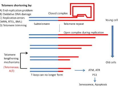
Figure 1.5 - Factors affecting telomere length dynamics in vertebrate cells
During DNA replication telomeres are in an open structure to allow DNA polymerase to
extend the entire length of the chromosome. However, various factors contribute to
telomere shortening that are propagated during replication such as the end-replication
problem, oxidative DNA damage, replication errors, and telomere trimming. As cells
divide and telomeres reach a critical length, they can no longer form a closed t-loop
structure which signals cell cycle checkpoints such as ATM, ATR and p53, causing cells
to enter a state of senescence or apoptosis. Telomere lengthening mechanisms, such as
telomerase and alternative lengthening of telomeres can extend the replicative capacity of
1.6.1.1
Oxidative DNA damage
While the end-replication problem sets the baseline of telomere shortening, oxidative
stress is the best recognized modifier. The minimal rate of telomere shortening observed
in culture is 10-20 bp per cell division in fibroblasts with high antioxidant capacity (Serra
et al., 2003; von Zglinicki, 2002) or those treated with an agent that reduces oxidative
stress in cells (Passos et al., 2007). This lower rate of attrition is likely that set by the
end-replication problem, which differs significantly from the average rate of telomere
attrition in human cells (50-200 bp). This difference is primarily due to oxidative stress
(von Zglinicki et al., 2000).
The major source of reactive oxygen species (ROS) in cells comes from mitochondria, as
a metabolic by-product. Burden of ROS in a cell induces various types of DNA damage
(oxidized bases, single and double strand breaks), which accumulates over time (Fraga et
al., 1990; Packer and Fuehr, 1977). Telomere shortening is largely accelerated by
accumulation of oxidative DNA damage; in particular, by single strand breaks (von
Zglinicki et al., 2000; von Zglinicki and Schewe, 1995). Those damages that are not
repaired or replaced before the next round of DNA replication will be lost due to stalling
of the replication fork. Therefore, telomere loss due to oxidative damage is dependent on
DNA replication (von Zglinicki, 2002).
Cells engage antioxidant defenses to protect against the harmful effects of oxidative
stress by neutralizing free radicals (von Zglinicki, 2002). The strength of these defenses
varies between cell types and species. Some strains of human fibroblasts have higher
antioxidant levels than others, reflective in lower telomere attrition rates (von Zglinicki,
2002). As mentioned in section 1.4, mouse cells are more prone to oxidative damage
than human cells (Hornsby, 2003). Replicative lifespan can be extended and telomere
shortening reduced by increasing the expression of antioxidant enzymes (Furumoto et al.,
1998; Serra et al., 2003). In contrast, cell lifespan can be decreased by increasing
oxidative stress or inhibition of antioxidant defenses (Kurz et al., 2004; Passos et al.,
2007). The degree of damage at a given level of oxidative stress is highly variable
(Von Zglinicki, 2003).
Telomeres are particularly sensitive to oxidative damage compared to other parts of the
genome due to the presence of guanine triplet base pairs (GGG), which are major targets
of ROS (Kruk et al., 1995; Oikawa et al., 2001; Petersen et al., 1998). In fact, human
telomeres showed 7 times more DNA breaks than control sequences when inserted into a
plasmid (Henle et al., 1999). Moreover, repair of damage at telomeres is significantly
less efficient than the rest of the genome, even compared to guanine-rich interstitial
sequences (Petersen et al., 1998). Therefore, telomeres contain a greater degree of
oxidative damage upon initiation of DNA replication than the bulk of the genome,
contributing significantly to their attrition (von Zglinicki et al., 1995). In fact, the
frequency of single-stranded breaks is directly proportional to the amount of telomere
loss at replication (Sitte et al., 1998). One theory for this increased damage at telomeres
is an evolutionary role as sentinels to detect levels of DNA damage procured by the cell
and shut it down before they become dangerously high (von Zglinicki et al., 2000).
1.6.1.2
Other factors that contribute to telomere shortening
Telomere shortening can be caused by DNA replication errors caused by mutations or
malfunction of various proteins such as RecQ protein-like helicases (WRN and BML)
and RTEL (Crabbe et al., 2004; Machwe et al., 2011; Sidorova, 2008; Uringa et al.,
2011). These helicases function in the resolution of higher order telomeric structures
(G-quadruplexes and t-loop) to allow for efficient replication of telomeres and prevent
stalling of the replication fork (Machwe et al., 2011; Postberg et al., 2012).
A recently discovered and less understood mechanism of telomere shortening is called
telomere trimming, which involves resolution of t-loops from telomeres by
recombination. This mechanism has been observed in human and mouse cells and is
believed to set an upper limit on telomere length and contribute to a length equilibrium
1.6.2
Factors involved in telomere length maintenance
The progressive attrition of telomeres caused by the end-replication problem and
oxidative damage posed a problem for certain cell types, especially in the germ line. If
telomeres continue to shorten across generations, species with linear chromosomes
(eukaryotes) would soon become extinct. Cells had to develop ways to overcome this
problem by mechanisms of telomere lengthening. The most common method,
discovered by Nobel Prize laureates Dr. Elizabeth Blackburn and Dr. Carole Greider, is
by the activity of a reverse transcriptase called telomerase (Greider and Blackburn, 1985,
1987). A second, less common method, alternative lengthening of telomeres (ALT), was
discovered in telomerase negative cancer cells (Bryan and Reddel, 1997; Reddel et al.,
1997).
1.6.2.1
Telomerase
Telomerase exists to circumvent the end-replication problem by catalyzing the addition
of telomeric repeats de novo to the ends of DNA (Blackburn, 2005). Telomerase is a
ribonucleoprotein composed of a core reverse transcriptase (TERT) and an RNA
component (TERC), which acts as a template for addition of telomeric repeats to the 3’
end of telomeres (Figure 1.6) (Blackburn, 2005).
Proper folding and stability of TERC is orchestrated by the nucleolar enzyme dyskerin
(DKC1 gene) (Gu et al., 2009). Both components of telomerase are expressed at low
levels, giving rise to clinical problems by haploinsufficiency in either TERT or TERC
(Du et al., 2007; Mason and Bessler, 2004), or mutations in DKC1 (Mason et al., 2005).
Expression and activity of telomerase are highly regulated at various levels, which
include transcription, splicing, post-translational modifications, assembly, and
Figure 1.6 - Elongation of telomeres by telomerase
Telomerase adds repeats six nucleotides at a time in three distinct steps that involve
substrate recognition and binding, elongation, and translocation or dissociation from
telomere ends. Image modified from (Nicholls et al., 2011)
Many proteins function in concert to orchestrate recruitment of telomerase, telomere
length itself being a signal. In fact, telomerase preferentially elongates the shortest
telomeres (Britt-Compton et al., 2009), which seems to be a function of shelterin proteins
(Wojtyla et al., 2011). Longer telomeres contain more shelterin proteins, which represses
access of telomerase to telomere ends. However, the rate of telomere elongation by
telomerase can be increased by inhibition of various shelterin components, such as POT1
or TRF1 (Wojtyla et al., 2011). The dynamics of telomerase repression and recruitment
are intricate and change in different conditions. In fact, the interplay between POT1 and
TPP1 acts both as a repressor of telomerase in one condition and a processivity factor in
another (Wang et al., 2007). Furthermore, a novel protein (MOV10 helicase) was
discovered that binds to both TERT and telomeric DNA. MOV10 is expressed
exclusively in human testis and ovaries, and is thought to maintain telomerase activity in
those tissues (Nakano et al., 2009).
Telomerase expression is ubiquitous in the developing embryo (Wright et al., 1996).
However, during somatic differentiation telomerase is down-regulated, which is
considered to have evolved as an anti-tumorigenesis mechanism (Forsyth et al., 2002). In
this way, somatic cells do not retain capacity to divide indefinitely. Conversely, stem
cells and germ cells maintain a degree of telomerase activity, which is highest in testes
(Blackburn, 2005). This is vital so that telomere loss is not passed on to offsprings
(Schaetzlein et al., 2004). Nonetheless, even somatic stem cells show telomere
shortening with age with increased demand to replenish aging cells in tissue (Allsopp et
al., 2001; Vaziri et al., 1994).
Telomerase expression in somatic cells also varies between species. Mice continue to
express telomerase to some degree in somatic tissues (Prowse and Greider, 1995),
whereas humans do not (Kim et al., 1994). Furthermore, recent evidence suggests that
decreased somatic telomerase expression correlates with increased body size of a species
(Gomes et al., 2011), proposing an evolutionary mechanism to offset the increased risk of
cancer due to increased body mass (Gorbunova and Seluanov, 2009; Seluanov et al.,
Ultimately, telomerase expression is highly associated with risk of cancer as expressed in
over 85% of human cancers (Kim et al., 1994). Therefore, there is a trade-off between
replicative capacity and risk of cancer in the regulation of telomerase activity, which
varies between species and cell types.
1.6.2.2
Alternative lengthening of telomeres (ALT)
Alternative lengthening of telomeres involves a recombination based mechanism that is
used in 15% of cancer cells lacking telomerase (Bryan and Reddel, 1997). However,
anti-telomerase treatment of cancer can provoke the use of ALT (Hu et al., 2012). Two
characteristic features used to detect ALT cells are the formation of ALT-associated
promyelocytic leukaemia (PML) body (ABP) and long and heterogeneous telomeres
(Nabetani and Ishikawa, 2011).
PML body is a nuclear aggregate of PML and other proteins, which is present in many
cell types and functions in various cellular processes (stress response, tumour formation,
DNA repair, etc.). However, APB’s are special as they include telomere DNA and
chromatin, which can be readily observed (Yeager et al., 1999). The molecular
mechanism of PML and how APB’s are related to the ALT pathway is currently
unknown. However, many proteins involved in metabolism, cell growth regulation, and
particularly homologous recombination localize at APB. RecQ-like DNA helicase BLM
is an APB protein that is required for growth of ALT cells (Bhattacharyya et al., 2009),
whereas WRN appears dispensable (Laud et al., 2005). The MRN complex and shelterin
components are also important for the formation of APB and maintenance of telomeres
by the ALT pathway (Nabetani and Ishikawa, 2011).
The long and heterogeneous nature of telomeres in ALT cells represents the critical
difference between ALT- and telomerase-dependent telomere lengthening. First, mean
telomere length of ALT cells is ~20 kb compared to <10 kb in telomerase-positive cells
(Bryan et al., 1995). Second, analysis of metaphase spreads by qFISH (discussed in
ALT cells in comparison to telomerase-positive cells (Perrem et al., 2001). Studies
using tagged telomeres support a recombination based mechanism based on observations
of telomere duplication from one chromosome to the next, as well as drastic elongation
and deletion events (Dunham et al., 2000; Murnane et al., 1994). Moreover, high levels
of telomere-sister-chromatid exchange (T-SCE) was observed in ALT cells but not
telomerase-positive cells (Bailey et al., 2004; Bechter et al., 2004; Londono-Vallejo et al.,
2004).
ALT cells also contain extrachromosomal telomere repeat (ECTR) in the form of
telomeric circles (t-circles), determined by 2D gel electrophoresis (Cesare and Griffith,
2004; Wang et al., 2004). However, t-circles can also be observed in cells with defects in
TRF2 or cells containing extensively long telomeres, such as Mus musculus mice (Pickett
et al., 2009). T-circles are formed by resolution of the t-loop structure by
intra-chromosomal recombination events, termed telomere trimming (section 1.6.1.2). Several
mechanisms of recombination are proposed to be used by the ALT pathway (Figure 1.7).
Unlike break induced replication (BIR) and rolling-circle replication (RCR), integration
of t-circles into chromosome ends do not cause an increase in total cellular telomeric
repeats (Nabetani and Ishikawa, 2011). RCR is probably the most efficient
recombination method of lengthening telomeric ends and can cause drastic telomere
lengthening. BIR and RCR are not exclusive of each other and are likely governed by the
same molecular mechanisms (Nabetani and Ishikawa, 2011).
Currently, little evidence suggests that non-neoplastic cells use the ALT pathway to
maintain their telomeres. However, one study provides evidence that ALT may occur in
endothelial, stromal and some epithelial cells upon acquisition of sufficient DNA damage
(Slatter et al., 2012). Nonetheless, telomerase is the dominant mechanism when it comes
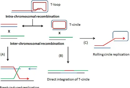
Figure 1.7 - Mechanisms of alternative lengthening of telomeres
Recombination in ALT cells may occur by intra-chromosomal recombination causing
drastic telomere shortening as seen by the formation of T-circles. Lengthening by
recombination can occur by three possible mechanisms: (A) invasion of chromosome
ends to induce break-induced replication, (B) integration of a T-circle into a chromosome
end, and (C) rolling-circle replication initiated by the 3’overhang invading proximal
1.7
Telomere length measurement
Several methods of measuring telomere lengths have been developed over the years, each
with their individual caveats (Aubert et al., 2012; Lin and Yan, 2005). The main two
groups of measurements are those that measure average telomere length (TRF and qPCR)
and those that measure telomere lengths of individual chromosomes (Q-FISH,
Flow-FISH, and STELA). All methods use binding of nucleic acid probes or primers specific
to telomeric repeats. Discrepancies between telomere length measurements of the
various methods arise from variability in the amount of extra-telomeric region (Figure
1.8) included in some techniques and less in others (Aubert et al., 2012). The two most
popular methods in the literature are telomere restriction fragment (TRF) analysis and
quantitative polymerase chain reaction (qPCR).
1.7.1
TRF analysis
TRF analysis by southern blotting was the first method to be described (Harley et al.,
1990) and represents the gold standard used to validate all other techniques to date
(Aubert et al., 2012; Kimura et al., 2010b; Lin and Yan, 2005). This technique uses a
pair of frequent cutting restriction enzymes (i.e. Hinf1 and Rsa1) to cut DNA into small
fragments, specifically excluding telomeric repeats as substrates. Frequent cutting
restriction enzymes are used to minimize the size and variation of extra-telomeric region
(Figure 1.8). The fragmented DNA is then separated by agarose gel electrophoresis,
transferred to a nitrocellulose or nylon gel and hybridized with a radioactive (32P) or
chemiluminescent (digoxienin) labeled probe complementary to telomere sequences.
Average telomere length of the resulting smear is estimated based on a DNA ladder and
normalization to a reference sample to correct for inter-experimental gel effect. The
length and intensity of the smear needs to be accounted for in the calculation of average
telomere length. Although this is a well established technique, variations between studies
have procured over time because the technique was not standardized in terms of
restriction enzymes used, DNA quantity and quality, and blot analysis. There are several
main pitfalls to this method. First, a relatively large amount of pure and unfragmented
DNA (0.5-10 µg) is required and small differences in telomere lengths are difficult to
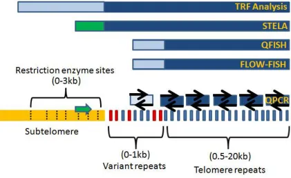
Figure 1.8 - Detection of extra-telomeric DNA with different methods of telomere
length measurement
In Figure 1.8 modified from (Aubert et al., 2012), extra-chromosomal region is described
as the proportion of DNA that is not considered pure telomere repeats (blue bars). In
gold is the subtelomere region that contains restriction enzyme sites (dashed lines) used
to digest DNA in TRF analysis. In red are the variant repeats that contain telomere-like
sequences. The green box for STELA is the primer used in the subtelomeric portion that
is later subtracted from the calculation of telomere repeats. The light blue boxes are
Furthermore, TRF analysis is time consuming (5-7 days), making it less ideal for large
clinical studies, although skilled technicians can process up to 130 samples per week
(Kimura et al., 2010b). Lastly, large variations in telomere length (up to 5%) can be seen
depending on the choice of restriction enzymes used and because of subtelomeric
polymorphisms. However, despite limitations of this technique, the errors of this method
are relatively small (CV=1.74%) (Aviv et al., 2011).
1.7.2
qFISH
Telomere length can also be measured by quantitative fluorescence in situ hybridization
(qFISH), which combines use of image cytometry and metaphase spreads through use of
peptide nucleic acid (PNA) probes (Aubert et al., 2012; Martens et al., 1998; Poon and
Lansdorp, 2001). These probes have a greater affinity than DNA oligonucleotides and
bind specifically to denatured telomere repeats. The fluorescent signal is acquired using
specific qFISH analysis software and telomere lengths are measured relative to standards
of known length. Karyotyping of metaphase spreads allows telomere lengths to be
matched with its corresponding chromosome arm. It’s the method of choice to determine
telomere length of specific chromosome ends. The analysis requires 15-20 metaphases
per sample to obtain reliable results due to the high variation in the technique. Some
advantages are that qFISH can detect chromosome fusion events and ends with virtually
no repeats (<0.5 kb). This technique is a good choice to study telomere biology in rare
cell types and can be used to ask more specific scientific questions because it requires so
few cells. However, some disadvantages are that qFISH cannot be used to measure
telomeres in cells that cannot divide such as senescent cells and highly aberrant cells.
Furthermore, the technique is labor intensive and takes a long time, and requires
specialized equipment.
1.7.3
Flow-FISH
Similar to qFISH, flow-FISH also uses PNA probes, however hybridization is done with
cells in suspension and median telomere length of individual cells is measured using flow
cytometry (Aubert et al., 2012; Martens et al., 2000). This technique can be used to
few cell surface markers that are retained after hybridization). A semi-automated
adaptation of this technique uses a 96-well plate and robotic microdispenser to reduce
tedious work and increase reproducibility. It is currently the method of choice for
measuring telomere length in specific subsets of blood cells. Some drawbacks of this
technique are that it requires a suspension of living cells, which is very fragile. This
technique is generally limited to use of fresh blood. Like qFISH this method requires
specialized equipment, is technically challenging and costly (Aubert et al., 2012; Martens
et al., 2000).
1.7.4
STELA
One of the newest techniques available is Single Telomere Length Analysis (STELA),
which uses PCR to amplify a specific chromosome arm of telomere (Aubert et al., 2012;
Baird et al., 2003). STELA takes advantage of the fact that all chromosome arms end in
a 3’ overhang, which is targeted as a template to anneal an oligonucleotide linker at the 5’
end of the telomere. A linker-specific primer and subtelomere specific primer is then
used to amplify a precise length of a single telomere tract. PCR amplicons are then
separated by gel electrophoresis, southern blotting and probed with specific subtelomere
sequences. Resulting banding patterns are intricate sets of discrete bands of individual
telomeres, which can be measured individually or pooled based on size according to a
DNA ladder of known lengths. One drawback of STELA is its limitation to well
characterized chromosome arms (XpYp, 2p, 11q, 12q and 17p) (Britt-Compton et al.,
2006) because not all chromosomes have adequate sequences for the design of unique
chromosome primers. Therefore, caution must be taken when analyzing results because a
subset of telomere measurements may not be reflective of the overall telomere status of
cells (Bendix et al., 2010). Although no specialized equipment is required, STELA is
technically challenging and requires intensive initial preparation and optimization (Baird
et al., 2003). Furthermore, STELA has an upper detection limit of 20kb, which limits its
use mostly to human samples, and it cannot be used on model organisms with long
telomeres such as Mus musculus strains of mice (Kipling and Cooke, 1990). Benefits of
changes in telomere length, and minimal starting material required (as few as 50 cells) to
produce reliable results (Aubert et al., 2012). Like qFISH, STELA is well fit to study
telomere lengths in rare cell populations, and it can also detect short telomere outliers,
those responsible for chromosome fusion and senescence.
1.7.5
qPCR
QPCR based telomere length measurement (Cawthon, 2002) has become widely used in
large clinical and epidemiological studies because of its rapid design, ease of use, and
low cost. This technique relies on specific amplification of telomeric repeats (T)
normalized to a single-copy gene (S) to produce an average telomere length (T/S ratio).
A sample calculation can be viewed in appendix B. A special set of telomere specific
primers were designed that contain mismatches every six base pairs, different in both the
forward and reverse primer. These mismatches are important to minimize primer dimer
formation and maximize primer-template hybridization. Telomere-specific primers bind
along telomeric repeats at each chromosome arm and amplify in fragments of at least 76
bp and up to ~500 bp long. SYBR green, a dye that fluoresces upon binding to double
stranded DNA, is used to monitor the amplification of DNA using Real-Time PCR. The
longer the average telomeres are in a given sample, the greater the fluorescent signal.
A setback of qPCR is in the variability between experiments. Proper controls are
required to minimize this variability. In every experiment, a set of inter-plate control
samples and a standard curve must be produced to offset the variability (Aubert et al.,
2012; Aviv et al., 2011). An important quality control of this technique is in the
preparation of DNA samples. It is critical that all DNA samples are of equivalent quality
so that there are no variations in amplification efficiencies. Another drawback of this
technique is that it is not standardized across different laboratories, and therefore results
between laboratories cannot be compared. A multiplex version of this technique has
recently been developed (Cawthon, 2009) that further reduces variation, which amplifies
telomere repeats and single-copy-gene in the same PCR reaction. Although qPCR
technique is prone to variation, with proper controls it can produce quick and reliable
results at a low cost, which is beneficial for most large-scale experimental designs.
both techniques, although TRF had slightly better inter-assay variation (Aviv et al.,
2011).
Choosing the proper technique depends solely on the experimental design and scientific
question being asked, as each technique has its advantages and disadvantages.
1.8
Telomere dynamics in the aging population
There are key differences between human and mouse telomere biology (Table 1.2). How
these differences relate in vivo toconsequences in telomere length shortening with age
and disease are currently not fully understood. However, genetically modified mice have
been useful to elucidate the role telomeres play in aging and chronic illness. Key
findings that link telomere biology to cardiovascular disease and cancer are further
discussed below.
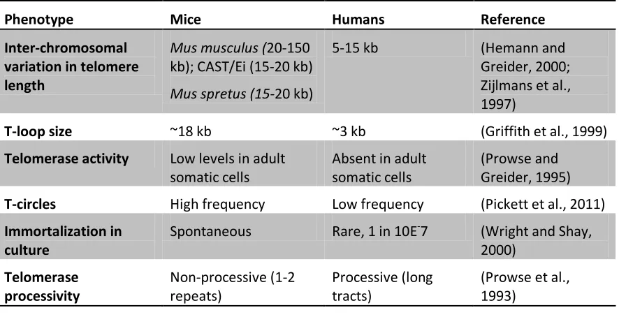
Table 1.2 - Differences between mouse and human telomere length dynamics
Phenotype Mice Humans Reference
Inter-chromosomal variation in telomere length
Mus musculus (20-150
kb); CAST/Ei (15-20 kb)
Mus spretus (15-20 kb)
5-15 kb (Hemann and
Greider, 2000; Zijlmans et al., 1997)
T-loop size ~18 kb ~3 kb (Griffith et al., 1999)
Telomerase activity Low levels in adult somatic cells
Absent in adult somatic cells
(Prowse and Greider, 1995)
T-circles High frequency Low frequency (Pickett et al., 2011)
Immortalization in culture
Spontaneous Rare, 1 in 10E-7 (Wright and Shay, 2000)
Telomerase processivity
Non-processive (1-2 repeats)
Processive (long tracts)
(Prowse et al., 1993)
1.8.1
Telomere length with age
Several studies have documented telomere length shortening with age in various tissue or
cell types both in vivo and in vitro. The concept of telomeres as a mitotic clock came
(HSCs) lose between 37 and 120 bp per population doubling (Harley et al., 1990; Vaziri
et al., 1994; Vaziri et al., 1993). However, unlike human primary fibroblasts, mouse
embryonic fibroblasts in culture do not show obvious telomere shortening with age,
likely attributed to telomerase activity present in cultured mouse cells (Sachsinger et al.,
2001). In telomerase knockout mice (TERT-/- or TERC-/-), telomeres do in fact shorten
in culture. Furthermore, by inducing expression of the catalytic subunit of telomerase
(TERT) in vitro to human differentiated cells, telomere length is maintained and
replicative senescence postponed (Bodnar et al., 1998; Ramirez et al., 2001; Yang et al.,
1999). One group studied telomere attrition in 14 different tissues of a large human
population ranging from 0 to 104 years of age. They reveal a significant reduction in
telomere length with age in all tissues studied except cerebral cortex and myocardium,
thought to be due to the quiescent nature of cells in those tissues (Takubo et al., 2002). In
mice, telomere length attrition has also been shown in several tissue types with age,
including spleen, skin, small intestine and brain (Coviello-McLaughlin and Prowse,
1997; Flores et al., 2008). Telomere loss per year in vivo for most tissues appears to be
within the range of telomere loss of one cell division in vitro. However, these results are
likely skewed by telomere length maintenance and heterogeneity in cell or tissue types
(Takubo et al., 2002). Several studies have calculated an average telomere shortening
rate in lymphocytes (53 bp/year), granulocytes (39 bp/year) (Hoffmann et al., 2009), and
pancreas (36 bp/year) (Ishii et al., 2006).
Telomere length attrition rates vary with age, showing a biphasic pattern in lymphocytes
(Frenck et al., 1998; Rufer et al., 1999). Most telomere shortening tends to happen in the
first few years of life, gradual shortening during adulthood, and accelerated shortening in
old age. From birth to age 4, an average telomere loss of 1kb is seen and thereafter
telomeres shorten by 100bp/year (Frenck et al., 1998). In specific subsets of blood cells,
Rufer et al. determined that 1.08kb of telomere sequence was lost in lymphocytes within
the first 1.5years, and 3.05kb loss in granulocytes in the first 0.5year of life (Rufer et al.,
1999). There are no gender differences in telomere length at birth in both human
leukocytes and mouse tissues (Coviello-McLaughlin and Prowse, 1997; Okuda et al.,
function of estrogen (Moller et al., 2009).
Telomere length dynamics with aging of hematopoietic stem cells (HSCs) is considered
to be one of the most reflective cell models of biologic aging. In fact, the most prominent
phenotypic consequence of defects in telomere length maintenance is bone marrow
failure as is seen in dyskerotosis congenital, Werner syndrome, and aplastic anemia.
Similarly in telomerase knockout mice, once telomeres are sufficiently short in late
generation mice, function of the hematopoietic system is compromised (Blasco, 2005b;
Herrera et al., 1999). Although telomerase is active in HSCs, telomeres shorten in culture
on average 1-2 kb per month (Engelhardt et al., 1997) as well as in vivo (Lee et al., 2003;
Vaziri et al., 1994). Seeing as telomere shortening outweighs telomerase activity, HSC
resources become exhausted with aging.
On the other hand, telomere lengthening with age is also observed. Telomere length in
spermatozoa is positively correlated with age such that offspring born with older fathers
tend to have longer telomeres at birth. A suggested mechanism of telomere lengthening
in germ cells is epigenetic modification and resistance to oxidative stress; however, it is
not yet fully understood (Kimura et al., 2008).
Longitudinal studies were necessary in order to understand the true impact of telomere
length with age. Using qPCR, two studies found a negative correlation of telomere
length with age over a 5 and 10 year period in 500 and 959 individuals, respectively.
However, in almost one third of individuals, no correlation with age has been found
(Aviv et al., 2009; Nordfjall et al., 2009). Furthermore, significant decline of leukocyte
telomeres seen early in life was confirmed in a study with baboons, whose telomeres
were measured longitudinally over the first few years of life (Baerlocher et al., 2007).
More recently, a study with birds demonstrated that telomere length at birth is correlated
with life expectancy (Heidinger et al., 2012). Similarly, a genetic variant of the RNA
component of telomerase (TERC) is also linked to leukocyte telomere length and
longevity (Soerensen et al., 2012). Therefore, longitudinal studies are important to
1.8.2
Telomere length and chronic illness
Age represents one of the greatest risk factors for chronic illness. Telomere length can
be considered a function of replicative history (age) and environmental stress, two
important parameters in the development of chronic illness (Oeseburg et al., 2010). A
clear link between telomere length and mortality in humans has been established
(Cawthon et al., 2003), but how telomere length correlates lifespan is still under debate.
Nonetheless, many studies provide evidence that telomere length is associated with
age-related illnesses, especially cardiovascular disease and cancer (Oeseburg et al., 2010).
1.8.2.1
Cardiovascular disease
Cardiovascular disease (CVD) is one of the main contributors to morbidity and mortality
in the aging population. Telomere length is a proposed biomarker of individuals at risk
for developing CVD. Several studies over the past decade have demonstrated the
relationship between short leukocyte telomere length and CVD (Table 1.3).
Table 1.3 - Short leukocyte telomere length and associated cardiovascular disorders
Cardiovascular disorder Participants Reference
Hypertension 96 cases; 98 controls
327 men from FHS
(Bhupatiraju et al., 2012)
(Demissie et al., 2006)
Left ventricular dysfunction 89 subjects (85 years old) (Collerton et al., 2007)
Atherosclerosis 10 cases; 20 controls
164 hypertensive men
1062 individuals from FHS
(Samani et al., 2001)
(Benetos et al., 2004)
(O'Donnell et al., 2008)
Myocardial infarction 203 cases; 180 controls
484 cases; 1058 controls
337 cases; 337 controls
(Brouilette et al., 2003)
(Brouilette et al., 2007)
(Zee et al., 2009)
Abdominal Aortic Aneurysms 190 cases; 183 controls (Atturu et al., 2010)
Congestive heart failure 620 cases; 182 controls (van der Harst et al., 2007)
Aortic dissection 72 cases; 72 controls (Yan et al., 2011)
Cardiovascular mortality 143 subjects (>60 years old)
780 CAD subjects
(Cawthon et al., 2003)
Two major risk factors to CVD, diabetes and hypertension, have both been linked to short
telomeres. One of the key studies linking short telomeres to insulin resistance and
hypertension was the Framingham Heart Study (Demissie et al., 2006), later confirmed in
a Chinese (Yang et al., 2009) and Indian population (Bhupatiraju et al., 2012).
Hypertensive individuals with short telomeres are more susceptible to develop
atherosclerosis (Yang et al., 2009), and display increased risk of cardiovascular mortality,
stroke or angina pectoris (Fyhrquist et al., 2011). Even mild activation of the
rennin-angiotensin system has been linked to shorter leukocyte telomeres (Demissie et al., 2006;
Vasan et al., 2008). Endothelial dysfunction, marked by cholesterol burden,
inflammatory markers, and increased oxidative stress, is considered an early marker of
atherogenesis (Asselbergs et al., 2005; van der Harst et al., 2006). Consistent with this,
senescent endothelial cells and short telomeres have been seen in atherosclerotic plaques
(Minamino et al., 2002; Ogami et al., 2004). The West of Scotland Primary Prevention
Study (WOSCOPS) determined that patients were more susceptible to develop coronary
heart disease if they were in the middle or lower tertile for telomere length. Patients with
shorter telomeres also benefited more from treatment with statins (Brouilette et al., 2007).
Furthermore, offspring of patients with coronary artery disease (CAD) have shorter
telomeres than those with healthy parents (Brouilette et al., 2008), and may explain part
of CAD heritability (Samani et al., 2007).
Several studies have linked short telomere length to glucose intolerance, insulin
resistance and type 2 diabetes (Adaikalakoteswari et al., 2005; Adaikalakoteswari et al.,
2007; Sampson et al., 2006). Even subclinical levels of insulin resistance were linked to
shortened telomeres in participants of the Framingham Heart Study (Demissie et al.,
2006). Patients with type 2 diabetes show signs of accelerated aging (accelerated
telomere attrition, increased oxidative damage, and decreased mitochondrial DNA)
compared to control subjects (Monickaraj et al., 2012).
Other cardiovascular risk factors are also associated with short telomeres. Arterial
Chinese stroke patients revealed that short telomere length is associated with ischemic
stroke and post-stroke death (Ding et al., 2012). Telomere length was determined in a
large prospective study of 19838 Danish participants followed for up to 19 years for
incident myocardial infarction, ischemic heart disease, and death. Short telomere length
was modestly correlated with increased risk of each outcome in the population (Weischer
et al., 2012).
Although an etiologic role for telomere length in CVD has yet to be established, a recent
study of 3271 Caucasians in the Cardiovascular Health Study aged ≥65 years old
revealed SNPs in OBFC1 (a gene linked to leukocyte telomere length) were associated
with CVD mortality in women (Burnett-Hartman et al., 2012a).
1.8.2.2
Cancer
A major factor influencing life expectancy is cancer, especially in long-lived species.
Short leukocyte telomere length is a risk for the development of many types of cancer,
including head and neck, breast, bladder, prostate, lung, kidney, ovarian (Blackburn,
2011; Martinez-Delgado et al., 2012; Wu et al., 2003).
As stated in section 1.4, dysfunction of the p53 pathway causes cells to continue to divide
until a state of crisis is reached. A series of breakage fusion bridge cycles between
chromosomes with dysfunctional telomeres causes genomic rearrangements that can
induce upregulation of oncogenes and promote tumorigenesis (Deng et al., 2008; Hackett
and Greider, 2002; Rudolph et al., 2001). Mice with enhanced p53 responses show
increased resistance to cancer, however, have shorter lifespan and display age-related
phenotypes sooner (Donehower, 2002; Matheu et al., 2007). In contrast, Li-Fraumeni
syndrome (LFS) is characterized by a germline mutation in TP53, predisposing LFS
patients to cancer. With each consecutive generation there is earlier onset of cancer
caused by progressive telomere shortening in the germ cells. Therefore, telomere length
has been used as a biological marker to monitor LFS patients (Tabori et al., 2007).
Tumor cells also utilize one of two mechanisms to maintain their telomeres, providing
Bacchetti, 1997) or a combination of both (Venturini et al., 2012). Similar to enhancing
p53, suppression of telomerase can reduce risk of cancer, but it also induces age-related
phenotypes. However, reactivation of telomerase after 4 weeks reverses the degenerative
phenotypes seen in telomerase-deficient mice (Bernardes de Jesus and Blasco, 2011;
Jaskelioff et al., 2011). Proliferative capacity of post-senescent cells can also be
extended by ectopic expression of human TERT (Counter et al., 1998). However,
continued expression of hTERT in culture promotes genomic instability, perhaps due to
accumulation of replication errors from an extended lifespan in culture (Roth et al., 2005;
Schreurs et al., 2005).
The link between cancer and lifespan is a fine balance between protection against
tumorigenesis and degeneration due to age. By combining the protective effects of
enhancing p53 activity and increasing telomerase expression, the lifespan of mice can be
extended and risk of tumorigenesis reduced (Donate and Blasco, 2011; Tomas-Loba et
al., 2008).
1.8.3
Telomere length (TL) variation in the population
Telomere lengths are highly variable between and within individuals. Within an
individual, telomeres vary in length between tissues, cells, and chromosomes (Lansdorp
et al., 1996; Oeseburg et al., 2010; Zijlmans et al., 1997). However, variability between
individuals is greater than that within each individual, which makes cross-sectional
studies noisy, requiring large cohorts (Aviv and Levy, 2011; Kimura et al., 2010a;
Takubo et al., 2002). At birth, telomeres vary in the population by 4-6 kb in blood
leukocytes (Akkad et al., 2006; Frenck et al., 1998; Okuda et al., 2002), which is
maintained throughout the human lifespan (Alter et al., 2007; Rufer et al., 1999).
Telomere length variation in the population is a result of inherited differences, and
differential rates of telomere shortening between individuals (Aviv, 2012; Chen et al.,
2011). Both genetic and environmental or lifestyle factors contribute to differences in the