Paediatrica Indonesiana
Original Article
p-ISSN 0030-9311; e-ISSN 2338-476X; Vol.57, No.3(2017). p. 138-43; doi: http://dx.doi.org/10.14238/pi57.3.2017.138-43
CD4
+
T-cell, CD8
+
T-cell, CD4
+
/CD8
+
ratio,
and apoptosis as a response to induction phase
chemotherapy in pediatric acute lymphoblastic
leukemia
May Fanny Tanzilia
1, Andi Cahyadi
2, Yetti Hernaningsih
3, Endang Retnowati
3,
I Dewa Gede Ugrasena
2Abstract
Background Acute lymphoblastic leukemia (ALL) is a neoplastic disease resulting from somatic mutation in the lymphoid progenitor cells, often occuring in children aged 2-5 years, predominantly in males. Results from the induction phase of chemtherapy are used to measure success, but the failure remission rate is still high. Increased apoptosis of cancer cells, as induced by CD4+
and CD8+ T-cells, is an indicator of prognosis and response to
chemotherapy.
Objective To assess for correlations between CD4+, CD8+, or
CD4+/CD8+ ratio to the chemotherapy induction phase response
(i.e., apoptosis) in pediatric ALL patients.
Methods This observational analytical cohort study was done in 25 pediatric ALL patients. Whole blood (3 mL) with EDTA anticoagulant were used to measure absolute counts of CD4+,
CD8+, and CD4+/CD8+ ratio. Peripheral blood mononuclear cells
(PBMC) were examined for apoptosis. The principle of CD4+,
CD8+ examination was bond between antigens on the surface of
the leukocyte in the blood with fluorochrome labeled antibodies in the reagents, while the principle of apoptosis examination was FITC Annexin V will bonds with phosphatidylserine that moves out of the cell when the cell undergoes apoptosis, then intercalation with propidium iodide (PI). All examination were detected by flow cytometry BD FACSCalibur.
Results Subjects were 25 newly-diagnosed, pediatric ALL patients (64% males and 36% females). Most subjects were 3 years of age (20%). Numbers of CD4+ and CD8+ cells, as well as CD4+/
CD8+ ratio were significantly decreased after chemotherapy.
However, apoptosis was not significantly different before and after chemotherapy (P=0.689), There were significant negative
From the Clinical Pathology Specialization Program1, Department of Child Health2, and Department of Clinical Pathology3, Airlangga University Medical School/Dr. Soetomo Hospital, Surabaya, East Java, Indonesia.
Reprint requests to: May Fanny Tanzilia, Sukolilo Dian Regency, Makmur 3/8, Surabaya 60111, East Java, Indonesia. Email: dr.mayfannytanzilia@ gmail.com.
correlations between apoptosis and CD4+ (P=0.002; rs=-0.584),
and CD8+ (rs=-0.556; P=0.004), before chemotherapy. In
addition, CD4+-delta and apoptosis-delta also had a significant
positive correlation (rs=0.478; P=0.016). However, no correlation was found between the CD4+/CD8+ ratio and apoptosis, before
or after chemotherapy.
Conclusion There are significantly lower mean CD4+, CD8+,
and CD4+/CD8+ ratio after chemotherapy than before. Also,
there are significant correlations between CD4+-delta and
apoptosis-delta, as well as between apoptosis and CD4+, CD8+,
and CD4+/CD8+ ratio, before chemotherapy. The CD4+, CD8+,
and CD4+/CD8+ ratio can be used to predict apoptosis before
chemotherapy. In addition, CD4+-delta can be used to predict
apoptosis-delta as a response to induction phase chemotherapy in pediatric ALL. [Paediatr Indones. 2017;57:138-43 doi: http:// dx.doi.org/10.14238/pi57.3.2017.138-43 ].
A
cute lymphoblastic leukemia (ALL) is a neoplastic disease resulting from somatic mutation in the lymphoid progenitor cells in the bone marrow, often occurring in children aged 2-5 years, and predominantly in males. Diagnosis of ALL is based on finding ≥ 25% lymphoblasts of blood smear or bone marrow aspiration (BMA) evaluation. Outcome criteria was made as follows, < 5% lymphoblasts was belonging to remission, and > 5% was belonging to non remission, including partial remission.1-5Failure of apoptosis and the immune response causes uncontrolled growth of cancer cells. The progression of ALL has been correlated with quantitative and qualitative (function) host immune responses. Cluster differentiation 8+ (CD8+) T-cell
are an effector cell resulting an apoptosis tumor cells by cytotoxic or cytolytic effects. Anti-tumor mechanisms of CD4+ T-cells are not fully understood.
The CD4+ T-cells have no cytotoxic or cytolytic
effects. Many cytokines produced by CD4+ T-cells are
needed for the development of CD8+ effector cells.
Tumor necrosis factor (TNF) and interferon-gamma (IFN-gamma) secreted by activated CD4+ T cells
will induce expression of major histocompatibility complex I (MHC I) and induce CD8+ T-cell
activity to lyse tumor cells.6-10 The induction phase
of chemotherapy is usually used as a measure of successful chemotherapy, but remission failure rates are still high.10 Increased apoptosis of cancer cells
induced by CD4+ or CD8+ T-cells can be used as
one indicator of prognosis and response to induction phase chemotherapy in pediatric ALL patients.9 The
aim of this study was to assess for correlations between apoptosis and CD4+, CD8+, and CD4+/CD8+ ratio,
before and after the induction phase of chemotherapy in pediatric ALL patients.
Methods
This observational, analytical, cohort study included 25 new cases of pediatric ALL patients who were diagnosed based on bone marrow aspiration at the Department of Pediatrics, Airlangga University Medical School/Dr. Soetomo Hospital, Surabaya, from July to December 2016. The inclusion criteria were new cases of pediatric ALL aged 1 month to 16 years
who had never received chemotherapy, and planned to undergo induction phase chemotherapy regularly for 6 weeks. The exclusion criteria were ALL patients with a history of HIV/AIDS or had infections as indicated by fever and other signs. Healthy patients control were obtained from healthy volunteers aged 1 month to 16 years who had investigated no history of certain diseases and seem good condition during the examination was done. This study was approved by the Ethics Committee of our institution and written informed consent was obtained from all patients’ parents.
Clinical characteristics including age, sex, hemoglobin (Hb) level, platelet count, and leukocyte count were obtained at diagnosis (before chemotherapy) and after induction phase chemotherapy. Whole blood (3 mL) with EDTA anticoagulant were used to measure absolute counts of CD4+, CD8+, and CD4+/CD8+
ratio. Reagents used for CD4+, CD8+ examination
was BD Multitest CD3FITC/CD8PE/CD45PerCP/ CD4APC. The principle of CD4+, CD8+examination
was to detect bonds between antigens on the surface of the leukocyte present in the blood with fluorochrome labeled antibodies present in the reagents. Peripheral blood mononuclear cells (PBMC) were examined for apoptosis. The FITC Annexin V and propidium iodide (PI) were used for apoptosis examination. The principle of apoptosis examination was FITC Annexin V will bonds with phosphatidylserine that moves out of the cell when the cell undergoes apoptosis, then intercalation with propidium iodide (PI). The FITC Annexin V can identify early apoptosis, late apoptosis and necrosis based on nucleus changes. Propidium Iodide (PI) was used to distinguish late apoptosis and necrosis with early apoptosis. All examination were detected by flow cytometry BD FACSCalibur performed before and after induction phase chemotherapy, at the Department of Clinical Pathology, Airlangga University Medical School/ Dr. Soetomo Hospital, Surabaya.
Comparison of Hb levels before and after induction phase chemotherapy was evaluated by paired T-test, while leukocyte counts, platelet counts, CD4+, CD8+ cells, CD4+/CD8+ ratio, and apoptosis
before and after induction phase chemotherapy were evaluated by Wilcoxon Signed Rank test. Correlations of absolute count of CD4+, CD8+ cells, CD4+/CD8+
correlation test. CD4+ -delta was defined as difference
between number of CD4+ before and after induction
phase chemotherapy, as well as CD8+-delta, CD4+/
CD8+-delta, and apoptosis delta. Results with P
values ≤ 0.05 were considered to be statistically significant.
Results
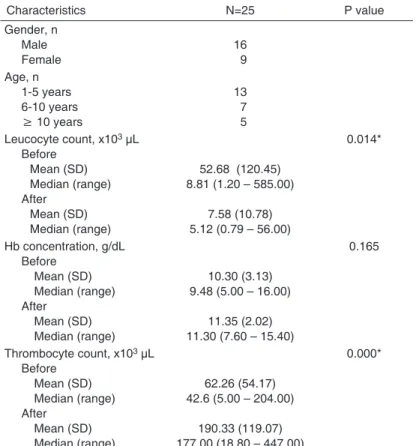
During the study period, 41 new cases of pediatric ALL patients fulfilled the inclusion criteria, but 16 patients dropped out, of whom 11 patients died and 5 patients did not complete the induction phase of chemotherapy. Hence, our cohort study of 25 new cases of pediatric ALL patients consisted of 16 males (64%) and 9 females (36%), with a 1.78:1 ratio (Table 1). Many of our subjects were 3 years of age (20%). Leucocyte count was significantly lower
(P=0.014) but platelet count was signigficantly higher (P=0.000), between before and after induction phase of chemotherapy. Mean numbers of CD4+ and CD8+
cells, as well as CD4+/CD8+ ratio were significantly
decreased after induction phase chemotherapy. Mean numbers of CD4+ before induction phase
chemotherapy was 3,060.24 (SD 4660.03), after induction phase chemotherapy was 887.64 (1531.33). Mean numbers of CD8+ before induction phase chemotherapy was 3,084.76 (SD 4535.51), after induction phase chemotherapy was 1,647.28 (SD 3644.99). Mean numbers of CD4+/CD8+ ratio
before induction phase chemotherapy was 1.12 (SD 0.67), after induction phase chemotherapy was 0.65 (SD 0.61). However, apoptosis did not significantly decrease after chemotherapy (P=0.689), before induction phase chemotherapy was 18.19 (SD 19.82) and after induction phase chemotherapy was 14.09 (SD 10.85) (Table 2).
Table 1. Clinical characteristics of study subjects before and after induction phase chemotherapy
Characteristics N=25 P value Gender, n Male Female 16 9 Age, n 1-5 years 6-10 years ≥ 10 years 13 7 5 Leucocyte count, x103 μL Before Mean (SD) Median (range) After Mean (SD) Median (range) 52.68 (120.45) 8.81 (1.20 – 585.00) 7.58 (10.78) 5.12 (0.79 – 56.00) 0.014* Hb concentration, g/dL Before Mean (SD) Median (range) After Mean (SD) Median (range) 10.30 (3.13) 9.48 (5.00 – 16.00) 11.35 (2.02) 11.30 (7.60 – 15.40) 0.165 Thrombocyte count, x103 μL Before Mean (SD) Median (range) After Mean (SD) Median (range) 62.26 (54.17) 42.6 (5.00 – 204.00) 190.33 (119.07) 177.00 (18.80 – 447.00) 0.000*
*significant if P ≤ 0.05, Before=before induction phase chemotherapy, After=after induction
A comparison of apoptosis was done in ALL patients and healthy control subjects. We found no significant differences in apoptosis between the two groups either before or after chemotherapy (Table 3).
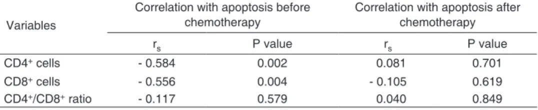
We also assessed for correlations between apoptosis, before and after chemotherapy, and CD4+, CD8+ absolute counts, and CD4+/CD8+
ratio. There were significant, negative correlations between apoptosis and CD4+, as well as apoptosis and
CD8+, before chemotherapy. However, there was no
significant correlation between CD4+/CD8+ ratio and
apoptosis, before or after chemotherapy (Table 4). There was a significant correlation between CD4+-delta and apoptosis-delta after induction phase
chemotherapy. However, we observed no significant correlation between apoptosis-delta and CD8+-delta
or CD4+/CD8+-delta (Table 5).
Discussion
Acute lymphoblastic leukemia is a hematological malignancy that often occurs in children, and comprises 25–30% of all pediatric malignancies. The highest incidence is in 2–5-year-olds, and predominantly in boys. Our cohort study of 25 new cases of pediatric ALL patients consisted of 16 (64%) males and 9 (36%) females, with 1.78 : 1 ratio. Many subjects were 3 years of age (20%) (Table 1).
There were significant differences in leukocyte (P=0.014) and thrombocyte (P=0.000) counts, before and after induction phase chemotherapy. Uncontrolled lymphocyte proliferation and defective apoptosis in ALL patients caused leukocytosis dominated by lymphoblasts. Infiltration of hematopoetic cells by leukemic cells accumulated in the bone marrow Table 2. Comparison between CD4+, CD8+ cells, CD4+/CD8+ ratio, and apoptosis, before and after
induction phase chemotherapy in pediatric ALL patients
Variables Time Mean (SD) Median (range) P value
CD4+, cells Before After 3,060.24 (4660.03)887.64 (1531.33) 1751.00 (210.0 – 23,016.0199.00 (4.0 – 5,966.0) 0.000* CD8+, cells Before After 3,084.76 (4535.51)1,647.28 (3644.99) 1820.00 (341.0 – 18,541.0)423.00 (52.0 – 17,859.0) 0.004* CD4+/CD8+ ratio Before After 1.12 (0.67)0.65 (0.61) 1.03 (0.33 – 2.91)0.44 (0.06 – 2.31) 0.004* Apoptosis Before After 18.19 (19.82)14.09 (10.85) 10.83 (0.26 – 80.58)10.34 (2.86 – 49.17) 0.689
*significant if P ≤ 0.05, Before=before induction phase chemotherapy, After=after induction phase chemotherapy
Table 3. Comparison of apoptosis between pediatric ALL patients and healthy children before and after induction phase chemotherapy
Variables Apoptosis P value
Mean (SD) Median (range)
ALL patients (before chemotherapy) 18.19 (19.82) 10.83 (0.26-80.58) 0.683 Healthy control patients 16.21 (3.52) 17.73 (12.19-18.71)
ALL patients (after chemotherapy) 14.09 (10.85) 10.34 (2.86-49.17) 0.316 Healthy control patients 16.21 (3.52) 17.73 (12.19-18.71)
Table 4. Correlation between apoptosis and CD4+, CD8+ cells, and CD4+/CD8+ ratio before and after induction phase chemotherapy
Variables
Correlation with apoptosis before
chemotherapy Correlation with apoptosis after chemotherapy
rs P value rs P value
CD4+ cells - 0.584 0.002 0.081 0.701
CD8+ cells - 0.556 0.004 - 0.105 0.619
causes anemia and thrombocytopenia.1,13 After
chemotherapy, leukocyte count decreased, while Hb level and thrombocyte count increased. Evaluation of bone marrow aspiration after induction phase chemotherapy showed that all the patients were in remission, as determined by leukemic cell clearance from the bone marrow, mainly in the first 2 weeks after induction phase chemotherapy.13
Mean CD4+, CD8+ cells, and CD4+/CD8+
ratio were significantly decreased after induction phase chemotherapy (Table 2). Verma et al. reported a significant decrease in B cells, T cells, and NK cells approximately 2 weeks after chemotherapy.14
Decreased CD4+/CD8+ ratio after chemotherapy in
this study (P=0.004) was due to the larger decrease in CD4+ cells than that in CD8+ cells. Recovery
of CD4+ cells after chemotherapy was slower than
recovery of CD8+ cells. Chemotherapy decreases
the CD4+ and CD8+ cells by increasing regulatory
T cells (T reg), which supress the immune response by downregulating IL-2, and upregulating IL-10 and TGF-beta. Although IL-10 and TGF-beta are strong, immunosupressive factors, IL-2 is an important immune regulating factor. It is produced by T helper cells, which increase T cell proliferation, NK cell activity, and B cell antibody secretion. Past studies have shown that IL-2 concentration in ALL patients is low.15,16
After chemotherapy, apoptosis was not significantly decreased (P=0.689). In contrast, Laane et al. found an increase in apoptosis after chemotherapy.17 Firstly, our results may have been
caused by: 1) sampling time outside the window of maximal effect on apoptosis time (24 - 72 hours). Liu et al. reported a significant increase in apoptosis of lymphoblasts > 24 hours after induction phase chemotherapy in pediatric ALL patients, rather than in the early hours after chemotherapy.18 Secondly,
chemotherapy can cause necrosis, so lymphoblasts may not have been detected as apoptotic bodies.19
Third, the subjects may have had chemotherapy drug resistance, although 100% of patients entered remission,20 and forth, anti-apoptotic proteins levels
(e.g., BCL-2, BCL-XL, etc.) in the patients were higher than pro-apoptotic proteins levels (e.g., BAX, BOK, BCL-Xs, BID, BAD, or Noxa).18 Lymphoblast cells in
ALL patients are more fragile than in healthy children, thus, apoptosis in ALL patients was not significantly
higher than apoptosis in the healthy control group (Table 3).
There was a negative, significant correlation between apoptosis and CD4+ and CD8+ cells before
chemotherapy (Table 4). This finding indicates that greater numbers of CD4+ and CD8+ cells result in
decreased apoptosis. We noted that not only the number of CD4+ and CD8+ cells, but their function,
was also important in immune response. The number and function of CD4+ and CD8+ cells before
chemotherapy were correlated with subjects’ response to chemotherapy and improved survival.10
Apoptosis had no significant correlation with CD4+/CD8+ ratio, either before or after
chemotherapy. Similarly, Dewyer et al. suggested that CD4+/CD8+ ratio did not correlate with
tumor response in induction phase chemotherapy.21
No correlation between CD4+ or CD8+ cells and
apoptosis after induction phase chemotherapy, may have been due to decreases of CD4+, CD8+ cells,
and apoptosis after chemotherapy.
There was a significant correlation between CD4+-delta (number of CD4+ cells before
chemotherapy minus the number of CD4+ cells after
chemotherapy) and apoptosis-delta (Table 5). We found that decreased CD4+ cells lead to decreased
apoptosis. The CD4+ cells had no cytotoxic or cytolytic
effect on tumor cells, but many cytokines produced by CD4+ cells are needed for the development of CD8+
into effector cells.6-10
The limitations of this study included the lack of an extended time to recognize the possibility of future relapse or resistance to chemotherapy drugs, apoptosis sample preparation was not accompanied by a specific marker for lymphocytes, so a series of monocytes may have been included, and the sampling time among patients after chemotherapy varied, potentially effecting the decreased apoptosis.
Comparing the values before and after induction phase of chemotherapy we conclude that: 1) CD4+
and CD8+ count cells not significantly higher, 2) there
is no difference in apoptosis, 3) there was a negative correlation between apoptosis and CD4+ and CD8+
cells before induction phase chemotherapy in pediatric ALL patients, but no correlation after induction phase chemotherapy, 4) there is no correlation between CD4+/CD8+ ratio and apoptosis, 5) there
apoptosis-delta, after induction phase chemotherapy. CD4+ and CD8+ cells, and CD4+/CD8+ ratio can
be used to predict apoptosis before chemotherapy, while CD4+-delta may be useful to predict apoptosis
after induction phase response to chemotherapy in ALL patients.
Suggestions for future research also include determining the best time of sampling after chemotherapy (24 hours, 36 hours, 48 hours, or 72 hours) to obtain results with significantly increased apoptosis, compared to what our study showed, and to assess the profile of the percentage of T reg and expression of cytokines in ALL patients before and after chemotherapy.
Conflict of Interest
None declared.References
1. Pui CH. Acute lymphoblastic leukemia. In: Williams Hematology. Kenneth Kaushansky, Marshall A Lichtman, Ernest Beutler, et al., editors. 8th ed. New York The
McGraw-Hill Companies, Inc.; 2010. p. 1409-30.
2. Wintrobe, MW. Acute lymphoblastic leukemia in children. In: Wintrobe’s Clinical Hematology. John P Greer, Daniel A Arber, Bertil Glader, et al., editors. 10th ed. Philadelphia: Lippincott Williams and Wilkins; 2009. p. 2209-60. 3. Stankovic T, Marston E. Molecular mechanisms involved in
chemoresistance in paediatric acute lymphoblastic leukaemia. Srp Arh Celok Lek. 2008;136:187-92.
4. Liang DC, Pui CH. Childhood acute lymphoblatic leukaemia. In: Postgraduate Hematology. A. Victor Hoffbrand, Daniel Catovsky, Edward GD Tuddenham, editors. 5th ed. Ljubljana: Blackwell Publishing; 2008; p. 542-7.
5. Permono B. Leukemia akut. Buku ajar hematologi onkologi anak. Jakarta: Badan Penerbit IDAI; 2010. p.236-47. 6. Simanjorang C, Kodim N, Tehuteru E. Perbedaan kesintasan 5
tahun pasien leukemia limfoblastik lkut dan mieloblastik akut pada anak di rumah sakit kanker “Dharmais”. Indonesian J Cancer. 2013;7:15-21.
7. Oluboyo AO, Meludu SC, Onyenekwe CC, Oluboyo BO, Chianakwanam GU, Emegakor C. Assessment of immune stability in breast cancer subjects. Eur Sci J. 2014;10:224-8.
8. Kresno SB. Ilmu dasar onkologi. 2nd ed. Jakarta: Fakultas
Kedokteran Universitas Indonesia; 2011. p. 156-283. 9. Wong RS. Apoptosis in cancer: from pathogenesis to
treatment. J Exp Clin Cancer Res. 2011;30:87.
10. Elzubeir AM, Angi AM, Rahoum HM, Osama A. Prognostic significance of immune function parameters (CD4, CD8 and CD4/CD8 ratio) in Sudanese patients with chronic lymphocytic leukemia. Int J Multidisciplinary Curr Res. 2016;4:650-3.
11. Technical Data Sheet BD Multitest CD3 FITC/ CD8 PE/ CD45 PerCP/ CD4 APC Reagent, BD Pharmingen 2012. 12. Technical Data Sheet FITC Annexin V Apoptosis Detection
Kit I, BD Pharmingen 2008.
13. Nguyen VT, Melville A, Nath S, Story C, Howell S, Sutton R, et al. Bone marrow recovery by morphometry during induction chemotherapy for acute lymphoblastic leukemia in children. PLoS One. 2015;10:1-10.
14. Verma R, Foster RE, Horgan K, Mounsey K, Nixon H, Smalle N, et al. Lymphocyte depletion and repopulation after chemotherapy for primary breast cancer. Breast Cancer Res. 2016;18:10.
15. Salem MP, El-Shanshory MR, El-Desouki NI, Abdou SH, Attia MA, Zidan AA, et al. Children with acute lymphoblastic leukemia show high numbers of CD4+ and CD8+ T-cells
which are reduced by conventional chemotherapy. Clin Cancer Investig J. 2015;4:603-9.
16. Wu CP, Qing X, Wu CY, Zhu H, Zhou HY. Immunophenotype and increased presence of CD4(+)CD25(+) regulatory T cells in patients with acute lymphoblastic leukemia. Oncol Lett. 2012;3:421-4.
17. Laane E, Panaretakis T, Pokrovskaja K, Buentke E, Corcoran M, Soderjall S, et al. Dexamethasone–induced apoptosis in acute lymphoblastic leukemia involves differential regulation of Bcl-2 family members. Haematologica. 2007;92:1460-9. 18. Liu T, Raetz E, Moos PJ, Perkins SL, Bruggers CS, Smith F,
et al. Diversity of the apoptotic response to chemotherapy in childhood leukemia. Leukemia. 2002;16:223-32.
19. Blagosklonny MV. Cell death beyond apoptosis. Leukemia. 2000;14:1502-8.
20. Niknafs B. Induction of apoptosis and non-apoptosis in human breast cancer cell line (MCF-7) by cisplatin and caffeine. Iran Biomed J. 2011;15;130-3.
21. Dewyer NA, Wolf GT, Light E, Worden F, Urba S, Eisbruch A,
et al. Circulating CD4-positive lymphocyte levels as predictor of response to induction chemotherapy in patients with advanced laryngeal cancer. Head Neck. 2014;36:9-14.