The Study of
MTHFR
Gene C677T Polymorphism in Human
Embryonic Blastomeres by Fluorescence PCR Method
Introduction
Pre-implantation genetic diagnosis (PGD) is a new technique for obtaining the genetic information of the embryo before transferring it to the uterus. In fact, PGD is a novel technology that enables individuals with certain genetic diseases to avoid the transmission of undesirable genes to their offspring (1). Many people who apply for PGD do not suffer from infertility. They ask for PGD because this serves to prevent a specific genetic disorder from passing to their children or to eradicate it from their families (2). In 1990, the first successful clinical application of the PGD method in human was reported in a study by Handyside et al (3). They used PCR method to amplify a specific repeat sequence on the Y chromosome for sex selection in the embryos. In addition, in 1992, the first live birth of PGD was reported for cystic fibrosis (4). Today, after more than 2 decades of the introduction of this technique, PGD is widely used and medical need for PGD services is significant.
PGD starts with IVF process that involves controlled ovarian stimulation, oocyte retrieval, fertilization in the laboratory, and embryo biopsy. In this method, the specialist removes one or 2 cells from the embryo and after genetic analysis, normal embryos are transferred to
the mother’s uterus (5,6). Today, most of the PGD centers perform cleavage stage biopsy on the third day after fertilization to obtain genetic material, while the embryo has between 6-8 cells. At this stage, the blastomeres are totipotent, and it seems that biopsy does not affect their metabolism and survival (7). PGD experiments have mainly focused on 2 methodologies: fluorescent in situ hybridization (FISH) and polymerase chain reaction (PCR) (8).
The normal coagulation processes in the mother and the placenta are critical for the maintenance of pregnancy and are associated with abortion (9). Recurrent miscarriage is a multifactorial disease in which genetic factors, especially thrombophilic factors, can be considered as one of the causes of the disease (10). Polymorphisms in the genes encoding enzymes involved in important metabolic pathways, such as MTHFR, are considered as one of the key factors involved in the formation of thrombophilia. It is important to highlight that MTHFR enzyme plays an important role in the metabolism of folate, methionine and homocysteine (11) and deficiency in folate metabolism might have adverse effects on many essential processes and even affects implantation and embryogenesis (12,13). The MTHFR enzyme converts 5, 10-methylenetetrahydrofolate Abstract
Objectives:The aim of the present study was to evaluate methylene tetrahydrofolate reductase (MTHFR) gene C677T polymorphism in human pre-implantation embryos.
Methods: Fifty embryos after biopsy at cleavage stage (which were inappropriate for assisted reproductive techniques) were analyzed in order to study MTHFR C677T polymorphism and sex determination. After cell lysis, in the first step, whole genome amplification (WGA) was performed by primer extension pre-amplification (PEP) PCR method. In the next step, fluorescence PCR with labeled primers was used to examine this polymorphism in the blastomeres. Finally, the Hinfl enzyme was employed to cut the fragments and the capillary electrophoresis was used to determine the size of PCR products.
Results: only 37 blastomeres among 50 samples were successfully amplified; 23 male and 14 female embryos were observed. The frequencies of CC, CT and TT genotypes in embryos were 67.56%, 24.32%, 8.10%, respectively. Additionally, the frequencies of 677C and 677T alleles were 80% and 20%, respectively.
Conclusions: In this study, the frequency of MTHFR c.677C˃T genotype was evaluated in human embryonic blastomeres before implantation. Therefore, this polymorphism may provide a new and valuable biomarker for evaluating the embryo usefulness and improving the In vitro fertilisation (IVF) process by choosing the best embryo. Our findings revealed that the detection rate of C677T polymorphism was 74% in the samples.
Keywords: PGD, PEP-PCR, MTHFR gene, C677T polymorphism, Recurrent miscarriage
Saeedeh Saeedi1, Mohammad Reza Mashayekhi1*, Masoud Maleki1
Open Access Original Article
Crescent Journal of Medical and Biological Sciences
Received 6 April 2017, Accepted 1 September 2017, Available online 30 September 2017
eISSN 2148-9696
to 5-methyltetrahydrofolate, which is the predominant form of folate in the blood stream. Then, homocysteine is converted to methionine by obtaining a methyl group and methionine synthase catalyzes the transfer of methyl group. The decrease in the activity of the MTHFR enzyme leads to the reduction of the substrate for methionine synthase, which leads to a stop in the formation of methionine with the increase of serum homocysteine levels (14).
The gene encoding the MTHFR enzyme is located on the distal end of the short arm of chromosome 1 (1p36.22) and has 11 exons and 10 introns. The length of the gene is about 2037 bp. The protein product of this gene has 656 amino acids with a molecular weight of 74.7 kDa (15).
In C677T polymorphism, substitution of cytosine with thymine in nucleotide 677 at exon 4 of this gene leads to the replacement of alanine with valine at codon 222 of the protein sequence. This point mutation leads to the formation of an unstable and thermolabile enzyme, and the level of homocysteine is increased due to the decreased enzymatic activity of the mutant gene. The enzyme activity is reduced 70% and 30% in homozygous and heterozygous individuals, respectively, compared to normal individuals (16).
The finding revealed that MTHFR is expressed in both human oocytes and pre-implantation embryos (17). With regard to the fact that MTHFR genotype can be easily determined in the embryo, C677T polymorphism can be selected as a new and valuable biomarker for evaluating the embryo ability and thus, improve In vitro fertilisation (IVF)process by selecting the best embryo and subsequently creating a viable pregnancy (13). Therefore, the study of the polymorphism of this gene in the IVF– derived embryos before implantation is recommended due to the importance of this gene in the embryo development and success of the implantation.
In this regard, MTHFR genotyping in pre-implantation embryos can increase the rate of implantation and can reduce abortion and eventually, reduces the need for prenatal diagnosis.
Objectives
Since folate is important in human fertility processes and insufficient absorption of folic acid affects reproductive functions and has harmful effects on embryonic implantation and preservation of pregnancy, our aim was
to investigate MTHFR gene C677T polymorphism in pre-implantation embryos.
Materials and Methods
In this study, 50 embryos (which were unusable for fertility treatment) were received from Infertility Treatment Center of Academic Jihad of Tabriz. In this center, intra-cytoplasmic sperm injection (ICSI) was used to reduce possible contamination with surplus sperm after oocyte retrieval. The embryo biopsy was performed in the morning of the third day after fertilization (cleavage stage biopsy). The biopsy was performed in Ca++ and Mg++ free medium and zona pellucida was removed using laser (OLYMPOS 1X71). Blastomeres were washed three times in buffer containing phosphate buffered saline (PBS) solution with 0.1% polyvinyl alcohol (PVA). Subsequently, each blastomere was transferred into 0.2 mL PCR tube containing 5 μL of alkaline lysis buffer (200 mM KOH, 50 mM DTT [Dithiothreitol]). The microtubes containing single cells were incubated at 65°C for 10 minutes and then, were cooled at 4°C. Finally, 5 μL of neutralization buffer (900 mMTris-HCl pH 8.3, 300 mMKCl and 200 mMHCl) was added to each tube. The first round PCR reactions were performed using PEP primer in a final volume of 50 μL (Table 1). In this step, 15-mer oligonucleotide primers were used for whole genome amplification (WGA) in blastomeres. These primers bind non-specifically to different regions of the genome and their sequence was as follows: 5’-NNN NNN NNNNNN NNN-3’.
PEP-PCR was set up as follows: Initial denaturation at 95°C for 5 minutes (1 cycle); denaturation at 92°C for 1 minutes, annealing at 40°C for 2 min, extension at 55°C for 4 minutes (45cycles); final extension at 72°C for 5 minutes (1 cycle).
After DNA amplification of the blastomere samples in the first stage of the PCR reaction, in the next round, PCR reactions were performed by fluorescence PCR with specific primers for SRY gene (for sex determination) and MTHFR (for evaluation of C677T polymorphism) in a final volume of 25 μL. The conditions of the PCR reaction are shown in Table 2.
Fluorescence PCR with specific primers for SRY gene and MTHFR was set up as follows: Initial denaturation at 95°C for 10 minutes (1 cycle); denaturation at 95°C for 30 seconds, annealing SRY and MTHFR at 59°C for 30 seconds, extension at 72◦C for 40 seconds (35 cycles); final
Table1. First Round PCR Reactions With PEP-Primer
Buffers+Single cell H2O Taq Polymerase (5u/µL) MgCl2(100mM) dNTP(10mM) PEP Primer(10 pmol/µL) Buffer 10x Total Volume
11 µL 24 µL 1 µL 2 µL 2 µL 5 µL 5 µL 50 µL
Table 2. Multiplex Fluorescence PCR
PCR Product H2O Taq Polymerase (5u/µL) MgCl2 (100mM) dNTP (10mM) Primers (10pmol/µl) Buffer 10x Total Volume
extension at 72◦C for 7 minutes (1 cycle).
The primers used at this stage were attached to 6-FAM (6-Carboxyfluorescein) fluorescent dye at the end of 5’ strands (Table 3).
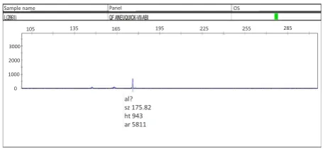
The accuracy of PCR was confirmed by agarose gel electrophoresis. After amplification of samples by multiplex fluorescence PCR, samples were digested with Hinfl (Thermo-ER0801) enzyme to determine the genotype of blastomeres. After enzymatic digestion, the homozygous wild type (CC), heterozygote (CT), mutant homozygote (TT) showed 1 band (198 bp), three bands (198, 175 and 23bp), 2 bands (175, 23 bp), respectively. The band of 23 bp was not visible due to its small size. Finally, the exact size of PCR products was determined using capillary electrophoresis and the results were analyzed using GeneMapper software version 4.0. Fluorescent-labeled primers were used to work with this kind of electrophoresis.
Results
In this study, the frequency of MTHFR gene C677T polymorphism was investigated in blastomeres. We found that only 37 samples among 50 blastomeres were successfully amplified. Twenty-three male and 14 female embryos were observed and 25, 9 and 3 samples were diagnosed as CC, CT and TT, respectively. As demonstrated in Table 4, the frequencies of C and T allele were 80% and 20%, respectively. The samples of the capillary electrophoresis results are illustrated in Figures 1, 2 and 3.
Indeed, only one distinct peak of 243 bp was observed in male embryos, while it was not detected in female embryos. Each of the peaks in the figures represents a single allele or amplified piece with specified size.
Discussion
The risk of developing a variety of abnormalities in the child can be increased with the mother’s age in couples with genetic diseases. These couples are often at the risk
of transmitting these diseases to their children. PGD is a screening method for couples who wish to have a healthy child and can determine the genotype of embryos before implantation to prevent transfer and implantation of affected embryos. To date, PGD technique allows these couples to have a child with no genetic disease. In recent years, the use of PGD technique has been considered in various diagnostic fields, and recent studies have shown that PGD can even play significant role in the management of recurrent miscarriage, especially in women over 35 years old, and these patients are the best candidates for PGD because it helps to improve fertility outcome (18).
Regarding our literature review, there are not enough
Table 3. The Sequence and Characteristics of Specific Primers Used for PCR
Name Sequence Melting Temperature PCR Product
SRY-Forward
SRY-Reverse
5′-6-FAM-TTTCGAACTCTGGCACCTT-3' 5′-CATGGGTCGCTTCACTCTA-3′
61°C
60°C 243bp
MTHFR-Forward
MTHFR-Reverse
5′-6-FAM-TGAAGGAGAAGGTGTCTGCGGGA-3′
5′-AGGACGGTGCGGTGAGAGTG-3′
65°C
64°C 198bp
Table 4. MTHFR C677T Genotype and Allele Frequencies in Embryos
Genotype n = 37 %
677CC 677CT 677TT
25 9 3
67.56 24.32 8.10
Allele C T
59 15
80 20
Figure 3. Female embryo with mutant genotype (TT).
Figure 1. An example of a Male Embryo With Normal Homozygous Genotype (CC).
Figure 2. Male Embryo With Heterozygous Genotype (CT).
al? sz 242.20 ht 483 ar 2794
al? sz 196.65 ht 298 ar 1750 900
600 300 0
0 110 220 330 440 550
al? sz 175.82 ht 943 ar 5811
0 1000 2000 3000
105 135 165 195 225 255 285
Panel OS
Sample name
al? sz 176.08 ht 746 ar 4528
al? sz 241.98 ht 1092 ar 6123 al?
sz 197.03 ht 513 ar 2996
3000 3000
2000 1000 0
135 165 195 225 255 285 315 345
Sample name Panel OS
conducted studies on the use of pre-implantation of genetic diagnosis method to study the MTHFR gene polymorphisms. More recently, there are reports that describe the effects of these polymorphisms in patients who use the IVF technique and also show the effect of some MTHFR variants on the embryo quality (morphological evaluation) and the implantation potential of the embryo (19,20). The study of the polymorphism of this gene in human embryos derived from IVF is recommended before implantation due to the importance of the MTHFR gene in the embryo development and success of the implantation.
In the study that was done by Galluzzi et al, non-invasive methods in PGD were used (21). In this research, after DNA extraction from blastocoel fluid samples and embryo culture media, MTHFR gene C677T polymorphism was investigated in these samples. They explained that amplification was successful only in 62.5% and 44.4% of the culture media and blastocoel fluid samples, respectively. In another study, the effect of embryo MTHFR genotype on the implantation was investigated by Enciso et al. (2016) (13). The results indicated that there was a significant risk of implantation failure in the euploid embryos homozygous for the 677T allele and MTHFRc.677C> T genotype affects the ability of an embryo to create a successful pregnancy. In fact, the data obtained from this study showed that homozygosity for the 677T allele could account for about 20% of the embryo implantation failure and consequently, determining the MTHFR genotype in IVF-derived embryos before transfer to the uterus can be helpful in ensuring the success of fertility treatments.
Our aim was to investigate MTHFR gene C677T polymorphism in IVF-derived pre-implantation embryos. We highlight here that Only 37 samples among 50 blastomeres were successfully amplified (74%), and the frequency of the C allele was significantly higher than T allele. It is noteworthy that due to inaccessibility to parent genotype in this study, the effect of parental genotype on the success rate of the implantation was not investigated and because these embryos were unusable for fertility treatment, it was virtually impossible to evaluate the success or failure of implantation.
Conclusions
PGD is a screening technique for couples who wish to have a healthy child and can be potentially used to genotype embryos before implantation to prevent the transfer of affected embryos to the uterus. In the present study, MTHFR gene C677T polymorphism was investigated in pre-implantation embryos because folate is important in human fertility processes and insufficient absorption of folic acid affects reproductive functions and has harmful effects on embryonic implantation and preservation of pregnancy.
Conflict of Interests
The authors declare that they have no conflicts of interest
concerning this article.
Ethical Issues
Surplus nontransferred embryos received from fertility treatment center were used for this research. The study protocol was approved by the Ethics Committee of Islamic Azad University of Tabriz (No. 23630503941005).
Financial Support
This research received no specific grant from any funding agency in the public, commercial, or not-for-profit sectors.
Acknowledgments
We gratefully acknowledge the distinguished staff of Infertility Treatment Center of Academic Jihad of Tabriz for providing the samples for this research.
References
1. Hardy K, Wright C, Rice S, et al. Future developments in assisted reproduction in humans. Reproduction. 2002;123(2):171-183. doi:10.1530/rep.0.1230171
2. Verpoest W. Preimplantation genetic diagnosis: design or too much design. Facts Views Vis Obgyn. 2009;1(3):208-222.
3. Handyside AH, Kontogianni EH, Hardy K, Winston RM. Pregnancies from biopsied human preimplantation embryos sexed by Y-specific DNA amplification. Nature. 1990;344(6268):768-770. doi:10.1038/344768a0
4. Handyside AH, Lesko JG, Tarin JJ, Winston RM, Hughes MR. Birth of a normal girl after in vitro fertilization and preimplantation diagnostic testing for cystic fibrosis. N Engl J Med. 1992;327(13):905-909. doi:10.1056/ nejm199209243271301
5. Ogilvie CM, Braude PR, Scriven PN. Preimplantation genetic diagnosis--an overview. J Histochem Cytochem. 2005;53(3):255-260. doi:10.1369/jhc.4B6395.2005 6. Pickering S, Polidoropoulos N, Caller J, Scriven P, Ogilvie
CM, Braude P. Strategies and outcomes of the first 100 cycles of preimplantation genetic diagnosis at the Guy’s and St. Thomas’ Center. Fertil Steril. 2003;79(1):81-90. doi:10.1016/S0015-0282(02)04540-5
7. Hardy K, Martin KL, Leese HJ, Winston RM, Handyside AH. Human preimplantation development in vitro is not adversely affected by biopsy at the 8-cell stage. Hum Reprod. 1990;5(6):708-714. doi:10.1093/oxfordjournals. humrep.a137173
8. Thornhill AR, Snow K. Molecular diagnostics in preimplantation genetic diagnosis. J Mol Diagn. 2002;4(1):11-29. doi:10.1016/s1525-1578(10)60676-9 9. Regan L, Rai R. Thrombophilia and pregnancy loss. J
Reprod Immunol. 2002;55(1):163-180. doi:10.1016/ S0165-0378(01)00144-9
10. Poursadegh Zonouzi A, Chaparzadeh N, Ghorbian S, et al. The association between thrombophilic gene mutations and recurrent pregnancy loss. J Assist Reprod Genet. 2013;30(10):1353-1359. doi:10.1007/s10815-013-0071-5
changes in activity of 5,10-methylenetetrahydrofolate reductase (MTHFR) and recurrent miscarriages]. Ginekol Pol. 2009;80(10):762-767.
12. Thaler CJ. Folate Metabolism and Human Reproduction. Geburtshilfe Frauenheilkd. 2014;74(9):845-851. doi:10.1055/s-0034-1383058
13. Enciso M, Sarasa J, Xanthopoulou L, et al. Polymorphisms in the MTHFR gene influence embryo viability and the incidence of aneuploidy. Hum Genet. 2016;135(5):555-568. doi:10.1007/s00439-016-1652-z
14. Spiroski I, Kedev S, Antov S, et al. Methylenetetrahydrofolate reductase (MTHFR-677 and MTHFR-1298) genotypes and haplotypes and plasma homocysteine levels in patients with occlusive artery disease and deep venous thrombosis. Acta Biochim Pol. 2008;55(3):587-594.
15. Goyette P, Pai A, Milos R, et al. Gene structure of human and mouse methylenetetrahydrofolate reductase (MTHFR). Mamm Genome. 1998;9(8):652-656. doi:10.1007/s003359900838
16. Frosst P, Blom HJ, Milos R, et al. A candidate genetic risk factor for vascular disease: a common mutation in methylenetetrahydrofolate reductase. Nat Genet.
1995;10(1):111-113. doi:10.1038/ng0595-111
17. Dobson AT, Davis RM, Rosen MP, et al. Methylenetetrahydrofolate reductase C677T and A1298C variants do not affect ongoing pregnancy rates following IVF. Hum Reprod. 2007;22(2):450-456. doi:10.1093/humrep/del396
18. Tempest HG, Simpson JL. Role of preimplantation genetic diagnosis (PGD) in current infertility practice. Int J Infertil Fetal Med. 2010;1(1):1-10. doi:10.5005/jp-journals-10016-1001
19. Laanpere M, Altmae S, Kaart T, Stavreus-Evers A, Nilsson TK, Salumets A. Folate-metabolizing gene variants and pregnancy outcome of IVF. Reprod Biomed Online. 2011;22(6):603-614. doi:10.1016/j.rbmo.2011.03.002 20. Soldo V, Cutura N, Zamurovic M. Defect of
methylenetetrahydrofolate reductase in a patient with ten habitual misscarriages: a case report. Clin Exp Obstet Gynecol. 2012;39(4):556-558.
21. Galluzzi L, Palini S, Stefani S, et al. Extracellular embryo genomic DNA and its potential for genotyping applications. Future Sci OA. 2015;1(4):Fso62. doi:10.4155/fso.15.62