International Conference on Frontiers in Life and Earth Science © 2018 IJSRST | Volume 5 | Issue 1 | Print ISSN: 2395-6011 | Online ISSN: 2395-602X
Effect of Lead Acetate Toxicity on Experimental Chick Embryo
Sawale Shweta*1, Kachhaway Archana R2
1Department of Biotechnology, Savitribai Phule Pune University, Pune,Maharashtra, India 2Department of Zoology,Shri Sishivinayak Mahila Mahavidyalaya,Karve Nagar, Pune, Maharashtra, India
ABSTRACT
Heavy metals are natural components of the Earth's crust and are important environmental pollutants which are mostly toxic even at very low concentrations. Lead is a common industrial and environment pollutant which is carcinogenic in animals. This study was conducted to understand the toxic effects of lead acetate on antioxidant enzymes and its bioaccumulation in liver and brain using a chick embryo as model. Chick embryos were divided into three groups, first group represented the healthy control and remaining two groups were injected as single dose of 20μl and 40μl/egg of lead acetate respectively, on 7th day of incubation with 1mg. concentration of lead acetate. On 11th and 14th day of incubation, control and treated chick embryos were sacrificed to collect liver and brain for biochemical assays. Different doses of lead acetate caused an increase or decrease of the activity of all investigated Enzymes, with a variable values, at almost all developmental stages. The lead that entered into the body is accumulated in organs and modifies the function of organ by changing the structure of cells of organs biochemical modifications in cells and functional variations of many of enzymes. Results of the present study clearly suggest that lead acetate induce toxic effects on the chick embryo.
Keywords : Lead Acetate, Antioxidant Enzymes, Liver, Brain
I.
INTRODUCTION
Heavy metals are generally defined as metals with relatively high densities, atomic weights, or atomic numbers. They are a unique class of toxicants, cannot be degraded because of their bioaccumulation. Heavy metal toxicity can result in damage to blood composition, lungs, kidneys, liver and other vital organs and some metals or their compounds may even cause cancer (International Occupational Safety and Health Information Centre 1999). Lead is the most common heavy metal pollutants listed by the Environment Protection Agency (EPA). Important sources of environmental lead contamination include mining, smelting, manufacturing and recycling activities, chemical fertilizers, and canned foods, foods grown around industrial areas, newsprint and colored advertisements. In addition, heavy rains may cause
lead in surface soil to migrate into ground water and eventually into water systems, subsequently transferred to humans through the food chain.
not appear to cause genetic damage directly, but may do so through several indirect mechanisms.
The avian embryo is a long-standing model for developmental biology research. Chick embryo has contributed enormously to experimental embryology and there is a vast amount of literature describing the development and their use as model system (Freeman and Vince 1974; Patten 1961).
II.
MATERIAL AND METHODS
Materials
Lead Acetate( PbA), Copper Sulphate (CuSO4), Sodium Carbonate, Sodium potassium carbonate, Phenyl Methyl sulfonyl Fluroid(PMSF), Hydrogen peroxide (H2O2), Amino-antipyrine(A.A.P), Tris HCL Buffer, Phosphate Buffer, Acetate Buffer, Triton 100, Formalin, Eosin etc. All other reagents used were of high quality analytical grade and was procured from the local companies.
Methodology
Source of Fertilized Eggs and Incubation Conditions Freshly laid zero day old fertilized eggs were purchased from Venketashwara hatcheries Pvt. Ltd. Pune, Maharashtra. The eggs were incubated horizontally and rotated (3h intervals) at 37.5±0.5˚C with a relative humidity of 65% in an egg incubator.
Experimental Design
Fertile eggs were divided into three groups: group A, Group B and Group C. Two groups A &B were administered as single dose of 20μL and 40μL/egg of lead acetate respectively, on day7 of incubation. Group C received no lead acetate and served as healthy control.Eggs were set-up in an upright position with the blunt end at the top.
On day 7, each egg was sterilized with 70% ethanol and egg shell was opened to obtain access to the air
by a parafine wax to ensure the embryo’s health until tissue collection and blood sampling takes place. The eggs were placed back into the humidified incubator. The eggs were further incubated until the date of examination. Eggs were injected by the air sac method according to Blankenship et al., (2003), on day 7 of incubation. On day 11th and 14th of incubation, the egg shell was broken at the air chamber and embryos were pulled out, from which liver and brain were collected. Tissue samples were washed in ice cold normal saline solution to remove blood and fat debris.
Tissue was minced with scissors and homogenized in 50mM Tris-HCl buffer pH 8.0 containing 0.25M sucrose and 1mM PMSF using a potter homogenizer centrifuged (3000xg for 10min). The resulting clear supernatant was used as enzyme source for antioxidant enzyme assays.
A.
Alkaline phosphatase assay:Buffer, 1 ml. (pH 10 for alkaline), is added to 1 ml. substrate(M / 100 Na2-phenyl phosphate), warmed at 37° C for three min. 0.1 ml. plasma added and mixed. The solution is incubated at 37° C for 15 min. for alkaline phosphatase. Then 0.8 ml. N/2 NaOH is added for alkaline phosphatase and 1.2 ml. M/2 NaHCO3 added for alkaline phosphatase. Then 1 ml. 0.6% A.A.P. is added and mixed. Finally 1 ml. 2.4% K3Fe (CN) 6 is added and mixed. Take the readings at 405nm.
B. Acid phosphtase assay:
First add 0.2 ml of the enzyme solutions to 1.2ml of the acetate buffer and 0.1 ml of Triton X-100 and add 0.5ml of the substrate solution. Incubate for 10 min and 20 min and stop the reaction by adding 2ml of the alkaline tris buffer. Read extinction at 405 nm and calculate the enzymatic activity by reference to a standard curve of p-Nitrophenol.
Catalase in tissues with relatively high activity,such as liver and brain, can be determined spectrophotometrically if complete lysis of all organelles and clear (or only slightly colored) solutions or extracts can be obtained. Normally, catalase activity of tissue samples is expressed on a milligram wet weight or milligram total N basis. A convenient method for the measurement of catalase activity in tissue extracts. Assay Conditions Wavelength, 240 nm; light path, 10 mm; final volume, 3.00 ml. Read the sample containing, 2.00 ml enzyme solution or hemolysate and 1 ml H202 at 20 ° (- room temperature) against a blank containing, 1 ml phosphate buffer instead of substrate and 2 ml enzyme solution or hemolysate. The reaction is started by addition of H202. The initial absorbance should be approximately A = 0.500. Mix well with a plastic paddle and follow the decrease in absorbance with a recorder for about 30 sec.
III.
OBSERVATIONS:
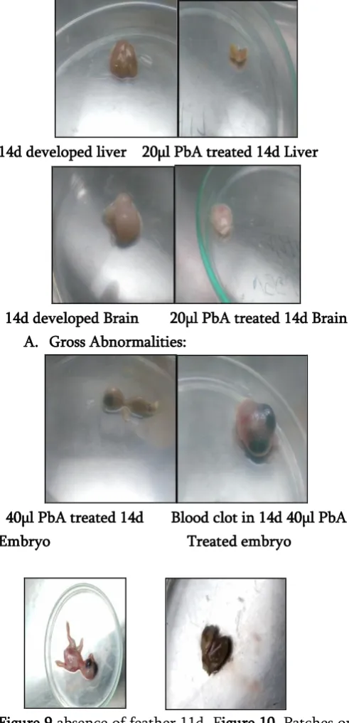
The observations show the various changes in chick embryo. As days pass mortality and change in weight of eggs were observed. Due to treatment of lead acetate change in size of control and treated chick embryo and its tissues were seen. Morphological deformities like blood clot on head region, patches on liver and absence of feathers were observed.
A.Normal Vs Affected.
14d developed embryo 20µl PbA treated embryo
14d developed liver 20µl PbA treated 14d Liver
14d developed Brain 20µl PbA treated 14d Brain A. Gross Abnormalities:
40µl PbA treated 14d Blood clot in 14d 40µl PbA Embryo Treated embryo
Figure 9 absence of feather 11d Figure 10. Patches on liver 11 d
20µl PbA treated embryo PbA treated embryo
IV.
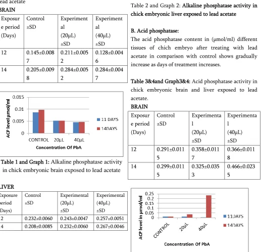
RESULT AND DISCUSSIONA. Alkaline phosphatse:
The alkaline phosphates content in (µmol/ml) brain tissue of chick embryo after treating with lead acetate gradually decreases as days of treatment increases when compare with control.
Table 1&2 and Graph 1&2: Alkaline phosphatase activity in chick embryonic brain and liver exposed to lead acetate
BRAIN Exposur e period (Days)
Control ±SD
Experiment al
(20µL) ±SD
Experiment al
(40µL) ±SD 12 0.145±0.008
7
0.211±0.005 2
0.128±0.004 6
14 0.205±0.009 8
0.284±0.005 2
0.284±0.004 7
Table 1 and Graph 1:
Alkaline phosphatase activity
in chick embryonic brain exposed to lead acetate
LIVER Exposure period (Days)
Control ±SD
Experimental (20µL) ±SD
Experimental (40µL) ±SD
12 0.232±0.0060 0.243±0.0047 0.257±0.0051 14 0.208±0.0085 0.232±0.0060 0.267±0.0046
Table 2 and Graph 2: Alkaline phosphatase activity in chick embryonic liver exposed to lead acetate
B. Acid phosphatase:
The acid phosphatase content in (µmol/ml) different tissues of chich embryo after treating with lead acetate in comparison with control shows gradually increase as days of treatment increases.
Table 3&4and Graph3&4: Acid phosphatase activity in chick embryonic brain and liver exposed to lead acetate.
BRAIN Exposur e period (Days)
Control ±SD
Experimenta l
(20µL) ±SD
Experimenta l
(40µL) ±SD 12 0.291±0.011
5
0.358±0.011 7
0.366±0.011 8
14 0.299±0.011 5
0.325±0.035 3
0.466±0.023 5
LIVER Exposur e period (Days) Control ±SD Experiment al (20µL) ±SD Experiment al (40µL) ±SD 12 0.400±0.020
3
0.408±0.023 5
0.466±0.031 8
14 0.491±0.011 8 0.456±0.023 5 0.566±0.031 1 Liver
Table 4 and Graph 4: Acid phosphatase activity in chick embryonic liver exposed to lead acetate
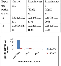
C. CATALASE ACTIVITY:
The effect of lead acetate on catalase activity of chick embryo liver was shown in graph 3. The catalase activity was significantly (p<0.05) decreased in 20µL and 40µL PbA treatment compared to control group.
BRAIN Exposure period (Days) Control ±SD Experiment al (20µL) ±SD Experime ntal (40µL) ±SD
12 0.87325
±0.0075
0.75425±0.0 0506
0.5365±0. 00332
14 2.1635±
0.04356
1.90525±0.0 379
0.5345±0. 00785
Table 5 and Graph 5: Catalase activity in chick embryonic liver exposed to lead acetate
LIVER Expos ure period (Days) Control ±SD Experimenta l (20µL) ±SD Experimenta l (40µL) ±SD 12 1.33825±0.2
521
0.98275±0.0 1176
0.59175±0.0 0665 14 0.895±0.027
68
0.82425±0.0 1628
0.26425±0.0 0723
Table 6 and Graph 6: Effect of lead acetate on catalase activity of 12d and 14 day old chick Embryonic Brain.
V.
DISSCUSIONPhosphatases are nonspecific phosphomonoestarse having pH specifically, which hydrolyze various phosphate esters and liberate phosphate from the substrate. Acid phosphatase (ACP) hydrolyses the phosphorous ester in acidic medium and autolysis process of the cell after its death. The employment of biochemical markers in monitoring pollution resulting from heavy metals has significantly shown the sensitivity of the tools and the potential risks facing man at the upper trophic level along the food chain . the present investigation shows toxicity of heavy metals on biosystems. Acid and alkaline phosphatase activities of control embryos are gradually changes as development proceeds from the 11th to 14th day of incubation. This findings are in accord with those of Romanoff (1988) and King and Liu (1974 a and b).
Catalase is one of the important antioxidant enzymes which help in converting the hydrogen peroxide to water and oxygen. Catalase is an efficient decomposer of H2O2 and is known to be susceptible to lead toxicity. (Sandhir and Gill 1995). The obtained results have displayed that treatment of chick embryo with lead acetate caused a reduction of liver catalase (Graph:4 and 5 ).Reduction in CAT impairs scavenging of hydrogen peroxide radicals. The reduction of catalase in liver and brain by Pb must be due to Pb induced inhibition of heme biosynthesis and since heme is a basic prosthetic molecule available with catalase for catalysis of peroxy radicals. Therefore the present results are in accordance with the earlier reports (Sandhir and Gill 1995).
VI. CONCLUSION
The environmental contamination by lead generated from human activities has become an evident problem during the last decades. Lead can penetrate to the human or animal organisms by inhalation, ingestion and by skin (Ryan and Terry, 1988;El-Fekiet al., 2000). The adverse effects of lead on health and productivity
animals. Berget al.,(1980) stated that contamination of the environment with lead has reached such a level that can affect the growth, productivity, and health of poultry as the toxicity of these metals (cadmium, lead) relies on binding the metallic cations with sulphydryl, amino and carboxylic groups of enzymes thus inhibiting enzymatic activities and disturbing energy metabolism.
In the present study, treatment with Pb-acetate has resulted in altered activity of alkaline phosphatase and acid phosphatase and the significant decrease of CAT levels. It shows changes in proteins content. It leads to excessive liver and brain damage and shows the tendency of bioaccumulation in the chick embryo. According to the results obtained in the present study, it appears that lead acetate administration to chick embryo would increase the toxicity and causing damage to vital tissue.
VI.
REFERENCES[1] A. Aebi H. (1984) :Catalase.Methods Enzymol., 105, 125-126.
[2] ATSDR- (2005): Draft toxicological profile for lead, US Department of health and human services, Atlanta,Georgia, USA. pp.102-225 [3] Clement, G.Y. and Paul, B.T
(2007).:N-Acetyl-L-Cysteine Affords Protection against Lead-Induced Cytotoxicity and Oxidative Stress in Human Liver Carcinoma (HepG2) Cells. International Journal of Environmental Research andPublic Health, 4, 132-137.
[5] Denizeau F, Marion M. (1989).:Genotoxic effects of heavy metals in rat hepatocytes. Cell Bio Toxicol 5:15-25.
[6] Freeman BM, Vince MA (1974): Development of the avian embryo London: Chapman and Hall, 1-156.
[7] Garaza, A., R. Vega and E. Soto, (2006): Cellular mechanisms of lead neuro toxicity.Med. Sci. Monitor., 12(3): 57-65.
[8] Kirkman, H. N., Rolfo, M., Ferraris, A. M., Gaetani, G. F. (1999): Mechanisms of protection of catalase by NADPH.J. Biol. Chem. 274, 13908-139
[9] Marcia,C. Carvalho, Evelise M. Nazari, Marcelo Farina,1 and Yara M. R. Muller,(2008): Behavioral, Morphological, and Biochemical Changes after In Ova Exposure to Methylmercury in Chicks toxicological sciences 106(1), 180–185.