Copyright © The Author(s). All Rights Reserved. Published by American Research Institute for Policy Development DOI: 10.15640/jns.v6n2a5 URL: https://doi.org/10.15640/jns.v6n2a5
Convolutional Neural Network Application in Biomedical Signals
Haya Alaskar
1Abstract
Recent improvements in big data and machine learning have enhanced the importance of biomedical signal and image-processing research. One part of machine learning evolution is deep learning networks. Deep learning networks are designed for the task of exploiting compositional structure in data. The golden age of the deep learning network in particular convolutional neural networks (CNNs) began in 2012. CNNs have rapidly become a methodology of optimal choice for analysing biomedical signals. CNNs have been successful in detecting and diagnosing an abnormality in biomedical signals. This paper has three distinct aims. The key primary aim is to provide state of the art knowledge about how deep learning evolved and revolutionized machine learning in the past few years. Second, to critically review the application of deep learning for different biomedical signals analysis and provide a holistic overview of current works of literature. Finally, to discuss the research opportunities with deep learning algorithms in the field of study that can serve as a starting point for new researchers to identify the future research direction in a concise manner.
Keywords: Convolutional neural network; Biomedical signals; Deep learning; Diagnosis. 1.Introduction
Once it was possible to record and load signals into a machine, researchers focused their efforts on designing systems for automated analysis. Initially, the electrocardiograph (electrocardiogram) in 1902 represent important information about the structure and function of the heart (AlGhatrif and Lindsay, 2012). Thirty-five years later, the first human electrocardiogram was introduced by Augustus Waller, a British physiologist. He used a capillary electrometer and electrodes located on a person’s chest (AlGhatrif and Lindsay, 2012). In 1938, Denny-Brown defined the fasciculation potentials and extracted them from fibrillations (Kazamel and Warren, 2017). In 1957, Lambert and Eaton defined the electrophysiologic structures of a myasthenic syndrome related to lung cancer. In terms of EEG signals, from 1929 and the late 1960s, EEGs were examined visually until digital tools were discovered (“History: From EEG to Quantitative EEG (QEEG),” 2017).
Since then, the importance of signal processing and analysing, identifying the differences between these signals, has become essential. These differences can help to classify normal and abnormal signals to identify diseases. In addition, time series analyses can be used to assess the progress of a patient's status over time (Machado, 1996), in order to extract useful information upon which clinicians can make determinations. However, in order to analysing biomedical signal in multiple dimensions different steps must be taken: pre-processing, feature extraction, classification (Alaskar et al., 2014; Alaskar and Hussain, 2018; Balli and Palaniappan, 2010; Chen et al., 2010; Liu et al., 2005; Orphanidou et al., 2018). Recently, machine learning techniques have proved to be successful in all science and engineering research, as well as the biomedical field (Begg et al., 2005; Gil and Manuel, 2009; Joshi et al., 2010; Khalaf et al., 2016; Mannini et al., 2016; Shoeb, 2009). One example of machine learning techniques and applications is supervised learning tools, which can also be called classification techniques. They are applied for abnormality detection and diagnosis (Alaskar and Hussain, 2018; Begg et al., 2005; Joshi et al., 2010; Shahbakhi et al., 2014; Shetty and Rao, 2016; Zheng et al., 2009).
In the literature, the machine learning approach receives great consideration from researchers and physicians. It has been implemented successfully on many occasions for biomedical signals analysis. The application of machine learning proved their success to support decision makers (Al-Askar, et al., 2016; Alaskar, 2014; Alasker et al., 2017; Miljkovic et al., 2016; Polat and Güneş, 2007). Therefore, instead of using systems that are entirely built by humans, systems that are built by computer can be used; this is called computer-aided diagnosis (CAD). CAD automatically distinguishes the class of biomedical signals using machine learning techniques. It trains machine learning tools on some data set. However, the data on time series signals are very high and a complex dimensional dataset. It is necessary to utilise some feature extraction methods that transform large time series signals into a small number of features that optimally discriminate (Übeyli, 2009; Wang et al., 1997). The feature extraction is an essential step to build an automated CAD.
On the scientist point of view, pre-processing and feature extraction in biomedical time series signals is not an easy task. There are a number of features set which can be related to different domains, such as the temporal domain and the frequency domain. Discovering the optimal features that are able to characterise the properties of signals requires time for investigation and examination (Chen et al., 2010; Verplancke et al., 2010). Furthermore, the selection of the best features is very important to improve the diagnosis and detection ability of classifiers. Unfortunately, the process of feature extraction and selection from signals is performed manually by researchers. And, as was mentioned earlier, it takes a number of examinations of features until the best features set is discovered. This usually takes a great deal of effort and is very time-consuming for researches.
Recently, Automated intelligent models can be built directly from the biomedical signals which can be done using deep learning. In the last few years, deep learning has transformed machine learning field. Deep learning networks have been attractive to many researchers since 2012. This type of learning netowrk has recorded success stories in computer vision. For example, Krizhevsky et al. (2012) won the ImageNet competition with their ALexNet design (Krizhevsky et al., 2012). In the following years, another development took place using deeper network architectures called GoogLeNet (Szegedy et al., 2015). Deep learning models by benefit of their high levels professional information processing offer much more effective representations of complex high dimensional data set.
To the best of our knowledge, this is the first paper that attempts to provide a concise survey of deep learning application in biomedical signals. The main aim of this survey is three-fold. First to document the background knowledge about how deep learning algorithm has evolved and revolutionized machine learning in the past few years. Second, to critically review the application of deep learning for different biomedical signal analysis and provide a holistic overview of current literatures. Finally, to discuss the research opportunities with deep learning algorithms in the field of study that can serve as starting point for new researchers to identify the future research direction in a concise manner. In the process of achieving these three aims of the paper, we have attempted to answer the following research questions:
Can deep learning be potential to analyze the biomedical signals?
How deep learning can be applied efficiently to analyze different type of biomedical signals?
How accurately deep learning detects abnormality pattern in biomedical signals? 2. Background
2.1 Deep Learning Network (DNN)
Deep learning is a branch of machine learning that originated from artificial neural network has shown considerable success in diverse fields in medicine, business, government sectors etc. It attempts to model data hierarchically and classifies patterns using multiple non-linear processing layers. There are several variants of deep learning such as Auto-encoders, Deep Belief Network, Deep Boltzmann Machines, Convolutional Neural Networks and Recurrent Neural Networks. Since recent literatures have demonstrated the success of CNN deep learning models in the application of biomedical signal analysis, the focus of this paper is limited to reviewing the past literatures related to CNN models.
2.2 Convolutional Neural Network (CNN)
However, the golden age of CNN started in the last decade. The first evolution of CNNs was when AlexNet won the ImageNet competition. Since then, only techniques use convolutional neural networks have won this competition (Goodfellow et al., 2016; He et al., 2015).
The CNN is a type of deep learning network which
contains one or more convolutional and max pooling layers followed by one or more fully connected layers,
which is referred to as the classification layer. An example of a CNN is shown in Fig. 1. The CNN is
different from the simple multi-layer network (MLP). MLPs only use input and output layers, and, at most, a
single hidden layer, where in the deep leaning network there are more than three layers including input and
output layers (Szegedy et al., 2015). Fig. 1 showed differentiates between a simple MLP and a CNN. Each
block in the CNN contains a number of layers.
The second key difference between the CNN and MLP is the combination of pooling layers in the former, where pixel values are aggregated using a transformation function. This will help to decrease the number of parameters in the network. Finally, fully-connected layers will be at the end of the network. In the fully connected layer, weights are no longer shared with the convolutional layer.
Figure 1: The differences architecture between a simple MLP and a CNN
The main advantages of CNNs are that they are self-learned and self-organised networks without the need for supervision (Savalia and Emamian, 2018). Furthermore, the task of pre-processing and feature extraction techniques are not required in CNNs. In contrast, the deep learning network can automatically recognise more complex features because of the number of convolutional layers it contains. This function of deep learning supports the ability of the network to handle large, high-dimensional data which contains a large number of features (Acharya et al., 2016). This makes the CNN beneficial and reduces liability during training and selecting of the best features that discriminate classes in the dataset.
Currently, a significant application of the CNN is in image classification, object or handwriting recognition and speech recognition (Dong et al., 2017; Goodfellow et al., 2016; Hinton et al., 2012; Khan and Yong, 2016; Szegedy et al., 2015). In addition, it plays an important role in the biomedical field for automated disease diagnosis (Acharya et al., 2018b, 2017c, 2016, 2016; Dong et al., 2017; Tajbakhsh et al., 2015). Also, Deep learning networks are also successfully used to applications requiring the processing of big amounts of data. Many applications that use the CNN have achieved greater efficiency and performance for real-time classification, as discussed below.
2.3 Biomedical Signals
The EEG records the signal from a large number of input channels. It can be from 12 to 256 electrodes. Each of these channels has a high temporal resolution. Analysing this number of signals requires the development of online algorithms that can deal with the varying types of data.
Another type of biomedical signal is the electrocardiogram (ECG). Researchers have attempted to record electrical activities of the heart in order to detect heart disease by using an ECG (Balli and Palaniappan, 2010; Chowdhury et al., 2013a; Derya Übeyli, 2010). The signals are recorded by placing an electrode in the chest. From these signals, the abnormal activity of the heart can be detected. The ECG is a beneficial diagnostic tool to detect various cardiovascular diseases. These signals contain information about the different forms of arrhythmias, namely Premature Ventricular Contraction (PVC), Atrial Premature Contraction (APC), Paced Beats (PB), Ventricular Couplet (VC) and Myocardial Infarction (MI).
Electromyogram (EMG) is the other type of biomedical signal. The EMG has been used to record the electrical activity of muscle contractions, and these signals have been recorded from different parts of the human body in order to understand the body’s behaviours under normal and pathological conditions (Alaskar et al., 2014, 2014; Alaskar, 2014; Konrad, 2005) (e.g. wrist, uterus, the human forearm, femoris muscle, Gait analysis, etc.) (Chowdhury et al., 2013b; Graupe, 2010; Ilbay et al., 2011; Miller, C, 2008; Mohamad O. Diab, Amira El-Merhie, Nour El-Halabi, 2010; Moslem et al., 2012).
2.4 Challenges of Biomedical signal analysis
Biomedical signals suffer from several causes of extensive noise, including device power interference, baseline drift, and noise between the skin and the electrode. In addition, motion artefacts can occur due to any muscular activity from the patient. Furthermore, inoffensive movement can be incorrectly recorded as arrhythmia in ECG signals. (Kim, 2018). The analysis of these signals needs to deal with noise and filtering signals. Also, it is challenging to visually analysis biomedical signals because these signals have small amplitude and duration.
On the other hand, the high dimensional biomedical signals make analysing them hard. Converting these signals into subsets of features is essential. Feature extraction is known as a conversion process of signals to features, and these features can characterise the properties of the signal (Übeyli, 2009; Wang et al., 1997). A number of techniques have been used to represent and extract features from biomedical signals in order to improve the classification (Forney and Anderson, 2011a; Subasia et al., 2005). The time domain, frequency domain, and time-frequency domain analysis measures have been utilised to represent signals (Chen et al., 2010; Fele-Zorz et al., 2008; La Rosa et al., 2007; Phinyomark et al., 2012). (Shaker, 2005). (Chiang, 2010; Fele-Žorž et al., 2008; Forney and Anderson, 2011b; Sarbaz et al., 2011).
Another method that can be used for such a process is wavelet transform, which is based on the time-frequency domain (Shaker, 2006). Wavelet transform allows the transformation of signals into smaller waves. Despite those variety of features domain, feature extraction methods are playing an important role in improving the discriminative performance of classifiers (Derya Übeyli, 2010; Moslem et al., 2011).
However, it should be considered that each feature represents a different classification power for different diagnosis. In addition, each disease might have some similarity in its features with other diseases, which can affect the network performance. (Savalia and Emamian, 2018). The examination of these different features can be a time-consuming, complex process and usually requires field expertise, and is limited in identifying abnormalities (Acharya et al., 2017c; Alaskar and Hussain, 2018; Alaskar, 2014; Dong et al., 2017). Additionally, the features should be invariant with respect to translations and scaling.
However, the requirement to discover automatically and intelligent tools to reduce the effort and time to examine all possible solution to finding optimal features is highly demand.
Deep learning is a promising technique for large-scale data analytics(Längkvist et al., 2012). During the last few decades, the use of deep learning networks to determine the character of biomedical signals’ properties has increased significantly. In the literature, they have been used in biomedical signal analysis such as EEG (Acharya et al., 2018b), ECG (Acharya et al., 2017c; Andreotti et al., 2017; Kim, 2018; Kiranyaz et al., 2016) and EMG(Biagetti et al., 2016; Xia et al., 2018).
Deep learning networks (DNN) can effectively replace the traditional hand-crafted features. Furthermore, once a large number of datasets is available, DNN models are a good method and usually surpass human agreement rates. The appearance of DNN has make the analysis of the biomedical signals simpler than before. This paper will give an overview of using DNN in biomedical signals.
Methodology
2.5 Search strategy and selection process
Database search through online databases such as google scholar is the most popular way of finding primary literature. But, here the search was extended to several other online sources such as PubMed, IEEE Xplore, ACM science direct etc., as the researchers working on biomedical signals could be from different research areas such as healthcare, medicine, signals processing.
3.2 Literature Sources
The investigation of the applications of deep learning in biomedical time series classification was addressed articles published in the domain were analyzed. Google scholar has been used to find the scientific papers.
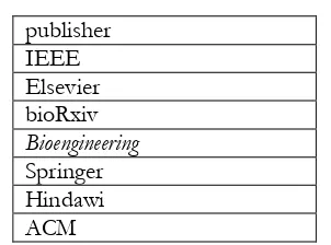
Table 1: the main sources of these articles. publisher
IEEE Elsevier bioRxiv Bioengineering Springer Hindawi ACM
Most of the selected articles were collected from these publishers, so that the credibility of this review paper is not compromised. However, there is a wide variety of other literature sources that are also adequate for this review. 2.6 Data Collection Process
The data collection process involved extensive research of papers that addressed the applications of deep learning in the biomedical field. These papers were downloaded and studied for the sake of obtaining sufficient information on the subject. The type of results in this paper were qualitative, and the main motivation is to provide survey of the applications of deep learning, and attempt to answer the research questions listed in the introduction section. Overall, the data collection process comprised three main phases:
Phase 1: Searching papers in reliable journals. This phase involved using some keywords.
Phase 2: Papers are selected and classified in line with the aim of the survey. Then the qualified papers are analyzed critically
Phase 3: Qualitative data attained and notes written in order to briefly introduced the data in the results section of this paper. Data was gathered in the form of what type of biomedical signals were used.
3 Literature Review
When it comes to deep learning and its application for biomedical signal diagnosis, there are two main approaches. The first approach is feature extraction and it is includes creating optimal features from CNN. The second approach is classification using CNN. The next three section will introduce the brief applications on deep learning. The last section will show the techniques the been used in order to fit the signals with deep leaning network.
Table 2: The published papers on the application of CNN on EEG, ECG and EMG.
Authors Medical data Deep learning Result
1. Ruffini et al., 2018 EEG CNN 80% accuracy
2. Yuan et al., 2017 EEG CNN 99.8% accuracy
3. Mirowski et al. 2018 EEG CNN 100% accuracy
4. Stober, et al., 2014 EEG CNN 55.4% accuracy
5. Acharya et al, 2018 EEG CNN 94% accuracy
6. Zheng et al.,2014 ECG (MC-DCNN 90.34 % accuracy
7. Geng et al., 2016 EMG CNN 99.0% accuracy
8. Wei et al., 2017 EMG CNN 85% accuracy
9. Ravi et al., 2017 EMG DNN 98%acuracy
10. Park et al., 2016 EMG CNN 90% accuracy
11. Acharya et al., 2017 ECG CNN 95.22% signals without noiz
93.53% with noise
12. Savalia et al., 2018 ECG CNN 88% accuracy
13. Kim 2018 ECG 88%
14. Nilanon et al., 2016 PCG CNN 82%
15. Al Rahhal et al., 2016 ECG SDAFs 99.85% accuracy
16. Hwang et al., 2018 ECG DeepECGNet 87%
17. Pourbabaee et al., 2017 ECG CNN 85.33%
18. Acharya et al., 2017 ECG CNN 99.13% sensitivity
19. Acharya et al., 2017 ECG CNN 94.03%
20. Fernando et al., 2017 ECG 83%
21. Sannino et al., 2018 ECG DNN 99.09% accuracy
22. Kamaleswara et al., 2018 ECG CNN 83% in F1 measurement
23. Ghiasi et al., 2017 ECG CNN 80% accuracy
24. Kiranyaz et al., 2015 ECG CNN 99 % accuracy
25. Acharya et al., 2017 ECG CNN 94.95% accuracy
26. Andreotti et al., 2017 ECG CNN 83% accuracy
27. Pourbabaee et al., 2017 ECG CNN 93% precision
28. Zhang et al., 2017 ECG CNN 93% accuracy
29. Moon et al., 2018 ECG CNN 99.72% accuracy
30. Acharya et al., 2018 ECG CNN 93% accuracy
31. Zhang et al., 2018 foetal ECG CNN 75.33% precision
32. Hasasneh et al., 2018 MEG CNN 91% sensitivity
33. Atzori et al., 2016 EMG CNN 66.59% accuracy
34. Wand et al., 2014 EMG CNN 70% accuracy
35. Xiong et al., 2017 ECG CNN 90% sensitivity
36. Vilamala et al., 2017 EEG CNN 89% accuracy
37. Tsinalis et al., 2016 EEG CNN 74 % accuracy
38. Luo et al., 2017 ECG CNN 97% accuracy
39. Vrbancic et al., 2018 EEG CNN 69.23 % accuracy
40. Xia et al., 2018 EMG Recurrent
CNN 90.3% in R
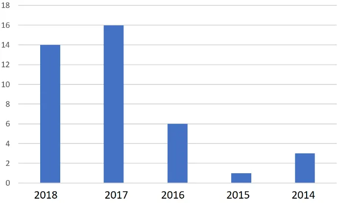
Figure 2: The numbers of papers from 2014 until 2018
From Fig. 2, it can be observed in 2015 there was just one publication comparing to previous year. This might relate to the lack of knowledge of how can deep learning efficiently employed in biomedical signals, however, 2016 until 2018 there was great effort to study and implanting convolutional neural network in this partiture type of data.
2.1 Application of CNN for ECG Analysis
During the last decades, investigations have been carried out to discover efficient and accurate ECG classification tools. For example, ICU monitoring tools are able to perform live diagnosis based on some ECG recordings. Unfortunately, the efficiency of this tool remains a problem (Kim, 2018)
In the literature, a number of studies have been placed to find automatic diagnostic tools for ECG signals(Acharya et al., 2017a; Kiranyaz et al., 2016; Moon et al., 2018).(Acharya et al., 2018a, 2017a) From the scientists’ point of view, the heart is very completed and different and new types of arrhythmia can affect the heart. Therefore, CNN has been extensively used for automated identification of ECG arrhythmia and it showed that it remains robust regardless of shifting and scaling invariance. For example, (Nilanon et al., 2016) showed that CNN is able to detect heart disease with higher accuracy. In this study PCG signals which record the sound of the heart beat is examined. CNN achieved, in this experiment, 82% accuracy (Nilanon et al., 2016). Another study, (Andreotti et al., 2017) employed the CNN to recognise four different arrhythmias from short segments of ECG recordings. Their result was 83% accuracy on test data. Where in (Ghiasi et al., 2017), CNN was used to distinguished atrial fibrillation. They achieved 80% accuracy on training datasets. Also, (Acharya et al., 2017b) used CNN to distinguish the four ECG classes: the normal (Nsr), Afib, Afl and Vfib ECG class. In this work, they used ECG signals of two seconds and five seconds’ duration without QRS detection. They obtained 98.09% on sensitivity and 93.13% on specificity for two seconds of ECG segments. In the five second duration, the sensitivity was 99.13% and specificity was 81.44%.(Acharya et al., 2017b)
Other study has used CNN with stacked denoising auto encoders (SDAEs). SDAE has been used to extract features from ECG signals (Al Rahhal et al., 2016). The dataset used in this paper was the MIT-BIH arrhythmia database as well as two other databases called INCART and SVDB. The highest result was 99.9% accuracy. The results obtained show that the proposed approach offers significant accuracy improvements faster online retraining compared to state-of-the-art methods. In addition, CNN used to automatically detect normal and MI ECG beats (Acharya et al., 2017c). In their experiment, they used two types of ECG signals, one with noise and the second without noise. They achieved an average accuracy of 93.53% in signals with noise and 95.22% in signals with noise removed. They concluded that CNNs correctly detect the MI ECG signals even with noise. Also, (Acharya et al., 2017d) developed a 9-layer CNN to automatically identify five different heartbeats in ECG signals. Their result was applied in original and noise of ECG signals. The CNN was trained and achieved an accuracy of 94.03% and 93.47% in the classification with and without noise ECG, respectively(Acharya et al., 2017d).
The automatic recognition of abnormal heartbeats from a large amount of ECG data was the main aims of this paper (Sannino and De Pietro, 2018). Deep learning was evaluated using MIT–BIH Arrhythmia Database. The results showed that deep learning is more efficient and competitive in terms of sensitivity and specificity which were 98.55% and 99.52% respectively. In this paper (Kamaleswaran et al., 2018) CNN implemented to early detect normal sinus rhythm, AF, other abnormal rhythms, and noise. CNN was trained on 8,528 single lead short ECG signals and 3,658 recordings for testing set. CNN achieved in detecting ordinary, AF and other rhythms with an average F1-score of 0.83.
Non-invasive foetal ECG was also analysis using CNN (Zhong et al., 2018). The study attempted to detect fetal QRS complex from raw NI-FECG signals. CNN was able to classify fetal QRS with and F-measure scores 77.85% , high precision 75.33%, and recall is 80.54%. In other study, CNN trained for each patient by using relatively small common and patient ECG signals (Kiranyaz et al., 2015). It can also be utilized to categorize long ECG records such as Holter registers. The experimental results prove that the CNN achieved a higher classification performance for the recognition of ventricular ectopic beats (VEB) and supraventricular ectopic beats (SVEB). CNN also used for PTB diagnostic ECG (Labati et al., 2018). The result accuracy was 69.23 % which is higher compared with traditional machine learning tools. Other paper attempted to convert ECG signals to wavelet domain in order to feed them to CNN(Zhang et al., 2017). CNN is directly learn features from the wavelet domain. The proposed approch is evaluated on eight ECG datasets with various behaviors and with different sensor placement methods. The experiment of this paper achieved 93.5% accuracy.
Furthermore, CNN was used to extract features and can replaced the hand-crafted features. For example. In, (Pourbabaee et al., 2016) CNN was used to extracted the features directly from the time domain ECG signals. Based on the paper results, the combined learned features has improved the performance of the patient screening system. In (Pourbabaee et al., 2017), they used the CNN-based feature learning mechanism where feature are extracted using CNN. These features were used to train standard classifiers, namely the K-nearest neighbour (KNN), SVM, and the multi-layered perceptron (MLP) networks. Also, end to end CNN structure that is used for both feature extraction and classification tasks. In this experiment, the focus is on patient screening and recognising patients with paroxysmal atrial fibrillation (PAF), which is sign of a life-threatening arrhythmia. They used the CNN with a large volume of raw ECG signals as inputs. Features of the PAF to be used by a classification model are obtained directly from the CNN. The study achieved high accuracy on end-to-end CNN classifier with 93% precision. The experimental results show the effectiveness and abilities of the learned features for PAF patient screening. In addition, the classification result significantly enhanced the patient screening system performance.
Another study (Zheng et al., 2014) proposed Multi-channels deep convolution neural network (MC-DCNN). They used ECG multivariate signals. Each channel is passed as input for proposed network to extract features. Each features of each channels combined and feed to MLP for classification performance. The experiments result showed that using MC-DCNN for features extraction outperforms the traditional methods.
2.2 Application of CNN for EEG Analysis
features from EEG signals. These features were used to train three classifiers such as regularized logistic regression, CNN, and SVM. The dataset used on this research on the standard Freiburg EEG dataset which recorded signals from 21 patients having medically intractable focal epilepsy. The CNN was achieved 100% of the seizures on average 60 minutes before the onset.
In addition, the CNN used REM Behaviour Disorder (RBD) diagnosis from the EEG (Ruffini et al., 2018). The EEG was recorded from RBD patients and healthy controls. In their work, they selected a few minutes of eyes-closed resting state from EEG signals. The input to the CNN was spectrograms of EEG channel data, i.e., 2D time-frequency maps. They studied a deep recurrent neural network using stacked long short-term memory network (LSTM) cells or gated-recurrent unit (GRU) cells. The proposed approach achieved 80% accuracy.
Most current research has focused on the recognition of abnormality of EEG signals. On the other hand, (Yuan and Cao, 2017) attempted to apply a deep learning network to EEG signals to prove clinical brain death diagnosis. The Short Time Fourier Transform technique was used as a time frequency analysis technique. Then, spectrogram images were created to characterise the features of EEG signals. For the experiment, EEG signals used in this paper were obtained from brain-damaged patients. These images were used as the training samples in the CNN. Finally, the trained network was applied to estimate the degree of similarity of the other patients’ EEG signals with those of the patients in a coma and with brain death symptoms. The result was 99.8% accuracy. This approach can be used to evaluate the condition of brain-damaged patients as well as for quasi-brain death diagnosis (Yuan and Cao, 2017). Another study focused on to distinguish EEG pattern of three classes namely normal, preictal and seizure pattern. (Acharya et al., 2018b). In this study, 13-convolutional layers were used to build the CNN in order to detect normal, preictal and seizure pattern. They achieved an accuracy of 88.67%, specificity of 90%, and sensitivity of 95.00%(Acharya et al., 2018b)
Also CNN has been used to discriminate different type of rhythm types and also to identify the rhythms themselves. In (Stober et al., 2014) EEG recordings of rhythm perception. The study used EEG data recorded within a rhythm perception study in Kigali, Rwanda which comprises 12 East African and 12 Western rhythmic. CNN was able to identify individual rhythms from the EEG with 24% accuracy.
2.3 Application of CNN for EMG Analysis
The rise in the use of wearable devices in recent years has increased the ability to capture a range of various physiological and functional data. This data can now be recorded continuously for applications in sports, welfare and healthcare. This huge amount of information needs effective tools and analysis techniques. Therefore, deep learning networks take the lead to deal with this high-dimensional data. In a paper by (Ravi et al., 2017), a deep learning network was developed to detect activity from inertial sensor data. They combined two sets of features. This combination overcame some of the limitations in a typical deep learning architecture. They used laboratory and real-world activity datasets. Spectral domain was utilized before the data is passed onto the deep learning network. The results demonstrated the ability of the proposed approach to detect different human activity, outperforming other methods with precision and the recall is above 85%. Furthermore, the experiment showed that the computation times for the proposed approach are steady with the limitations of real-time processing on smartphones and a wearable sensor (Ravi et al., 2017).
On the other hand, deep learning has been used for EMG hand movement recognition (Park and Lee, 2016). They used a CNN model to find six hand movements via EMG signals. The results show a good classification accuracy for CNN compared to SVM. The best accuracy was reached using CNN with 90% accuracy. In addition, a CNN was utilised for natural control of robotic hands by EMG signals (Atzori et al., 2016). The CNN was tested to classify an average of 50 hand movements in 67 intact subjects and 11 transradial amputees. With this model the reported accuracy was 66.59%. Also, there is study focusing on classification of phones and phonetic features by using facial EMG signals (Wand and Schultz, 2014). The study focused on EMG-based silent speech recognizer. They aimed to extract EMG activities for certain phones, present and absent phonetic features. They achieved a good accuracy in one of their experiments which was nearly 70% to detect Rounding word.
3. Discussion
The main motivation of this paper was to review various studies and papers in the field of deep learning implementation in biomedical signals. After analyzing more than 90 articles, 40 were further examined, and the results of each article were addressed. The aim of this survey was to introduce the variety of deep learning application in the biomedical signals field. Numbers of founding has been conducted firstly, most of papers have been published the last three years. In addition, the most commonly utilized deep learning network is convolutional neural networks CNN especially the pretrained CNNs. Furthermore, spectrogram images were most regularly used to train CNN. Previously reported studies mainly addressed medical signals analysis and diagnosis with the applications of expert-designed features.
It can be observed that there is a large variety in the type of signals that is used to train and apply deep neural networks. EEG, ECG and EMG. All of studies have investigate the application of deep learning for medical signals diagnosis and compared with state of art. Each of this study has confirmed and proved that deep learning a CNN can automatically extracted the optimal features and also learn to distinguish among different classes. Most of these articles results are comparable to state-of-the-art methods has the ability to analysis most of biomedical signals. CNN proven to be highly successful in analysing different type of medical signals such as analysing neurophysiological dynamics of sleep using EEG, identifying type of movement in EMG or type of activity etc.
Most of the articles obtained their best result without any human intervention. Furthermore, they did not need to have domain knowledge for the analysis of medical signals. Based on the results of each articles, CNN and deep learning in general can be considered as a sound basis for further optimization towards a competitive, fully automated classification method for classify medical signals. Therefore, CNN was replaced most of traditional machine learning application. This is the approach resulted by numbers of papers in this survey.
There is no doubt that deep learning application in the medical field will further investigate as it has already attained outstanding results in medical data analysis, and more precisely, in biomedical signals detection and diagnosis. Consequently, convolutional neural network, more generally deep learning, will positively increase the efficiency and quality of healthcare. It will reduce the risk of misclassification and furthermore, increase the early diagnosis of serious diseases. It also can be used to support computer aided diagnosis CAD.
4. Conclusion
The paper content is organized to describe the background knowledge on how deep learning was conceived, evolved and has revolutionized the field of biomedical signal analysis. Then the state of art applications of CNN deep learning model for different types of biomedical signals such as ECG, EMG, EEG are illustrated. Finally, 40 literatures were found to be relevant to the field of the study and most of these were recently published since 2016. These literatures were critical reviewed to provide a holistic overview on the performance of deep learning models for biomedical signal analysis. The review results of these literatures have confirmed deep learning as the potential technique for biomedical signal analysis. Thus, the brief nature of this survey can temperately contribute to the current body of literature and is expected to shed some light on research challenges and future opportunities in the field of biomedical signal analysis.
References
Acharya, U.R., Fujita, H., Adam, M., Lih, O.S., Hong, T.J., Sudarshan, V.K., Koh, J.E., 2016. Automated characterization of arrhythmias using nonlinear features from tachycardia ECG beats, in: 2016 IEEE International Conference on Systems, Man, and Cybernetics (SMC). IEEE, pp. 000533–000538.
Acharya, U.R., Fujita, H., Lih, O.S., Adam, M., Tan, J.H., Chua, C.K., 2017a. Automated detection of coronary artery disease using different durations of ECG segments with convolutional neural network. Knowl.-Based Syst. 132, 62–71.
Acharya, U.R., Fujita, H., Lih, O.S., Hagiwara, Y., Tan, J.H., Adam, M., 2017b. Automated detection of arrhythmias using different intervals of tachycardia ECG segments with convolutional neural network. Inf. Sci. 405, 81–90. Acharya, U.R., Fujita, H., Oh, S.L., Hagiwara, Y., Tan, J.H., Adam, M., 2017c. Application of deep convolutional
Acharya, U.R., Fujita, H., Oh, S.L., Raghavendra, U., Tan, J.H., Adam, M., Gertych, A., Hagiwara, Y., 2018a. Automated identification of shockable and non-shockable life-threatening ventricular arrhythmias using convolutional neural network. Future Gener. Comput. Syst. 79, 952–959.
Acharya, U.R., Oh, S.L., Hagiwara, Y., Tan, J.H., Adam, M., Gertych, A., San Tan, R., 2017d. A deep convolutional neural network model to classify heartbeats. Comput. Biol. Med. 89, 389–396.
Acharya, U.R., Oh, S.L., Hagiwara, Y., Tan, J.H., Adeli, H., 2018b. Deep convolutional neural network for the automated detection and diagnosis of seizure using EEG signals. Comput. Biol. Med. 100, 270–278.
Acharya, U.R., Oh, S.L., Hagiwara, Y., Tan, J.H., Adeli, H., Subha, D.P., 2018c. Automated EEG-based screening of depression using deep convolutional neural network. Comput. Methods Programs Biomed. 161, 103–113. Al Rahhal, M.M., Bazi, Y., AlHichri, H., Alajlan, N., Melgani, F., Yager, R.R., 2016. Deep learning approach for active
classification of electrocardiogram signals. Inf. Sci. 345, 340–354.
Alaskar, H., Hussain, A.J., 2018. Data Mining to Support the Discrimination of Amyotrophic Lateral Sclerosis Diseases Based on Gait Analysis, in: International Conference on Intelligent Computing. Springer, pp. 760–766. Alaskar, H., Hussain, A.J., Paul, F.H., Al-Jumeily, D., Tawfik, H., Hamdan, H., 2014. Feature Analysis of Uterine Electrohystography Signal Using Dynamic Self-organised Multilayer Network Inspired by the Immune Algorithm, in: International Conference on Intelligent Computing. Springer, pp. 206–212.
Al-Askar, H., Radi, N., MacDermott, A., 2016. Recurrent Neural Networks in Medical Data Analysis and Classification, in: Applied Computing in Medicine and Health. Elsevier.
Alaskar, H.M., 2014. Dynamic self-organised neural network inspired by the immune algorithm for financial time series prediction and medical data classification (PhD Thesis). Liverpool John Moores University.
Alasker, H., Alharkan, S., Alharkan, W., Zaki, A., Riza, L.S., 2017. Detection of kidney disease using various intelligent classifiers, in: Science in Information Technology (ICSITech), 2017 3rd International Conference On. IEEE, pp. 681–684.
AlGhatrif, M., Lindsay, J., 2012. A brief review: history to understand fundamentals of electrocardiography. J. Community Hosp. Intern. Med. Perspect. 2, 14383.
Andreotti, F., Carr, O., Pimentel, M.A., Mahdi, A., De Vos, M., 2017. Comparing Feature-Based Classifiers and Convolutional Neural Networks to Detect Arrhythmia from Short Segments of ECG. Computing 44, 1. Atzori, M., Cognolato, M., Müller, H., 2016. Deep learning with convolutional neural networks applied to
electromyography data: A resource for the classification of movements for prosthetic hands. Front. Neurorobotics 10, 9.
Balli, T., Palaniappan, R., 2010. Classification of biological signals using linear and nonlinear features. Physiol. Meas. 31, 903–20. https://doi.org/10.1088/0967-3334/31/7/003
Begg, R.K., Palaniswami, M., Owen, B., 2005. Support vector machines for automated gait classification. IEEE Trans. Biomed. Eng. 52, 828–838.
Biagetti, G., Crippa, P., Orcioni, S., Turchetti, C., 2016. Surface EMG fatigue analysis by means of homomorphic deconvolution, in: Mobile Networks for Biometric Data Analysis. Springer, pp. 173–188.
Chen, X., Zhu, X., Zhang, D., 2010. A discriminant bispectrum feature for surface electromyogram signal classification. Med. Eng. Phys. 32, 126–35. https://doi.org/10.1016/j.medengphy.2009.10.016
Chiang, A., 2010. Automated Quantification of Human Alpha Rhythms by. The University of Sydney.
Chowdhury, R.H., Reaz, M.B.I., Ali, M.A.B.M., Bakar, A. a a, Chellappan, K., Chang, T.G., 2013a. Surface electromyography signal processing and classification techniques. Sensors 13, 12431–66. https://doi.org/10.3390/s130912431
Chowdhury, R.H., Reaz, M.B.I., Ali, M.A.B.M., Bakar, A. a a, Chellappan, K., Chang, T.G., 2013b. Surface electromyography signal processing and classification techniques. Sensors 13, 12431–66.
https://doi.org/10.3390/s130912431
Derya Übeyli, E., 2010. Recurrent neural networks employing Lyapunov exponents for analysis of ECG signals. Expert Syst. Appl. 37, 1192–1199. https://doi.org/10.1016/j.eswa.2009.06.022
Desai, U., Martis, R.J., Acharya, U.R., Nayak, C.G., Seshikala, G., SHETTY K, R., 2016. Diagnosis of multiclass tachycardia beats using recurrence quantification analysis and ensemble classifiers. J. Mech. Med. Biol. 16, 1640005.
Fele-Zorz, G., Kavsek, G., Novak-Antolic, Z., Jager, F., 2008. A comparison of various linear and non-linear signal processingtechniques to separate uterine EMG records of term and pre-termdelivery groups. Med. Biol. Eng. Comput. 46, 911–922.
Fele-Žorž, G., Kavšek, G., Novak-Antolič, Ž., Jager, F., 2008. A comparison of various linear and non-linear signal processing techniques to separate uterine EMG records of term and pre-term delivery groups. Med. Biol. Eng. Comput. 46, 911–922.
Forney, E.M., Anderson, C.W., 2011a. Classification of EEG during imagined mental tasks by forecasting with Elman Recurrent Neural Networks. 2011 Int. Jt. Conf. Neural Netw. 2749–2755.
https://doi.org/10.1109/IJCNN.2011.6033579
Forney, E.M., Anderson, C.W., 2011b. Classification of EEG during imagined mental tasks by forecasting with Elman recurrent neural networks, in: Neural Networks (IJCNN), The 2011 International Joint Conference On. IEEE, pp. 2749–2755.
Ghiasi, S., Abdollahpur, M., Madani, N., Kiani, K., Ghaffari, A., 2017. Atrial Fibrillation Detection Using Feature Based Algorithm and Deep Convolutional Neural Network. Computing 44, 1.
Gil, D., Manuel, D.J., 2009. Diagnosing parkinson by using artificial neural networks and support vector machines. Glob. J. Comput. Sci. Technol. 9.
Goodfellow, I., Bengio, Y., Courville, A., 2016. Deep Learning (Book in preparation) -. MIT press.
Graupe, D., 2010. Recognition and prediction of individual and combined muscular activation modes via surface EMG analysis 1, 131–138.
He, K., Zhang, X., Ren, S., Sun, J., 2015. Delving deep into rectifiers: Surpassing human-level performance on imagenet classification, in: Proceedings of the IEEE International Conference on Computer Vision. pp. 1026–1034.
Hinton, G., Deng, L., Yu, D., Dahl, G.E., Mohamed, A., Jaitly, N., Senior, A., Vanhoucke, V., Nguyen, P., Sainath, T.N., 2012. Deep neural networks for acoustic modeling in speech recognition: The shared views of four research groups. IEEE Signal Process. Mag. 29, 82–97.
History: From EEG to Quantitative EEG (QEEG) [WWW Document], 2017. . Brain Clin. URL https://www.brainclinics.com/history-of-the-eeg-and-qeeg (accessed 10.21.18).
Hwang, B., You, J., Vaessen, T., Myin-Germeys, I., Park, C., Zhang, B.-T., 2018. Deep ECGNet: An Optimal Deep Learning Framework for Monitoring Mental Stress Using Ultra Short-Term ECG Signals. ℡EMEDICINE E-Health.
Ilbay, K., Übeyli, E.D., Ilbay, G., Budak, F., 2011. A New Application of Recurrent Neural Networks for EMG-Based Diagnosis of Carpal Tunnel Syndrome, in: Cardot, H. (Ed.), Recurrent Neural Network for Temporal Data Processing. InTech. https://doi.org/10.5772/631
Joshi, S., Shenoy, D., Rrashmi, P.L., Venugopal, K.R., Patnaik, L.M., 2010. Classification of Alzheimer’s disease and Parkinson’s disease by using machine learning and neural network methods, in: Machine Learning and Computing (ICMLC), 2010 Second International Conference On. IEEE, pp. 218–222.
Kamaleswaran, R., Mahajan, R., Akbilgic, O., 2018. A robust deep convolutional neural network for the classification of abnormal cardiac rhythm using single lead electrocardiograms of variable length. Physiol. Meas. 39, 035006.
Kazamel, M., Warren, P.P., 2017. History of electromyography and nerve conduction studies: A tribute to the founding fathers. J. Clin. Neurosci. 43, 54–60.
Khalaf, M., Hussain, A.J., Al-Jumeily, D., Keight, R., Keenan, R., Fergus, P., Al-Askar, H., Shaw, A., Idowu, I.O., 2016. Training neural networks as experimental models: classifying biomedical datasets for sickle cell disease, in: International Conference on Intelligent Computing. Springer, pp. 784–795.
Khan, S.A., Yong, S.-P., 2016. An Evaluation of Convolutional Neural Nets for Medical Image Anatomy Classification, in: Advances in Machine Learning and Signal Processing. Springer, pp. 293–303.
Khemphila, A., Boonjing, V., 2012. Parkinsons Disease Classification using Neural Network and Feature selection. Eng. Technol. 15–18.
Kim, K., 2018. Arrhythmia Classification in Multi-Channel ECG Signals Using Deep Neural Networks.
Kiranyaz, S., Ince, T., Hamila, R., Gabbouj, M., 2015. Convolutional Neural Networks for patient-specific ECG classification, in: Engineering in Medicine and Biology Society (EMBC), 2015 37th Annual International Conference of the IEEE. IEEE, pp. 2608–2611.
Konrad, P., 2005. The ABC of EMG: A Practical Introduction to Kinesiological Electromyography. Noraxon INC USA 1.0 April.
Krizhevsky, A., Sutskever, I., Hinton, G.E., 2012. Imagenet classification with deep convolutional neural networks, in: Advances in Neural Information Processing Systems. pp. 1097–1105.
La Rosa, P.S., Nehorai, a., Eswaran, H., Lowery, C., Preissl, H., 2007. Detection of uterine MMG contractions using a multiple change-point estimator and K-means cluster algorithm. Int. Congr. Ser. 1300, 745–748. https://doi.org/10.1016/j.ics.2007.01.049
Labati, R.D., Muñoz, E., Piuri, V., Sassi, R., Scotti, F., 2018. Deep-ECG: Convolutional Neural Networks for ECG biometric recognition. Pattern Recognit. Lett.
Längkvist, M., Karlsson, L., Loutfi, A., 2012. Sleep stage classification using unsupervised feature learning. Adv. Artif. Neural Syst. 2012, 5.
Lehnertz, K., Andrzejak, R.G., Arnhold, J., Kreuz, T., Mormann, F., Rieke, C., Widman, G., Elger, C.E., 2001. Nonlinear EEG Analysis in Epilepsy: Its Possible Use for Interictal Focus Localization, Seizure Anticipation, and Prevention. J. Clin. Neurophysiol. 18, 209–222.
Liu, B., Wang, M., Yu, H., Yu, L., Liu, Z., 2005. Study of Feature Classification Methods in BCI Based on Neural Networks., in: Conference Proceedings : ... Annual International Conference of the IEEE Engineering in Medicine and Biology Society. IEEE Engineering in Medicine and Biology Society. Conference. China, pp. 2932–5. https://doi.org/10.1109/IEMBS.2005.1617088
Machado, L.O., 1996. Medical Application of Artificial Network Connectionist Models of Survival. Stanford University.
Mannini, A., Trojaniello, D., Cereatti, A., Sabatini, A.M., 2016. A machine learning framework for gait classification using inertial sensors: application to elderly, post-stroke and huntington’s disease patients. Sensors 16, 134. Miljkovic, D., Aleksovski, D., Podpečan, V., Lavrač, N., Malle, B., Holzinger, A., 2016. Machine Learning and Data
Mining Methods for Managing Parkinson’s Disease, in: Machine Learning for Health Informatics. Springer, pp. 209–220.
Miller, C, J., 2008. Real-Time Feature Extraction and Classification of Prehensile. 2008. San Diego State University. Mirowski, P.W., LeCun, Y., Madhavan, D., Kuzniecky, R., 2008. Comparing SVM and convolutional networks for
epileptic seizure prediction from intracranial EEG. Presented at the IEEE Workshop on Machine Learning for Signal Processing, IEEE. https://doi.org/10.1109/MLSP.2008.4685487
Mohamad O. Diab, Amira El-Merhie, Nour El-Halabi, L.K., 2010. Classification of uterine EMG signals using supervised classification method. J. Biomed. Sci. Eng. 03, 837–842. https://doi.org/10.4236/jbise.2010.39113 Moon, S.-E., Jang, S., Lee, J.-S., 2018. Convolutional neural network approach for EEG-based emotion recognition using brain connectivity and its spatial information, in: 2018 IEEE International Conference on Acoustics, Speech and Signal Processing (ICASSP). IEEE, pp. 2556–2560.
Moslem, B., Karlsson, B., Diab, M.O., Khalil, M., Marque, C., 2011. Classification performance of the frequency-related parameters derived from uterine EMG signals. Conf. Proc. Annu. Int. Conf. IEEE Eng. Med. Biol. Soc. IEEE Eng. Med. Biol. Soc. Conf. 2011, 3371–4. https://doi.org/10.1109/IEMBS.2011.6090913
Moslem, B., Khalil, M., Diab, M.O., Marque, C., 2012. Classification of multichannel uterine EMG signals by using a weighted majority voting decision fusion rule. 2012 16th IEEE Mediterr. Electrotech. Conf. 331–334. https://doi.org/10.1109/MELCON.2012.6196442
Nilanon, T., Yao, J., Hao, J., Purushotham, S., Liu, Y., 2016. Normal/abnormal heart sound recordings classification using convolutional neural network, in: Computing in Cardiology Conference (CinC), 2016. IEEE, pp. 585–588. Orphanidou, N.K., Hussain, A., Keight, R., Lishoa, P., Hind, J., Al-Askar, H., 2018. Predicting Freezing of Gait in
Parkinsons Disease Patients Using Machine Learning, in: 2018 IEEE Congress on Evolutionary Computation (CEC). IEEE, pp. 1–8.
Park, K.-H., Lee, S.-W., 2016. Movement intention decoding based on deep learning for multiuser myoelectric interfaces, in: Brain-Computer Interface (BCI), 2016 4th International Winter Conference On. IEEE, pp. 1–2. Phinyomark, A., Chujit, G., Phukpattaranont, P., Limsakul, C., 2012. A preliminary study assessing time-domain EMG
Polat, K., Güneş, S., 2007. Breast cancer diagnosis using least square support vector machine. Digit. Signal Process. 17, 694–701.
Pourbabaee, B., Roshtkhari, M.J., Khorasani, K., 2017. Deep convolutional neural networks and learning ecg features for screening paroxysmal atrial fibrillation patients. IEEE Trans. Syst. Man Cybern. Syst. 1–10.
Pourbabaee, B., Roshtkhari, M.J., Khorasani, K., 2016. Feature leaning with deep convolutional neural networks for screening patients with paroxysmal atrial fibrillation, in: Neural Networks (IJCNN), 2016 International Joint Conference On. IEEE, pp. 5057–5064.
Ravi, D., Wong, C., Lo, B., Yang, G.-Z., 2017. A deep learning approach to on-node sensor data analytics for mobile or wearable devices. IEEE J. Biomed. Health Inform. 21, 56–64.
Ruffini, G., Soria, D.I., Dubreuil, L., Castellano, M., Gagnon, J.-F., Montplaisir, J., Soria-Frisch, A., 2018. Deep learning with EEG spectrograms in rapid eye movement behavior disorder. bioRxiv 240267.
Sannino, G., De Pietro, G., 2018. A deep learning approach for ECG-based heartbeat classification for arrhythmia detection. Future Gener. Comput. Syst. 86, 446–455.
Sarbaz, Y., Gharibzadeh, S., Towhidkhah, F., Banaie, M., Jafari, A., 2011. A Gray-Box Neural Network Model of Parkinson’s Disease Using Gait Signal 2, 33–42.
Savalia, S., Emamian, V., 2018. Cardiac Arrhythmia Classification by Multi-Layer Perceptron and Convolution Neural Networks. Bioengineering 5, 35.
Shahbakhi, M., Far, D.T., Tahami, E., 2014. Speech analysis for diagnosis of parkinson’s disease using genetic algorithm and support vector machine. J. Biomed. Sci. Eng. 7, 147–156.
Shaker, M.M., 2006. EEG waves classifier using wavelet transform and Fourier transform. brain 2, 3.
Shaker, M.M., 2005. EEG Waves Classifier using Wavelet Transform and Fourier Transform. Int. J. Biol. Life Sci. 1, 85–90.
Shetty, S., Rao, Y.S., 2016. SVM based machine learning approach to identify Parkinson’s disease using gait analysis, in: Inventive Computation Technologies (ICICT), International Conference On. IEEE, pp. 1–5.
Shoeb, A.H., 2009. Application of Machine Learning to Epileptic Seizure Onset Detection and Treatment. Masssachusetts Institute of Technology.
Stober, S., Cameron, D.J., Grahn, J.A., 2014. Using Convolutional Neural Networks to Recognize Rhythm Stimuli from Electroencephalography Recordings, in: Advances in Neural Information Processing Systems. pp. 1449–1457.
Subasia, A., Kiymika, M., Alkana, A., Koklukayab, E., 2005. NEURAL NETWORK CLASSIFICATION OF EEG SIGNALS BY USING AR. Math. Comput. Appl. 10, 57–70.
Szegedy, C., Liu, W., Jia, Y., Sermanet, P., Reed, S., Anguelov, D., Erhan, D., Vanhoucke, V., Rabinovich, A., 2015. Going deeper with convolutions. Cvpr.
Szkoła, J., Pancerz, K., Warchoł, J., 2011. Recurrent neural networks in computer-based clinical decision support for laryngopathies: an experimental study. Comput. Intell. Neurosci. 2011, 289398.
https://doi.org/10.1155/2011/289398
Tajbakhsh, N., Gurudu, S.R., Liang, J., 2015. Automatic polyp detection in colonoscopy videos using an ensemble of convolutional neural networks, in: Biomedical Imaging (ISBI), 2015 IEEE 12th International Symposium On. IEEE, pp. 79–83.
Übeyli, E.D., 2009. Analysis of EEG signals by implementing eigenvector methods/recurrent neural networks. Digit. Signal Process. 19, 134–143. https://doi.org/10.1016/j.dsp.2008.07.007
Verplancke, T., Van Looy, S., Steurbaut, K., Benoit, D., De Turck, F., De Moor, G., Decruyenaere, J., 2010. A novel time series analysis approach for prediction of dialysis in critically ill patients using echo-state networks. BMC Med. Inform. Decis. Mak. 10, 4.
Wand, M., Schultz, T., 2014. Pattern learning with deep neural networks in EMG-based speech recognition, in: Engineering in Medicine and Biology Society (EMBC), 2014 36th Annual International Conference of the IEEE. IEEE, pp. 4200–4203.
Wang, K., Lee, C., Member, S., Juang, B., 1997. Selective Feature Extraction via Signal Decomposition. IEEE Signal Process. Lett. 4, 8–11.
Wei, W., Wong, Y., Du, Y., Hu, Y., Kankanhalli, M., Geng, W., 2017. A multi-stream convolutional neural network for sEMG-based gesture recognition in muscle-computer interface. Pattern Recognit. Lett.
Yuan, L., Cao, J., 2017. Patients’ EEG Data Analysis via Spectrogram Image with a Convolution Neural Network, in: International Conference on Intelligent Decision Technologies. Springer, pp. 13–21.
Zhang, Q., Zhou, D., Zeng, X., 2017. HeartID: a multiresolution convolutional neural network for ECG-based biometric human identification in smart health applications. IEEE Access 5, 11805–11816.
Zheng, H., Yang, M., Wang, H., McClean, S., 2009. Machine learning and statistical approaches to support the discrimination of neuro-degenerative diseases based on gait analysis, in: Intelligent Patient Management. Springer, pp. 57–70.
Zheng, Y., Liu, Q., Chen, E., Ge, Y., Zhao, J.L., 2014. Time series classification using multi-channels deep convolutional neural networks, in: International Conference on Web-Age Information Management. Springer, pp. 298–310.