Kati. World Journal of Pharmaceutical and Medical Research
RETENTION METHODS FOR EXTRA-ORAL PROSTHESIS. REVIEW ARTICLE
Firas Abd Kati*
Lecturer, Department of Optical techniques, College of Health & Medical Technology, Baghdad, Middle Technical University, Baghdad, Iraq.
Article Received on 20/12/2018 Article Revised on 10/01/2019 Article Accepted on 31/01/2019
METHODS OF RETENTION
1. Adhesives
The use of adhesives materials can provide good retention for extra-oral prostheses. There are two types of adhesives, water and solvent based adhesives. Overall, water based adhesives are easier to apply especially in cleaning the prosthesis and skin than solvent base adhesives. On the other hand, solvent based adhesives provide better retention than water based adhesives.[1,2] To maintain the fine edge of prosthesis, water and solvent based adhesives can be used together, where a thin layer of water based adhesive such as Epithane 3 (Factor 2, Lakeside, USA) is applied to the fitting surface of prosthesis. This layer is allowed to cure. Then a thin layer of solvent based adhesive such as Secure B-400 (Technovent, Newport, UK) is applied on to cured Epithane 3 and left to dry for 1-2 minutes. After that, the prosthesis is attached to the skin. This method facilitates removing solvent and water based solvents from the prosthesis. Adhesives can be supplied as a spray such as Hollister (Factor II, Lakeside, USA). It is very difficult to control by patients. Therefore, the selection of spray adhesive is critical for use by patients.[1,2]
Furthermore, it is recommended to use tissue conditioners such as Comfeel applicator (Coloplast, Peterborough, UK) when applying the adhesives. This technique will prevent many problems such as soft tissue reactions. The purpose of using tissue conditioners is to keep the skin from adhesive and to provide good bonding between adhesive and skin.Overall, it is very important that patient must understand the process of application and removal of adhesives. The success of extra-oral prosthesis retained by adhesives relies on selection of the correct adhesives and patient’s dexterity.[1,2]
2. Natural anatomical undercuts
Anatomical undercuts can be used for the retention of extra-oral prostheses. For patients who have received a partial rhinectomy, there are enough spaces in the nasal cavity and maxillary sinus. These spaces can assist in the retention of nasal prosthesis. Patients with lateral nasal defects have natural undercuts that can be used to retain nasal prosthesis without using adhesives. Figure 1 demonstrates the lateral nasal defect and anatomically nasal prosthesis.[3]
Figure 1: Lateral nasal defect and anatomically nasal Prosthesis.[3]
Figure 2 demonstrates lateral nasal-canthal prosthesis with projections providing vertical and lateral resistance to displacement.[3]
ISSN 2455-3301
WJPMR
AND MEDICAL RESEARCH
www.wjpmr.com
*Corresponding Author: Firas Abd Kati
Lecturer, Department of Optical techniques, College of Health & Medical Technology, Baghdad, Middle Technical University, Baghdad, Iraq.
ABSTRACT
Various methods of retention are used to retain extra-oral prostheses. These methods include use of medical-grade skin adhesives, natural anatomical undercuts, mechanical retention using spectacles and osseointegrated implants. The chosen method of retention depends upon the size of the defect, soft tissue movement, age and manual dexterity, position and number of implants.
Figure 2: Lateral nasal-canthal prosthesis with projections and prosthesis in situ.[3]
For patients with total rhinectomy, natural undercuts offer less chance for retention of nasal prosthesis if the maxillary sinuses are not exposed. Hence, maxillary sinuses can assist for retention of nasal prosthesis if they are open as shown in the Figure3.[3]
Figure 3: Nasal defect and anatomically retained nasal prosthesis.[3]
Intranasal anatomical retention provides good retention and aesthetic in the beginning but movement of adjacent tissue during eating, smiling and speaking affects the stability of prosthesis. Therefore, this method is preferred when there is a little movement and is recommended in partial defects.[1]
In case of partial removal of ear, tissue remnants can provide retention of auricular prosthesis with using adhesive. However, this method is not recommended because of mobility of the remnant tissue.[1,3]
In case of total missing ear, open external auditory canal can assist in retention of auricular prosthesis but this will affect the hearing. Hence, this method is contraindicated. Figure 4 illustrates silicon rubber extension, which engages undercuts in the external auditory canal.[3]
Figure 4: Silicon rubber extension which engages undercuts in the external auditory canal.[3]
3. Mechanical retention
Spectacles can be used to retain extra-oral prosthesis. This method is considered as basic and user friendly method especially for elderly patients with limited dexterity. In the past, this method has been used to retain auricular prosthesis, however it has many problems. One of these problems is related to the pressure that is result from the eyeglass and prosthesis to the nose. Another problem is associated with stability of prosthesis during movement of the tissue.[1,2] For patients with partial or total rhinectomy, spectacles can be used to retain nasal prosthesis that is fixed to the frame by using clear acrylic resin. This method is preferred in patients who have limited dexterity. The most common problem that is associated with method is the movement of eyeglass frame, where this movement transfers to the prosthesis and affects the stability of prosthesis. To overcome this problem, a lock that is placed behind the ear can retain the eyeglass frame and prosthesis. Another important issue is that the prosthesis is attached to the frame and this means the patient is not able to remove his/her eyeglass without prosthesis.[1,2] Figure 5 illustrates the defect site and nasal prosthesis.[2]
Figure 5: Defect site and nasal prosthesis.[2]
mould. The silicone orbital prosthesis is checked on the patient. The spectacles are then checked on the patient for fit and comfort. A clear acrylic resin template is made to attach the prosthesis with spectacles at the bridge of the nose and spectacle arm as shown in the Figure 6. Ear locks can be used to prevent displacement of eyeglass frame and provide stability of prosthesis.[4]
Figure 6: Spectacles retention.[4]
4. Osseointegrated implants
The use of implants to retain extra-oral prosthesis is considered as the best method compared to other traditional methods of retention.[5-7]
4.1. Advantages and disadvantages
Overall, osseointegrated implants bring many benefits. Firstly, it provides more stability and retention than other methods of retention. The use of adhesives and removers are not required and this prevents tear of the edge of prosthesis and skin irritation that are result of application and removal of adhesives when cleaning the prosthesis. The most important point that the service life of silicone prosthesis will be increased.[8] On the other hand, the patient should aware all issues or problems which may result with this type of retention. These include:
Soft tissue complications which result from insufficient care of prosthesis and the tissue around the abutments.[9-10]
Patient with implant retained extra–oral prosthesis is often required to care of his / her prosthesis and tissue around the implants to prevent soft tissue inflammations. The patient is instructed to use soap with water and at least once a day.[10]
Regular clinical appointments.[11]
In case of implant failure, an additional surgery is indicated to place farther implant in order to support the prosthesis.[1]
4.2. Types of implant retention
4.2.1. Bar construction and retentive clips
This method gives an even good force distribution on the implants. For this reason, it is preferred to use with auricular and large orbital prostheses.[5,12] Figure 7 demonstrates a bar retained auricular prosthesis.
Figure 7: Retentive bar and ear prosthesis in situ.[12]
4.2.2. Magnetic retention
This method of retention is preferred when there is not enough space for a bar – clips construction. This method allows the patient to insert and remove the prosthesis easily and the patient is able to clean around the abutments properly.[5] Figures (8, 9 and 10) show magnetic retention in the auricular, nasal and orbital prostheses.[12-14]
Figure 8: Magnetic retention and magnet keepers incorporated into prosthesis.[12]
Figure 9: Three magnets in the nasal defect and nasal prosthesis in situ (Ethunandan et al., 2010).[14]
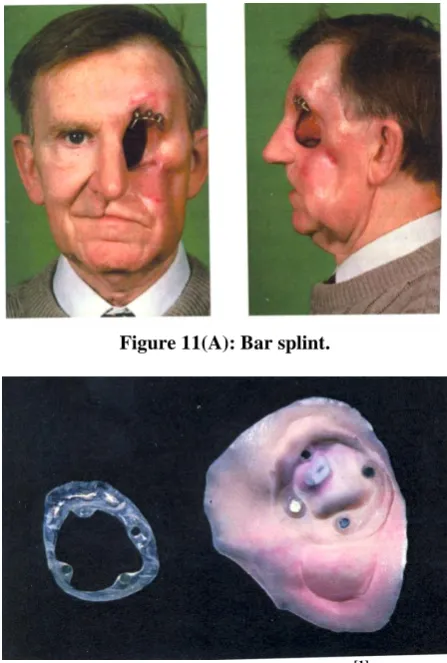
4.3.3 Bar splint / magnet retention
This method is used to retain large prosthesis such as hemi-facial prosthesis, where a number of implants are placed in the upper part of defects. This type provides satisfactory retention. Figure 11 (A & B) demonstrates bar splint and magnet retention.[1]
Figure 11(A): Bar splint.
Figure 11(B): Magnet retention.[1]
4.3.4. Ball attachments
This method is recommended to use in case of shallow defects especially orbital defects where there is a little space. Three implants can produce optimum retention and stability for prosthesis.[5] Figure 12 shows the orbital defect with ball attachment and the prosthesis.[15]
Figure 12: Orbital defect with ball abutments and prosthesis in situ.[15]
4.3.5. Combined direct adhesive / magnetic retention
This technique involves use of adhesive and magnets to retain extra-oral prosthesis. The advantage of use this technique is to prevent using the adhesive on the fitting
surface of silicone prosthesis. This method involves the construction of acrylic base which contains a number of magna- caps. This base is placed on the tissue of the defect area and fixed with adhesive to obtain retention. The prosthesis is then attached to the magna-acrylic base by using magnets that are also incorporated into acrylic base which is attached on the fitting surface of prosthesis using primer. The most important thing with this method is that the location of acrylic plate should be in accurate position so that the location of prosthesis will be in exist position. This method of retention can be used to retain extra-oral prostheses such as orbital, auricular, nasal and hemi facial cases. As well as it can be used in partial cases such as partial nasal and partial auricular defects. Figure 13 (A &B) demonstrates this type of retention.[1]
Figure 13(A): Magnets on the defect.[1]
Figure 13(B): Hemi facial prosthesis.[1]
CONCLUSION
spectacles and osseointegrated implants. The choice of retention method relies on many factors such as the size of the defect, soft tissue movement, age and ability of the patient, position and number of implants. Every method has its advantages and disadvantages. Implant retention is the best method to retain extra-oral prosthesis in comparison to other methods.
REFERENCES
1. Thomas K. The art of clinical anaplastology. Published by Thomas Keith in 2006.
2. Thomas K. The Prosthetic rehabilitation. Quintessence publishing Co, Ltd., London, 1994. 3. Parel S. Diminishing dependence on adhesives for
retention of facial prostheses. Journal of Prosthetic Dentistry, 1980; 63(5): 553-560.
4. Raizada K, Rani D., Ocular prosthesis. Contact Lens and Anterior Eye, 2007; 30(3): 152-162.
5. Reyes R, Tjellstrom A, Granstrom G. Evaluation of implant losses and skin reactions around extra- oral bone-anchored implants: A 0- to 8-year follow-up. Otolaryngology–Head and Neck Surgery, 2000; 122(2): 272-276.
6. Brånemark P. The ossessointegation Book: from calvarium to calcaneus. Quintessenz Verlags-GmbH, Berlin, 2000.
7. Aydin C, Karakoca S, Yilmaz H, Yilmaz C. Implant-retained auricular prostheses: an assessment of implant success and prosthetic complications. International Journal of Prosthodontics, 2008; 21(3): 241-244.
8. Karakoca S, Aydin C, Yilmaz H, Bal B. Retrospective study of treatment outcomes with implant-retained extraoral prostheses: Survival rates and prosthetic complications. Journal of Prosthetic Dentistry, 2010; 103(2): 118-126.
9. Holgers k, Tjellström A, Bjursten L, Erlandsson,B. Soft tissue reactions around percutaneous implants: A clinical study on skin-penetrating titanium implants used for bone-anchored auricular prostheses. International Journal of Oral and Maxillofacial Implants, 1987; 2(1): 35-39.
10. Reisberg D, Habakuk S. Hygiene procedures for implant-retained facial prostheses. The Journal of Prosthetic Dentistry, 1995; 74(5): 449- 502.
11. Wang R, Andres C. Hemifacial microsomia and treatment options for auricular replacement: A review of the literature. The Journal of Prosthetic Dentistry, 1999; 82(2): 197-204.
12. Wright R, Zemnick C, Wazen J, Asher E. Osseointegrated implants and auricular defects: A case series study. Journal of Prosthodontics, 2008; 17(6): 468–475.
13. Thomas K. Freestanding magnetic retention for extra-oral prosthesis with osseointegrated implants. Journal of Prosthetic Dentistry, 1995; 73(2): 162-165.
14. Ethunandan M, Downie I, Flood T. Implant-retained nasal prosthesis for reconstruction of large rhinectomy defects: the salisbury experience.
International Journal of Oral and Maxillofacial Surgery, 2010; 39(4): 343–349.
![Figure 1: Lateral nasal defect and anatomically nasal Prosthesis.[3]](https://thumb-us.123doks.com/thumbv2/123dok_us/8365127.1673131/1.595.312.535.484.597/figure-lateral-nasal-defect-anatomically-nasal-prosthesis.webp)
![Figure 2: Lateral nasal-canthal prosthesis with projections and prosthesis in situ.[3]](https://thumb-us.123doks.com/thumbv2/123dok_us/8365127.1673131/2.595.320.522.75.247/figure-lateral-nasal-canthal-prosthesis-projections-prosthesis-situ.webp)
![Figure 7: Retentive bar and ear prosthesis in situ.[12]](https://thumb-us.123doks.com/thumbv2/123dok_us/8365127.1673131/3.595.312.537.631.743/figure-retentive-bar-ear-prosthesis-situ.webp)