Pseudothrombocytopenia in a Preterm Neonate
Robert D. Christensen, MD*; Martha C. Sola, MD‡; Lisa M. Rimsza, MD§; Michael J. McMahan, MD
㛳
; and
Darlene A. Calhoun, DO*
ABSTRACT. Severe and prolonged thrombocytopenia is not uncommon among ill preterm infants. Pseudothrom-bocytopenia, which has the appearance of severe and prolonged thrombocytopenia, has not been described in this population. We observed a preterm neonate who had EDTA-independent pseudothrombocytopenia and con-clude that this condition should be considered when severe and prolonged thrombocytopenia occurs in a ne-onate in the absence of clinical signs of platelet-type hemorrhage.Pediatrics2004;114:273–275; thrombocytope-nia, pseudothrombocytopethrombocytope-nia, neonate, preterm.
P
seudothrombocytopenia is a rare condition in
which platelet counts are reported to be low,
often
⬍
10 000 per
L, yet no hemorrhagic
ten-dencies are present.
1–6This condition involves
plate-let aggregation in vitro and is generally associated
with using the anticoagulant
ethylenediaminetet-raacetic acid (EDTA) in the blood-collection tube.
1–6Shreiner and Bell first reported
pseudothrombocy-topenia in 1973,
1and subsequently both
EDTA-pendant and -independent varieties have been
de-scribed.
2–6Chiurazzi et al
7reported a term neonate
with a transient, presumably transplacentally
ac-quired variety of EDTA-dependant
pseudothrombo-cytopenia, but we know of no cases among preterm
infants. In fact, we are aware of no cases in neonates
other than the Chiurazzi et al case.
We recently observed a preterm neonate who had
severe and prolonged thrombocytopenia, in whom
many platelet counts were
⬍
10 000 per
L, and
several were 0 per
L; however, no clinical bleeding
problems were seen, and the bleeding time was
nor-mal. Platelet counts in citrate-containing and in
EDTA-containing tubes were identical, and
numer-ous platelet clumps were observed on the blood film.
These results, taken together with the clinical history
and lack of thrombocytopenia in the mother, suggest
that our patient had EDTA-independent
pseudo-thrombocytopenia and thus fundamentally differs
from the Chiurazzi et al
7case.
CASE REPORTS
The patient was delivered at 26 weeks’ gestation to a healthy 22-year-old primigravida by caesarian section at Arnold Palmer Hospital for Women and Children for suspected abruption and preterm labor. Apgar scores were 2 and 7 at 1 and 5 minutes, respectively, and birth weight was 886 g. The male infant received exogenous surfactant in the delivery room. His complete blood count on admission to the neonatal intensive care unit revealed a platelet count of 8000 perL. The hematocrit was 41.4%, and the leukocyte count was 5800 perL, with 39% segmented neutro-phils and 13% bands. No dysmorphic features were observed, and no petechiae, purpura, or bleeding were noted. Because of the low platelet count, a platelet transfusion, consisting of 15 mL/kg non-packed, single-donor platelets, was given.
Over the following week, platelet counts ranged from 17 000 to 47 000 perL, and 4 additional platelet transfusions were given. However, no petechiae, purpura, or clinical bleeding were seen. The prothrombin time was normal at 13.7 seconds, the fibrinogen was normal at 226 mg/L, and a heparin platelet antibody study was negative. Cultures of urine for cytomegalovirus and blood cultures for bacteria were negative. IgM titers for cytomegalovi-rus, rubella, and toxoplasmosis were negative. The mother’s and infant’s serum were negative for antiplatelet antibodies. The mother’s platelet count was normal, and she had no history of thrombocytopenia or abnormal bleeding.
Because of the lack of any bleeding manifestation, it was de-cided to administer platelet transfusions only if the platelet count was⬍10 000 perL rather than the usual “trigger” level in this neonatal center of⬍25 000 perL.8On 2 occasions the platelet
count was reported as 0 perL. During the second week of life, the pathologist noted clumping of the platelets on the blood film. At 13 weeks of age, the patient underwent surgical repair of bilateral inguinal hernias. He received a platelet transfusion the day before surgery, which resulted in a platelet count of 43 000 per L on the day of surgery and 14 000 perL the day after surgery. Platelets were available for transfusion during the surgery but were not given because no abnormal bleeding was observed.
A Surgicutt newborn bleeding time test, done when the platelet count was 2000 perL, was normal for age and hematocrit (24%) at 4 minutes, 15 seconds.9Electronic platelet counts were done on
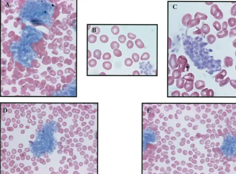
blood simultaneously drawn into an EDTA-containing tube and into a citrate-containing tube. Both were 2000 perL. A blood film made directly from an automated lance wound of the heel dem-onstrated few scattered platelets of normal size and morphology but many platelet clumps, particularly at the edge of the blood film (Fig 1). The nonclumped platelets appeared normal in size and shape.
After a diagnosis of pseudothrombocytopenia was made, the patient had no additional platelet counts or platelet transfusions. At 17 weeks, he was discharged from the hospital on 0.1 L of O2
per min. Two cranial ultrasound studies were done during the hospitalization; one at 1 week and one at 6 weeks of life, and both were normal with no evidence of hemorrhage. Before this was recognized to be pseudothrombocytopenia, he had 90 platelet counts and 29 platelet transfusions. Three months after discharge, he was still receiving 0.1 L of O2per min. He had 2 brief
hospi-talizations for upper respiratory infections. His growth has been normal, and he has had no petechiae, purpura, epistaxis, or any hemorrhagic or thrombotic problems. He has no additional plate-let counts.
From the *Division of Neonatology, Department of Pediatrics, University of South Florida College of Medicine and All Children’s Hospital, St Peters-burg, Florida; ‡Division of Neonatology, Department of Pediatrics, Univer-sity of Florida College of Medicine, Gainesville, Florida;㛳Division of Neo-natology, Arnold Palmer Hospital for Women and Children, Orlando, Florida; and §Division of Hematopathology, Department of Pathology, University of Arizona College of Medicine, Tucson, Arizona.
Received for publication Jul 21, 2003; accepted Nov 13, 2003.
Address correspondence to Robert D. Christensen, MD, All Children’s Hospital, 801 Sixth St S, Box 9360, St Petersburg, FL 33701. E-mail: [email protected]
PEDIATRICS (ISSN 0031 4005). Copyright © 2004 by the American Acad-emy of Pediatrics.
PEDIATRICS Vol. 114 No. 1 July 2004 273
at Viet Nam:AAP Sponsored on August 29, 2020
www.aappublications.org/news
DISCUSSION
Severe thrombocytopenia in a preterm neonate
generally triggers a platelet transfusion.
8,10,11How-ever, if it were known that the case was actually a
pseudothrombocytopenia, and therefore the risk for
serious bleeding was not increased, platelet
transfu-sions would not be given.
1–6We observed a preterm
neonate who had severe and prolonged
thrombocy-topenia but had no clinical bleeding problems. Even
when the platelet count was recorded as 0 per
L,
no petechiae, purpura, or other hemorrhagic
prob-lems were seen. Moreover, the patient had no
intra-ventricular hemorrhage, although he was born at 26
weeks’ gestation with hyaline membrane disease and
a platelet count of 8000 per
L. No abnormal bleeding
was observed after bilateral herniorrhaphy with a
platelet count of 14 000 per
L. This lack of hemorrhage
is concordant with the normal bleeding time but
dis-cordant with the repeatedly very low platelet counts.
The clinical history and multiple platelet clumps
seen on the blood film indicate
pseudothrombocyto-penia. The appearance of platelet clumps when
blood was drawn into citrate-containing tubes, or
into EDTA or into no anticoagulant, excludes the
usual type of EDTA-dependent
pseudothrombo-cytopenia and suggests the diagnosis of
non–EDTA-dependent pseudothrombocytopenia. It is thought
that EDTA-dependent pseudothrombocytopenia is
mediated by antibody.
1–6However, this mechanism
is unlikely in our case, which is more likely due to a
specific platelet abnormality. Our case differs from
the transplacentally acquired case described by
Chiurazzi et al
7in that our patient’s mother was not
affected, he did not remit in 1 month, and his
plate-lets clumped in citrate as well as in EDTA.
To our knowledge, no previous cases of
pseudo-thrombocytopenia have been reported among
pre-term neonates, and thus this is certainly a rare
con-dition. However, the present case is instructive,
affirming the adage that physicians should treat the
patient and not the laboratory value. Furthermore,
the case illustrates that pseudothrombocytopenia can
indeed occur in the neonatal intensive care unit and
should be considered when severe and prolonged
thrombocytopenia occurs in the absence of clinical
signs of platelet-type hemorrhage.
ACKNOWLEDGMENTS
This work was supported by National Institutes of Health grants HL-61798 and HL-69990.
We thank Dr John Lazarchick (Department of Pathology and Laboratory Medicine, Medical University of South Carolina, Fig 1. A compilation of several photomicrographs that were taken from a single blood film made without anticoagulant and stained with Wright/Giemsa (A, D, and E: original magnification⫻400; B and C: original magnification⫻1000).
Charleston) for helpful discussions of the case and Aaron Barley-corn and Dr Wenge Lu for technical assistance.
REFERENCES
1. Shreiner DP, Bell WR. Pseudothrombocytopenia: manifestation of a new type of platelet agglutinin.Blood.1973;42:541–549
2. Mant MJ, Doery JC, Gauldie J, Sims H. Pseudothrombocytopenia due to platelet aggregation and degranulation in blood collected in EDTA. Scand J Haematol.1975;15:161–170
3. Onder O, Weinstein W, Hoyer LW. Pseudothrombocytopenia caused by platelet agglutinins that are reactive in blood anticoagulated with che-lating agents.Blood.1980;56:177–182
4. Bartels PC, Schoori M, Llombarts AJ. Screening for abnormalities in platelet distribution histograms in pseudothrombocytopenia. Scand J Clin Lab Invest.1997;57:629 – 636
5. Berkman N, Michaeli Y, Or R, Eldon A. EDTA-dependent pseudo-thrombocytopenia: a clinical study of 18 patients and review of the literature.Am J Hematol.1991;36:195–201
6. Bizzaro N. EDTA-dependent pseudothrombocytopenia: a clinical and epidemiological study of 112 cases, with 10-year follow-up.Am J He-matol.1995;50:103–109
7. Chiurazzi F, Villa MR, Rotoli B. Transplacental transmission of EDTA-dependent pseudothrombocytopenia.Haematologica.1999;84:664 8. Calhoun DA, Christensen RD, Edstrom CS, et al. Consistent approaches
to procedures and practices in neonatal hematology. Clin Perinatol. 2000;27:733–754
9. Sola MC, Edwards TJ, Del Vecchio A, Suttner D, Hutson AD, Chris-tensen RD. A low hematocrit prolongs the bleeding time of very low birth-weight infants in the first week of life. J Perinatol.2001;201: 368 –371
10. Sola MC, Del Vecchio A, Rimsza LM. Evaluation and treatment of thrombocytopenia in the neonatal intensive care unit.Clin Perinatol. 2000;27:655– 680
11. Del Vecchio A, Sola MC, Garcia MG, et al. Platelet transfusions in the neonatal intensive care unit: factors predicting patients who will require multiple transfusions.Transfusion.2001;41:803– 808
EXPERIENCE AND REASON 275
at Viet Nam:AAP Sponsored on August 29, 2020
www.aappublications.org/news
DOI: 10.1542/peds.114.1.273
2004;114;273
Pediatrics
Darlene A. Calhoun
Robert D. Christensen, Martha C. Sola, Lisa M. Rimsza, Michael J. McMahan and
Pseudothrombocytopenia in a Preterm Neonate
Services
Updated Information &
http://pediatrics.aappublications.org/content/114/1/273
including high resolution figures, can be found at:
References
http://pediatrics.aappublications.org/content/114/1/273#BIBL
This article cites 11 articles, 3 of which you can access for free at:
Subspecialty Collections
http://www.aappublications.org/cgi/collection/blood_disorders_sub
Blood Disorders
sub
http://www.aappublications.org/cgi/collection/hematology:oncology_
Hematology/Oncology
http://www.aappublications.org/cgi/collection/neonatology_sub
Neonatology
sub
http://www.aappublications.org/cgi/collection/fetus:newborn_infant_
Fetus/Newborn Infant
following collection(s):
This article, along with others on similar topics, appears in the
Permissions & Licensing
http://www.aappublications.org/site/misc/Permissions.xhtml
in its entirety can be found online at:
Information about reproducing this article in parts (figures, tables) or
Reprints
http://www.aappublications.org/site/misc/reprints.xhtml
Information about ordering reprints can be found online:
at Viet Nam:AAP Sponsored on August 29, 2020
www.aappublications.org/news
DOI: 10.1542/peds.114.1.273
2004;114;273
Pediatrics
Darlene A. Calhoun
Robert D. Christensen, Martha C. Sola, Lisa M. Rimsza, Michael J. McMahan and
Pseudothrombocytopenia in a Preterm Neonate
http://pediatrics.aappublications.org/content/114/1/273
located on the World Wide Web at:
The online version of this article, along with updated information and services, is
by the American Academy of Pediatrics. All rights reserved. Print ISSN: 1073-0397.
the American Academy of Pediatrics, 345 Park Avenue, Itasca, Illinois, 60143. Copyright © 2004
has been published continuously since 1948. Pediatrics is owned, published, and trademarked by
Pediatrics is the official journal of the American Academy of Pediatrics. A monthly publication, it
at Viet Nam:AAP Sponsored on August 29, 2020
www.aappublications.org/news