University of Pennsylvania
ScholarlyCommons
Publicly Accessible Penn Dissertations
2016
Self-Assembling Peptide Nanomaterials: Molecular
Dynamics Studies, Computational Designs And
Crystal Structure Characterizations
Huixi Zhang
University of Pennsylvania, [email protected]
Follow this and additional works at:https://repository.upenn.edu/edissertations
Part of theChemistry Commons
This paper is posted at ScholarlyCommons.https://repository.upenn.edu/edissertations/2662 For more information, please [email protected].
Recommended Citation
Zhang, Huixi, "Self-Assembling Peptide Nanomaterials: Molecular Dynamics Studies, Computational Designs And Crystal Structure Characterizations" (2016).Publicly Accessible Penn Dissertations. 2662.
Self-Assembling Peptide Nanomaterials: Molecular Dynamics Studies,
Computational Designs And Crystal Structure Characterizations
Abstract
Peptides present complicated three-dimensional folds encoded in primary amino acid sequences of no more than 50 residues, providing cost-effective routes to the development of self-assembling nanomaterials.� The complexity and subtlety of the molecular interactions in such systems make it interesting to study and to understand the fundamental principles that determine the self-assembly of nanostructures and morphologies in solution. Such principles can then be applied to design novel self-assembling nanomaterials of precisely defined local structures and to controllably engineer new advanced functions into the materials. We first report the rational engineering of complementary hydrophobic interactions to control β-fibril type peptide self-assemblies that form hydrogel networks. Complementary to the experimental observations of the two distinct branching morphologies present in the two β-fibril systems that share a similar sequence pattern, we investigated on network branching, hydrogel properties by molecular dynamics simulations to provide a molecular picture of the assemblies. Next, we present the theory-guided computational design of novel peptides that adopt predetermined local nanostructures and symmetries upon solution assembly. Using such an approach, we discovered a non-natural, single peptide tetra-helical motif that can be used as a common building block for distinct predefined material nanostructures. The crystal structure of one designed peptide assembly demonstrates the atomistic match of the motif structure to the prediction, as well as provides fundamental feedback to the methods used to design and evaluate the computationally designed peptide candidates. This study could potentially improve the success rate of future designs of peptide-based self-assembling nanomaterials.
Degree Type Dissertation
Degree Name
Doctor of Philosophy (PhD)
Graduate Group Chemistry
First Advisor Jeffery G. Saven
Subject Categories Chemistry
SELF-ASSEMBLING PEPTIDE NANOMATERIALS: MOLECULAR DYNAMICS
STUDIES, COMPUTATIONAL DESIGNS AND CRYSTAL STRUCTURE
CHARACTERIZATIONS
Huixi Violet Zhang A DISSERTATION
in Chemistry
Presented to the Faculties of the University of Pennsylvania in
Partial Fulfillment of the Requirements for the Degree of Doctor of Philosophy
2016
Supervisor of Dissertation ________________________
Dr. Jeffery G. Saven Professor of Chemistry
Graduate Group Chairperson ________________________ Dr. Gary A. Molander
Hirschmann-Makineni Professor of Chemistry
Dissertation Committee
Dr. Tobias Baumgart, Associate Professor of Chemistry Dr. Ivan J. Dmochowski, Professor of Chemistry
ii
ACKNOWLEDGMENT
Throughout the past five years, I have come to realize that the journey of a Ph.D. is a daunting one for the most part. It is full of frustrations, struggles and loneliness. Yet it is an invaluable experience that makes one grow in all aspects and evolve into a new person who thinks deeply and fundamentally about the knowledge in this world. This journey would be much worse, or at least greatly different, without many people. I am not confident that I could make this far if these people haven’t appeared and shared a part of the past five years with me. I am greatly thankful for their companion, support and guidance along the way.
I would like to thank foremost my research advisor, Professor Jeffery G. Saven, who is the most brilliant and integrative scientist I personally know. He is such an inspiration and a role model for me as a researcher and a person. I am deeply influenced by his fundamental and precise way of thinking. He gave me the freedom to explore the scientific challenges in my research project, yet offered much-needed advices and wisdom when I felt stuck and wasn’t able to move forward myself. He trained me with high standards to be an independent researcher who could think through a detailed problem and understand the broad impact of the research at the same time. I am extremely blessed to be the graduate student of such a nice and smart person.
I am also very grateful for my wonderful experimental collaborators at the University of Delaware, Professor Darrin Pochan, Professor Kristi Kiick, Dr. Frank Pochan, Michael Haider, Yu Tian and Dr. Sameer Sathaye. They had faith and trust in my work. Their expertise in material characterizations contribute tremendously to the successful publications of the collaborative research presented in the thesis.
Additionally, I greatly appreciate the time and advices my thesis committee: Prof. Ivan Dmochowski, Prof. Tobias Baumgart and Prof. Amish Patel have provided for my research projects. I also thank Ivan for the opportunity of my 1st-year rotation in his lab and the recommendation letters for various travel award applications.
iii
was my mentor when I first joined and passed along a lot of helpful research experience to me, as well as Matthew (Will) Eibling, who is a very capable experimentalist and played a critical role in getting the crystal structures of the peptides that I designed. My dear friends, Jose Villages and Krisha Vijayendran, who have endured my occasional complaints about the Ph.D misery and always cheered me up.
Last but not least, I want to thank my friends and family for the emotional support and
love that keep me fighting in this journey and survive until now. When I am stressed, my caring
Christian friends always send prayers and let me feel God’s covenants and almighty love. Their
comforting words have been a powerful source of positive energy for me to stay out the dark
holes of depression. My loving parents, although they don’t understand at all my research, they
have always believed I should get the best education as possible and have provided
unconditional support for that, which I am deeply grateful. My husband, John, who himself has a
doctorate degree as well, can understand all my pains and struggles throughout the journey, and
has offered most helpful, constructive, yet loving suggestions and endless support for me. I want
iv ABSTRACT
SELF-ASSEMBLING PEPTIDE NANOMATERIALS: MOLECULAR DYNAMICS STUDIES,
COMPUTATIONAL DESIGNS AND CRYSTAL STRUCTURE CHARACTERIZATIONS
Huixi Violet Zhang Jeffery G. Saven
v
TABLE OF CONTENTS
ACKNOWLEDGMENT ... II
ABSTRACT ... IV
LIST OF TABLES ... VII
LIST OF FIGURES ... VII
CHAPTER 1 INTRODUCTION ... 1
1.1 General background ... 1
1.1.1 Existence of biomolecular-derived assemblies in nature ... 1
1.1.2 Overview and history of designed self-assemblies ... 2
1.2 Motivations of designing peptide-based self-assemblies ... 4
1.2.1 Advantages of designed peptide self-assemblies ... 5
1.2.2 Computational design vs. rational design ... 7
1.3 Thesis Contributions ... 10
CHAPTER 2 ENGINEERING COMPLEMENTARY HYDROPHOBIC INTERACTIONS TO CONTROL Β-HAIRPIN PEPTIDE SELF-ASSEMBLY, NETWORK BRANCHING, AND HYDROGEL PROPERTIES ... 14
2.1 Abstract ... 14
2.2 Introduction ... 15
2.3 Materials and methods ... 20
2.4 Computational modeling ... 24
2.4.1 Preparation of initial peptide and fibril structures ... 24
2.4.2 Molecular simulations ... 25
2.5 Results and discussion ... 27
2.5.1 MAX1 and LNK1 hydrogel assembly ... 27
2.5.2 Rheological behavior of hydrogel assembly ... 30
2.5.3 Simulation trajectories of MAX1 and LNK1: Local molecular structures ... 36
vi
CHAPTER 3 COMPUTATIONALLY DESIGNED PEPTIDES FOR
SELF-ASSEMBLY OF NANOSTRUCTURED LATTICES ... 45
3.1 Abstract ... 45
3.2 Introduction ... 46
3.3 Results ... 48
3.3.1 Computational design ... 48
3.3.2 Solution assembly and characterization ... 56
3.3.3 Covalent modifications and solution modulations ... 60
3.4 Discussion ... 65
3.4.1 Computational investigation of the lattices formed by P422_1 and P222_1 ... 65
3.5 Conclusion ... 67
3.6 Materials and methods ... 67
CHAPTER 4 CRYSTAL STRUCTURE OF A COMPUTATIONALLY DESIGNED LATTICE-FORMING PEPTIDE ... 73
4.1 Abstract ... 73
4.2 Introduction ... 73
4.3 Materials and methods ... 76
4.4 Results ... 82
4.4.1 Computational design of P422_1 Peptide ... 82
4.4.2 Solution phase assembly, crystallization and molecular structure ... 83
4.4.3 Comparison between the crystal structure and the design ... 87
4.4.4 Consistent lattice structure between the crystal and the solution assembly ... 93
4.4.5 Molecular modeling consistent with the crystal structure ... 95
4.5 Discussion ... 96
4.5.1 The successful design of the helical bundle motif ... 96
4.5.2 Hydrophobic Met-Trp-Met motif ... 96
4.5.3 Lattice association energy discrepancy between the design and the crystal ... 97
4.5.4 Feedback to improve the design program: solvent model for peptide lattice ... 98
4.6 Conclusion ... 99
CHAPTER 5 CONCLUSION ... 100
vii
LIST OF TABLES
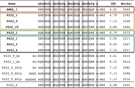
Table 3.1 Table of computationally determined peptides for solution assembly... 50
Table 3.2 Lattice parameters of the self-assembling peptides………... 60
Table 4.1 Crystallographic statistics………. 81
LIST OF FIGURES
Figure 2.1 Ribbon representations of MAX1 and LNK1………... 20
Figure 2.2 Circular dichroism data of MAX1 and LNK1…………... 28
Figure 2.3 Mean residual ellipticity values as a function of incident wavelength
(200-250 nm) at different temperatures(°C) ……….………... 29
Figure 2.4 Transmission electron micrographs of MAX1 and LNK1………... 30
Figure 2.5 Oscillatory time sweep measurements before and after application of
steady-state shear... 33
Figure 2.6 Oscillatory frequency sweep measurements and transmission electron
micrographs of MAX1 and LNK1 after application of injection shear treatment... 35
Figure 2.7 Renderings of representative equilibrium structures of MAX1 and LNK1
octamer after 70 ns………... 36
Figure 2.8 Crossing angle analysis of MAX1 and LNK1 models………... 41
Figure 3.1 Computationally designed, helical, homotetramer assemblies…………. 49
Figure 3.2 Representative backbone configurations of the helix bundle motif
building-block……….. 52
Figure 3.3 Side and top view of the selected low-energy helix bundle……….. 54
viii
Figure 3.5 Analytical ultracentrifugation data and analysis of BNDL_1…………... 58
Figure 3.6 Cast film TEM examples of morphology control with manipulation of
solution assembly conditions and peptide primary sequence... 62
Figure 3.7 High mag TEM of P222_9 ambiently cooled to room temperature... 63
Figure 3.8 High mag TEM of P222_9_Ac and P422_1_Ac ambiently cooled……. 64
Figure 3.9 High mag TEM of P222_9_6Gly ambiently cooled ……… 64
Figure 3.10 Putative structure of assembly of P422_1 helix bundles packed with P4
symmetry……….. 66
Figure 3.11 Putative assembly structures of P222_1 assemblies with P2 symmetry... 66
Figure 4.1 The designed model structure of P422_1……….. 83
Figure 4.2 P422_1 Single Crystals………. 84
Figure 4.3 The analysis of P422_1 crystal structure……….. 86
Figure 4.4 Comparison of the helical bundle between the crystal structure and the
design……….. 88
Figure 4.5 The interfaces between neighboring bundles in the crystal……….. 90
Figure 4.6 Comparison of the interfaces between the crystal structure and the
design……….. 92
Figure 4.7 The superposition of the measured and the calculated SAXS data of
P422_1……….... 94
Figure 4.8 The four-fold-like symmetry related surface holes on the ab plane of the
1 Chapter 1 Introduction
1.1 General background
1.1.1 Existence of biomolecular-derived assemblies in nature
Natural biomolecular assemblies and derivatives are highly-ordered materials that
are complex in structure and rich in function. For example, ferritin is a hollow protein
assembled from 24 subunits, and is critical in iron storage and mineralization in living
systems(1); viruses use highly-symmetrical, protein-coated vesicles for encapsulation and
protection of genetic information, immunological evasion, target binding, and
oligonucleotide delivery(2); S-layer proteins assemble as a major cell-wall component for
protein protection, scaffolding and nutrient uptake in many bacteria(3); laminins undergo
receptor-directed assembly on cell surfaces that are involved in activities such as cell
differentiation, movement and signaling and are important for tissue survival(4, 5);
assembly of nacre defines growth and ordering of inorganic/organic hybrid phases
essential for the superior mechanic and color features of shells(6). Despite the large
variety and unique material properties, assembling processes in natural systems
oftentimes are dependent on the environment or require external energy input. Synthetic
systems can be engineered to overcome the barriers and requirements of assembling
processes in nature. Therefore, it is of great interest to study and engineer biological and
synthetic self-assemblies that are robust in spontaneous assembling processes for
2
1.1.2Overview and history of designed self-assemblies
Engineered nanomaterial self-assemblies are targeted to achieve ordered systems
from disordered components with minimal human or machine intervention(7). Design
approaches have employed synthetic molecules and inorganic-organic hybrids as building
blocks targeting a broad range of applications in nanoelectronics, nanomachines,
photovoltaics, molecular-level data storage and catalysis(8, 9). Although numerous
examples of self-assembling nanomaterials have been discovered since as early as 1950s,
the design of complex, multi-dimensional, highly-ordered self-assemblies has become
successfully realized only in the past two decades. The Murray group pioneered the
design of diverse self-assembling binary and ternary nanoparticle superlattices via
synthetic small-molecules(10–14). Additionally, synthetic polymers are largely employed
in the designs of hierarchical self-assemblies(15), such as dendronized polymers and
block copolymers amphiphiles. The Percec group are experts in dendronized polymer
self-assemblies and have discovered the isomeric libraries of quasi-equivalent primary
structures composing self-assembling dendrons and dendrimers that decode the 3D
assembly architectures(9, 16, 17). The “self-assembly” of block copolymers is usually
realizable through the careful tuning of the assembling environment such as
solvents/non-solvents(18), pH and temperature, as well as block composition and length. For example,
successful approaches to achieve block copolymer assemblies include “phase
inversion”(19), living crystallization-driven self-assembly (CDSA)(18, 20), and kinetic
controls(21, 22).
However, synthetic molecules, especially block copolymers, rarely have
3
DNA, RNA, peptides, and proteins, naturally have the three-dimensional structures and
functions decoded in their primary sequences, therefore providing a sequence-controlled,
alternative approach for novel designs of advanced self-assembling nanomaterials. DNA
tiles and origami are established as a nanomaterials field, where both empirical
knowledge and computer prediction allow the design of proper base pairing and
hybridizations to build targeted, complex nanostructures(23–25). Although the DNA
nanostructures can be highly-ordered, there are still limitations in their finite assembly
size and dimensionalities in all cases(23). Only the DNA tensegrity triangle has produced
substantial 3D crystals thus far(26). DNA’s primarily function in nature is to encode
genetic information. This relatively low-level chemical and functional complexity is
another limitation of DNA self-assemblies compared to proteins- and peptides-based
assemblies. Additional attachments to DNA self-assemblies are necessary to achieve
functional diversity. For example, the Mirkin group has further functionalized DNAs into
spherical nucleic acid (SNA) self-assemblies through multivalent hybridization with
nanoparticles, creating superlattices of various architectures(27–29), and further
producing precisely engineered optical properties(30).
Because of such functional limitations, nature chooses proteins, rather DNAs, for
most of its functional molecular assemblies. Proteins and peptides have an expanded
building-block library of twenty natural amino acids rather than four bases used in DNAs.
For the past decade, more research efforts have been spent in the area of de-novo design
of peptide- and protein-based self-assemblies than that of DNA assemblies. The designed
assemblies span various geometrical, mechanical, and functional properties, and range
4
nanostructures, such as one-dimensional tubes(32), rods(33) and fibrils(31, 34),
two-dimensional sheets (35–38) and three-dimensional cages(39–43) and crystals (44–46).
Because proteins are rich in functions, it has become possible to utilize well-designed
complex nanostructures to spatially distribute chemical functionalities in the controlled
amino acid sequences, although the process remains more challenging for synthetic
polymers and DNAs. Recent advances include a self-assembling biomolecular
hydrogenase enzyme that has improved catalytic activity and resistance to protease, heat
and oxygen due to the ordered encapsulation(47). Additionally, controlled and precise
patterning of hybrid organic–inorganic structures has been successfully realized via
designed solution-phase protein self-assemblies, where metal ions(48), and functional
small molecules(45) co-assemble into hierarchical, highly-ordered nanostructures. Lastly,
the Mayo group and the Mirkin group reported designed self-assembly hybrids between
proteins and DNAs(49, 50) that lay the foundation for the engineering of a new class of
bio-nanomaterials.
1.2 Motivations of designing peptide-based self-assemblies
Proteins and peptides are structurally-complex building blocks that nature
provides for self-assembling nanomaterial. Their structural complexity comes from the
rich primary sequence of amino acids, well-defined secondary structures such as
α-helices and β-sheets, specific intramolecular interactions in tertiary structures, and from
the intermolecular interactions which guide the ordered, sometimes symmetrical,
quaternary structures. Such innate structural complexity of proteins and peptides offers
5
1.2.1 Advantages of designed peptide self-assemblies
Protein- and peptide-based self-assemblies and their derivatives have advantages
over synthetic organic polymers and DNA in multiple aspects: 1) controlled structural
output through primary sequence; 2) a large pool of amino acid monomers, including
unnatural amino acids; 3) accessibility to post-assembling chemical modifications; 4)
high bio-compatibility in-vivo. For peptides, which typically have a short primary
sequence, there are additional advantages, including straightforward synthesis through
standard solid-phase techniques, easier to design into self-assemblies than large proteins,
and highly compatible to nonbiological modifications.
Given these advantages, a variety of biomedical, chemical and energy related
applications of peptide-based self-assemblies have been reported. Hydrogel networks
cross-linked by peptide assemblies are promising artificial extracellular matrices for cell
culture, tissue engineering and regenerative medicine(31, 51, 52). These hydrogel
networks could also serve as carriers for controlled delivery and release of drugs(53, 54)
and functional proteins(55). Peptide-based self-assemblies rich in arginine and lysine
residues have also been proved to have antimicrobial or antibacterial effects, which could
be applied to treat infections(56, 57). Enzyme-responsive peptide self-assemblies have
shown efficacy in cancer therapeutics(58–60). Fusion peptide self-assemblies with
enzymes have assisted in improving protease and esterase catalytic activity(61) and
offered a simple and cost-effective strategy to stabilize and reuse carbonic anhydrase(62).
Besides that, peptide-inorganic hybrid self-assemblies have been designed towards the
peptide-6
DDP(Zn)-TiO2 hybrid assemblies capable of photoinduced charge separation with spatial
control of the donor-acceptor pair by tuning the peptide sequence(63).
Many past successful designs of highly-ordered self-assemblies rely on scaffolds
from natural proteins, where the interior/core structure of the protein is adopted to
preserve its globular fold (tertiary structure), and only interfaces between proteins are
designed and modified(35–38, 43, 46, 64). The de novo designs that use arbitrary protein
backbones and non-natural sequences without existing references to nature still remain
very challenging for predetermined molecular- and nano-structures. The Baker group
tackles the problem in the realm of novel isolated proteins such as tandem protein repeat
architectures and the TIM-barrel fold through geometric and chemical rules(65, 66).
Peptides have fewer degrees of freedom than proteins due to short primary sequences and
are a reasonable starting point to engineer novel tertiary folds and quaternary assembly
architectures from scratch, and design sequences accordingly using rational and
computational approaches. However, the reported successful designs of ordered peptide
self-assemblies with high control and precision are limited to peptide oligomers, like
helical barrels(67), and a cage-shaped nanostructure(68). Therefore, there is still large
room of improvement regarding designing novel and non-natural peptide-based
self-assemblies with predefined nanostructures. Some of the chapters in this thesis will
present results to address the engineering of lattice-forming peptides of non-natural
sequences and backbones by computational design approaches. Next, we will review
rational and computational approaches used in the designs that will appear in the later
7
1.2.2 Computational design vs. rational design
The design methodology for protein- and peptide-based self-assemblies can be
grouped into two categories: rational approaches and computational approaches. Rational
designs combine biomimicry and chemical intuitions to engineer self-assembling
biomaterials. In some cases, natural self-assembling sequence patterns are adopted, such
as amyloid beta and precursor proteins(61, 69, 70), leucine-zippers(71, 72) and laminin-
and collagen-derived motifs(73, 74). Other times peptide amphiphiles are designed based
on well-understood chemical interactions between the polar and nonpolar parts of the
sequences(75, 76). Self-assemblies designed by those strategies usually adopt irregular
nanostructures lack of long-term periodicity because of the less-controlled, nonspecific
associations between the flexible peptide building blocks. These structures are typically
observed in peptide-based hydrogels. To have a better control of the assembly
nanostructures and to engineer defined long-term order, an alternative rational design
strategy employs natural proteins of known structure and symmetry, and fuse them
together through mutagenesis(77),(39), or through simple covalent linkages such as
disulfide bonds. Although these attempts have successfully produced the defined
nanostructures, the selection of assembly building blocks is still constrained by a small
portion of known proteins in nature. Thus the accessible nanostructures are limited. On
the other hand, the computational approach, guided by theory and biomolecular models
of the defined assembly nanostructures such as rods, sheets, cages and lattices, can
achieve precise control and fine tuning of the building-block geometry and orientations.
Furthermore, it is possible to achieve atomistic-level precision with the designed
8
specifically, given an assembly nanostructure, computational design identifies energy
minima in a large conformational and sequence landscape where noncovalent,
interatomic interaction energetics are calculated within the building-block proteins and at
the protein-protein interfaces. However, such interactions are subtle to design as the
energetics are approximated. Therefore, finding sequences that will fold and
self-assemble into a particular nanostructure with predetermined order and symmetry in the
conformational and the sequence energy-landscape is a non-trivial task.
One critical step in the computational design of protein- and peptide-based
self-assemblies is to find such primary sequences able to fold into target tertiary structures
and assemble into targeted nanostructures of higher order. The target tertiary structure
can be taken from the backbone of natural proteins or created using mathematical
models(78, 79). Additional constraints can be imposed on the design, such as symmetric
approximations(80) between the neighboring monomer building blocks, to improve the
computational efficiency when designing infinite lattices or symmetrical cages. There is
typically no unique answer to what such sequences should be, since nature gives many
examples where sequences sharing no similarities fold into nearly identical
structures(81),(82). In order to find those sequences, approximations of folding free
energy, derived from appropriate physical models, can be used as an objective function
and minimized by optimization techniques. Due to the tremendous number of amino acid
combinations possible in only a medium-sized protein sequence, and the various
conformations each amino acid could possess, it is crucial to have computationally
9
accurately. Lazaridis & Karplus(83) and Mendes et al(84). have categorized the energy
models into three types: 1) Statistical effective energy functions, such as the
environmental energy model(85, 86) and the helix propensity model(87); 2) Empirical
effective energy functions achieved by machine-learning(88–92); 3) Physical effective
energy functions, which use atom-level force field such as CHARMM(93), AMBER(94)
and OPLS(95). A combination of these three types of energy models are usually
incorporated in the computational approach at different design stages.
Another important aspect of computational design is the balance between
computational efficiency and sampling thoroughness when facing the huge phase space
of possible sequences. Heuristic techniques are efficient for sampling the whole structural
and/or sequence space; for example, Monte Carlo simulated annealing(96)(97), and
genetic algorithms(98, 99) have been widely used in computational designs. The
drawback of heuristic search is that finding the global minimum of the energy objective
function is not guaranteed. Instead, the objective function could be trapped in one of the
local minima. To overcome this issue, another set of algorithms applying dead-end
elimination or its generations(100–102) have been developed to search through a subset
of the whole phase space and to identify the designed sequence at global minimum.
However, one has to keep in mind that dead-end elimination is very computationally
expensive. An alternative to explicit sampling is to apply computational design via a
statistical-mechanics based probabilistic approach, which characterizes the energetics of
an ensemble of designed sequences in a given nanostructure using the mean-field energy
10
peptide sites are estimated by minimizing the free energy or maximizing the entropy of
the sequence ensemble using nonlinear optimization techniques that identifies local
energy minima. Additional Monte Carlo sampling or the grid search techniques for
backbone structures with different nanostructure configurations can be combined into the
probabilistic approach to design peptide assemblies of defined symmetry and crystalline
order(44).
1.3 Thesis Contributions
This section is a summary of the set of key results and findings of the following chapters:
Chapters 2, 3, and 4, in this thesis.
In section 2.1-2.2, we will introduce de-novo, rationally designed, self-assembling
peptides MAX1 and LNK1. MAX1 and LNK1, despite their similar sequence patterns,
are capable of self-assembling into distinct β-fibril nanostructures respectively and form
hydrogel networks. In section 2.3, experimental characterizations for the two peptide
self-assemblies are detailed. In section 2.4, computational and modeling details for the
two peptide self-assemblies are described. Section 2.5-2.6 shows the key results of the
computational and experimental work followed by discussion. In brief, transmission
electron microscopy (TEM) reveals branched fibrils of MAX1 and rigid rod-like fibrils of
LNK1 with no branches (section 2.5.1). MAX1 and LNK1 hydrogel assembly exhibit
very different rhelogical behaviors (section 2.5.2). We constructed molecular models of
the two peptide self-assemblies and use molecular dynamics (MD) simulations to obtain
dynamic and molecular structural information and to understand the driving force for
11
hydrophobic patterning featuring a hydrophobic face and a hydrophilic face, only the
identities of the hydrophobic amino acids differ. MD simulations and quantitative
analysis of the fibril models are presented to provide molecular insights into the fibril
structures and the fluctuations of the two self-assemblies (section 2.5.3), which can
inform the rational design of new peptide-based materials.
Section 3.1-3.6 presents a set of computationally designed peptide self-assemblies
with four predetermined nanostructures, an isolated helical bundle and three crystalline
nanosheets with different local symmetries. Section 3.3.1 describes the computational
design details of the 29-residue peptides guided by statistical-mechanics based theory.
We encoded the information required for multiple, distinct nanostructures into a novel
sequence of a single bundle motif as a common building block for four distinct,
predetermined material nanostructures. These predetermined structures were specified by
the design of the exterior sequences of the helical bundles motifs. Section 3.3.2 -3.3.3
shows the TEM and small angle neutron scattering (SANS) characterizations of the
nanomaterials composed of the designed peptides upon solution assembly. The peptides
self-assemble into distinct nanostructures (non-assembling bundles and nanosheets) with
various degrees of agreement to the predefined local symmetry and repeating-unit size.
Furthermore, the material solution assembly process is robust with respect to both
variation of solution conditions (pH and temperature) and covalent modification of the
computationally designed peptides. Section 3.4 presents additional modeling to address
and discuss the possible lattice configurations formed in two of the nanosheet materials
12
Lastly, in section 4.1-4.7, we expand our study on one of the computationally
designed self-assembling peptides from Chapter 3, and report the crystal structure and
more solution phase characterizations of the lattice nanostructure. Section 4.4.1 recaps
the computational design strategy in brief, followed by detailed description of X-ray
crystallographic and small angle x-ray scattering (SAXS) characterizations of the peptide
lattice in section 4.4.2. In section 4.4.3, we compare the crystal structure of the peptide
lattice to the design model. The crystal structure reveals that the helical bundle motif
matches to the design with atomistic precision. Such motif is applicable as a common
building block to design different peptide lattices. Section 4.4.4 investigates the lattice
structures assembled under different solution conditions and shows the consistency
between these structures and robustness of the assembly. Additional molecular modeling
and energetics calculated on peptide assembly consistent with the crystal structure
configuration are reported in section 4.4.5. We discuss in section 4.5 the successful
design of the helical bundle motif, and the possible causes of disagreement between the
designed and the observed lattice structure from a thermodynamic perspective. In
particular, we emphasize the critical role of specific hydrophobic interactions in
stabilizing the peptide bundle-bundle interfaces in the tightly-packed crystal assembly.
This discussion also suggests fundamental ways to potentially improve the success rate of
future computational design of protein- and peptide- based lattices.
List of Publications:
13
H. V. Zhang, M. Eibling, Y. Tian, K. L. Kiick, D. J. Pochan, J. G. Saven, “Crystal Structure of a Computationally Designed Lattice-forming Peptide”. In preparation. (2016)
S. Sathaye*, H. V. Zhang*, C. Sonmez, J. P. Schneider, C. M. MacDermaid, V. D. Christopher, J. G. Saven, D. J. Pochan, “Engineering Complementary Hydrophobic Interactions to Control β-Hairpin Peptide Self-Assembly, Network Branching, and Hydrogel Properties”,
14
CHAPTER 2 Engineering Complementary Hydrophobic Interactions to
Control β-Hairpin Peptide Self-Assembly, Network Branching, and Hydrogel
Properties
Adapted with permission from Biomacromolecules. 15, 3891–900 (2014). Copyright
(2014) American Chemical Society.
2.1 Abstract
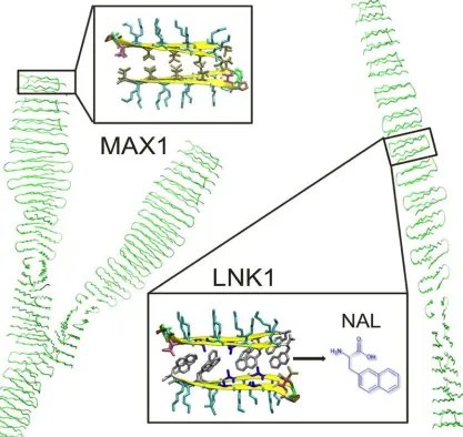
The MAX1 β-hairpin peptide (VKVKVKVK-VD
PPT-KVKVKVKV-NH2) has
been shown to form nanofibrils having a cross-section of two folded peptides forming a
hydrophobic, valine-rich core, and the polymerized fibril exhibits primarily β-sheet
hydrogen bonding(105–111). These nanofibrils form hydrogel networks through fibril
entanglements as well as fibril branching(112). Fibrillar branching in MAX1 hydrogel
networks provides the ability to flow under applied shear stress and immediately reform a
hydrogel solid on cessation of shear. New β-hairpins were designed to limit branching
during nanofibril growth because of steric specificity in the assembled fibril hydrophobic
core. The nonturn valines of MAX1 were substituted by 2-naphthylalanine (Nal) and
alanine (A) residues, with much larger and smaller side chain volumes, respectively, to
obtain LNK1: (Nal)K(Nal)KAKAK-VDPPT-KAKAK(Nal)K(Nal)-NH2. LNK1 was
targeted to self-associate with a specific “lock and key” complementary packing in the
hydrophobic core in order to accommodate the Nal and Ala residue side chains. The
experimentally observable manifestation of reduced fibrillar branching in the LNK1
peptide is the lack of solid hydrogel formation after shear in stark contrast to the MAX1
15
interpeptide interactions within the assembly that is consistent with the branching
propensity of MAX1 vs. LNK1 and in agreement with experimental observations.
2.2 Introduction
Specific molecular recognition interactions within peptide and protein molecules
have been used widely for designing smart, responsive hydrogel materials. Prominent
examples of such efforts include materials based on specific interactions between coiled
coil domains such as leucine zipper domains, interactions between ditryptophan (WW)
and proline-rich domains, standard linear peptides based on purely α-helical structures in
addition to tetratricopeptide repeat (TPR)−peptide interactions(113–118). A specific type
of protein−protein interaction, named the “Lock and Key” mechanism, involves
recognition between specific molecules with complementary steric binding domains. This
specific steric packing has been studied extensively in proteins but not toward designed
materials development(119–122). The lock and key analogy was first put forward by
Emil Fischer more than 100 years ago specifically to describe specificity in
enzyme−substrate interactions(123). An example of widely studied, shape-dependent
lock and key type interactions is the ligand protein interaction between the vitamin biotin
and the egg white glycoprotein avidin, which is of tremendous interest in
biotechnological applications(124). Similarly, Holzinger et al. have reported
complexation between biotin and β-cyclodextrin as a representation of a new bio-receptor
immobilization affinity system(125). Among the related shape-dependent recognition
patterns involving proteins, interactions between proteins and DNA based on DNA local
16
shape variations (various helical topologies and deformations) have also been widely
reported(126). Solution-assembled fibrillar networks based primarily on hydrophobic and
hydrogen bonding interactions between β-hairpin peptides have been studied
extensively(105, 108, 110). In this work, we discuss experimental and simulation studies
of the hydrogel network behavior of β-hairpin peptide-based hydrogels and the impact of
designed hydrophobic and steric interactions at the cores of the fibrils forming the
networks.
Hydrogen bonding-dominated assemblies of peptides into linear nanostructures
include natural nanostructures formed by amyloid and amyloid-like assembly of proteins
and polypeptides, as well as synthetic peptides demonstrating uniform, linear, and
unbranched morphologies such as nanofibrils, nanotapes, nanoribbons, nanobelts,
nanotubes and many hydrogel networks based on such morphologies(127–135). A
prominent example of hierarchical fibrillar self-assembly of peptides based on
hydrophobic interactions and hydrogen bonding has been reported by the Pochan and
Schneider research groups. These groups have studied the self-assembly of MAX1
(VKVKVKVK-VDPPT-KVKVKVKV-NH2) and related peptide sequences(105, 108,
110). MAX1 is an amphiphilic 20 amino acid residue peptide with alternating
hydrophobic valine and hydrophilic lysine residues with a -VDPPT- turn sequence in the
middle. Found in random coil conformations in aqueous solution with neutral to low pH
due to repulsion between positively charged lysine side chains, folding into the β-hairpin
conformation, and consequent intermolecular assembly can be triggered by modulating
17
strength (e.g., ∼ 150 mM NaCl)(108, 111, 112), and increasing temperature (T =37
°C)(106). The higher pH and high ionic strength conditions deprotonate lysines or screen
interactions between lysines, respectively, thus allowing the turn sequence to force the
peptide arms to arrange in an antiparallel conformation, the β-hairpin conformation. The
β-hairpin is stabilized by significant hydrogen bonding between the beta-strands of the
peptide as well as the conformation of the turn sequence that anchors the arms.
Additionally, folding and assembly can be affected by a rise in temperature that induces
the hydrophobic interactions both within and between the peptides both promoting
folding and intermolecular assembly. The hierarchical self-assembly of these β-hairpins
into uniform fibrils takes place due to several interactions. Facial hydrophobic
interactions between the valine faces of two hairpins collapsed together form the
cross-section of a growing fibril (Figure 2.1). Additionally, lateral intermolecular hydrogen
bonding and additional hydrophobic interactions between folded hairpins define the axis
of the growing fibrils(110). After assembly, MAX1 forms self-standing hydrogel
networks that are purely physically cross-linked. The facial hydrophobic collapse at the
core of the growing fibrils sometimes results in formation of a defect characterized by
potentially incomplete burial of the hydrophobic valine side chains and sliding of the
layers at the bilayer interface in a manner that disrupts extension of the linear fibril. Such
defects lead to the nucleation of a branch point in the fibril growth leading to two
daughter fibrils extending from the branch point(112). These branch points contribute to
physical cross-linking of the hydrogel network in addition to fibrillar entanglement. The
18
consequent branch point/new crosslink point that is formed, can be partially attributed to
the lack of specificity in the facial hydrophobic interactions between peptides due to the
uniform steric volumes of the valine side chains. In this paper, we attempt to introduce
“lock and key” type specificity in the facial hydrophobic interactions of the MAX1
peptide in an attempt to significantly limit the formation of branching cross-links formed
as a result of nonspecific hydrophobic collapse.
Several variants of MAX1 have been designed and studied. These variants have
different primary sequences and have been developed to incorporate different
functionalities such as faster gelation kinetics(136, 137), photo-cross-linkable hydrophilic
side chains(138), inherently antibacterial properties(139), and swapped positions of
hydrophobic and hydrophilic residues(110). They all undergo hierarchical self-assembly
in a manner very similar to MAX1 resulting into a uniform fibrillar nanostructure. These
functional variants have been designed by varying the hydrophilic side chains of MAX1.
Each of these peptides has a nonspecific valine hydrophobic face like MAX1. Thus,
designed modifications to the hydrophobic face of MAX1 offer relatively unexplored,
fertile ground to the study of self-assembly and network behavior of the resulting
peptides.
Herein a designed peptide, (Nal)K(Nal)KAKAK-VD
PPT-KAKAK(Nal)K(Nal)-NH2, is presented and studied, wherein four valine residues of MAX1
(VKVKVKVK-VDPPT-KVKVKVKV-NH2) have been replaced with non-natural 2-naphthylalanine
(Nal) amino acid residues, whose side chains possess larger steric volumes than valine.
19
has a smaller side-chain steric volume than valine. Thus, the hydrophobic surfaces of two
LNK1 hairpins can pack specifically into a lock and key type structure in the
hydrophobic core. Such complementary steric interactions are often associated with
well-defined structures in proteins (Figure 2.1). This is in stark contrast to the simple
hydrophobic collapse in MAX1 and the less specific interactions associated with an
interface that comprises only valine residues. Thus, fibrils formed from LNK1 peptide
self-assembly are intended to be unbranched (Figure 2.1) as compared to the branched
fibrils of MAX1 formed from incomplete, defective collapse of non- specific valine faces
during β-hairpin assembly. We hypothesize that LNK1 fibrils form percolated networks
only by fibril entanglement as opposed to the hydrogel networks of MAX1 that form a
network due to fibril branching as well as entanglement. Due to the hypothesized severe
inhibition in the branching in the LNK1 fibrillar hydrogel networks, these materials are
expected to have a significantly different response to shear treatment as compared to the
MAX1 network hydrogels. Specifically, the LNK1 hydrogel networks are expected to
undergo flow and fibril fracture and disentanglement when subject to shear treatment but
lack hydrogel reformation ability post-shear due to fibril collapse and permanent loss of a
percolated network. We report on the assembly of the LNK1 peptide, the local fibril
nanostructure, and ultimate hydrogel network structure via a combination of (i) physical
characterization techniques such as circular dichroism (CD), transmission electron
microscopy (TEM) and oscillatory rheological measurements as well as (ii) molecular
20
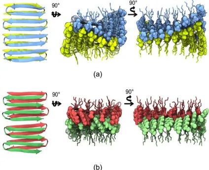
Figure 2.1. Ribbon representations of MAX1 and LNK1. MAX1 can form branched nanofibrils. LNK1 is restrained to form only linear fibrils. The inserts are views of fibrillar cross sections. In MAX1, valine residues form a nonspecific, “flat” hydrophobic interface. In LNK1, specific hydrophobic steric interactions appear as well as the interior hydrophobic interface between complementary naphthylalanine and alanine side chains.
Adapted with permission from Biomacromolecules (2014).
2.3 Materials and methods
Peptide Synthesis. Peptides MAX1 and LNK1 were designed at the University of
21
USA) where matrix-assisted laser desorption ionization time-of-flight (MALDI-TOF)
mass spectral analysis data confirming molecular weights of purified peptides were
obtained. Both peptides were prepared using Fmoc-based solid phase peptide synthesis as
described elsewhere(140).
Hydrogel Preparation. A 1 mg aqueous solution of either MAX1 or LNK1 peptide in
100 μL of deionized chilled water (5 °C) is prepared leading to a 1% (w/v) aqueous
solution. An equal amount of chilled (5 °C) buffer solution pH 9 (250 mM boric acid, 20
mM NaCl) is added to both aqueous solutions to give buffered solutions of either MAX1
or LNK1. Then the temperature of the solutions is raised to 30 °C to obtain 0.5% (w/v)
hydrogels with effective solution conditions pH 9 (125 mM boric acid, 10 mM NaCl).
The same procedure is used to obtain networks from MAX1 and LNK1 peptides using a
different solution condition using a pH 7 buffer (100 mM bis-tris propane, 300 mM
NaCl), ultimately leading to pH 7 (50 mM bis-tris propane, 150 mM NaCl). Briefly, an
equal volume of chilled (5 °C) pH 7 (100 mM bis-tris propane, 300 mM NaCl) buffer
solution is added to a chilled (5 °C) 1% (w/v) aqueous solution of either MAX1 or
LNK1. The temperature is immediately raised to 30 °C to obtain 0.5% (w/v) hydrogels.
Circular Dichroism. CD spectra were collected using an AVIV Model 420 (AVIV
Biomedical, Inc. Lakewood, NJ, USA) CD spectrophotometer. Solutions of MAX1 and
LNK1 (150 μM) at pH 9 and 10 mM NaCl were prepared by adding equal volumes of
chilled (5 °C) buffer solution of pH 9 (250 mM boric acid, 20 mM NaCl) to 300 μM
deionized peptide solution. The random coil to β-hairpin folding transition temperatures
22
incident radiation fixed at 218 nm, the signature wavelength at which a significant drop
in mean residue ellipticity indicates the formation of β-sheet secondary structure. Mean
residue ellipticity [θ] was calculated from the equation [θ]= θobs/(10l × c × n), where θobs
is the measured ellipticity (millidegrees), l is the path length of the cell (cm), c is the
peptide concentration (molar), and n is the number of residues on the peptide sequence.
Temperature scans were performed with 2 °C increments and 5 min equilibration time at
each temperature.
Oscillatory Rheology. Oscillatory rheology measurements were performed on an ARG2
rheometer (TA Instruments, New Castle, DE, USA) using 20 mm diameter stainless steel
parallel plate geometry. For the initial time sweep measurements, the samples for both
LNK1 and MAX1 were prepared as follows. Buffered peptide solutions were prepared in
ice-chilled conditions by adding 120 μL of chilled (5 °C) pH 9 (250 mM boric acid, 20
mM NaCl) buffer to 120 μL of 1% (w/ v) of peptide solution in chilled (5 °C) deionized
water. The chilled (5 °C) buffered peptide solution was quickly transferred to the Peltier
plate of the ARG2 rheometer equilibrated at 5 °C, and the upper plate was lowered to a
gap height of 500 μm. The upper plate and the Peltier plate were equilibrated to 35 °C
prior to carrying out the rheological experiments. Oscillatory time sweep measurement
steps carried out for 90 min each, before and after subjecting the hydrogel networks to a
steady-state shear of 1000/s for 120 s, were carried out for both MAX1 and LNK1.
Throughout the oscillatory time sweep measurements, the oscillatory frequency was
maintained at 6 rad/s and oscillatory strain at 1%. The gap height was maintained at 500
23
of network properties of both MAX1 and LNK1 networks was carried out using
oscillatory frequency sweep measurements at a constant 1% oscillatory strain. Prior to the
frequency sweep measurements both hydrogel networks were allowed to assemble inside
syringes by pulling buffered solutions (pH 9 125 mM boric acid, 10 mM NaCl) of both
peptides into syringes then maintained at 35 °C. The hydrogels were then subjected to a
multiple injection treatment that involved a sequential injection of MAX1 or LNK1
networks formed inside a syringe, seven times through a 27−1/ 2 G needle. For future
reference to this method within this thesis, it shall be referred to as the “Injection Shear”
treatment.
Transmission Electron Microscopy. Transmission electron microscopy was carried out
on a 120 kV Tecnai-12 Electron Microscope (FEI Company, Hillsboro, OR, USA).
MAX1 or LNK1 hydrogel was prepared at 0.5% (w/v) with final hydrogel conditions of
pH 7 (50 mM bis-tris propane, 150 mM NaCl). To observe the fibrillar width, particularly
the local nanostructure of LNK1 vs MAX1 networks, 10 μL of gel was diluted to a
concentration of 0.1% (w/v), and a drop was placed on a 300 mesh copper-coated grid
(Electron Microscopy Sciences, Hatfield, PA, USA) held by a pair of tweezers. Excess
fluid was blotted off with filter paper. Then, immediately, 3 μL of a 1% (w/v) of uranyl
acetate solution in water was placed on the grid and blotted off after 40 s. The grid was
left to dry for an hour and used for imaging. For the preparation of the sample for MAX1
and LNK1 networks after being subject to the injection shear treatment as described
above, a small piece of the treated hydrogel without dilution was placed on a 300 mesh
24
1% (w/v) of uranyl acetate aqueous solution was placed on the grid and blotted off after
40s to stain the sample. This method was applied to the network samples after injection
shear treatment to capture their morphology without dilution and additional mixing
effects.
2.4 Computational modeling
2.4.1 Preparation of initial peptide and fibril structures
The initial structures of both MAX1 and LNK1 were constructed as octamers of
β-hairpins, forming two layers of β-sheets (a bilayer) with hydrophilic lysine residues
exposed on the fibril exterior and the hydrophobic residues (valine in MAX1;
2-naphthylalanine and alanine in LNK1) buried within the bilayer at the interface between
the two β-sheets. The construction of the initial model involved three-steps. First, the
coordinates of the backbone atoms of two 8-residue β-strands were generated de novo,
consistent with trans amide bonds and a pleated β-sheet (φ = −135°, ψ = 135°). Individual
antiparallel β-strands were then positioned at hydrogen-bonding distance from each other
at heavy atom donor−acceptor distances of 3.1 Å. A β-turn (VDPPT) between the two
beta strands was added using cyclic-coordinate descent algorithm for loop
modeling(141), complemented by a neighbor-dependent Ramachandran
distribution(142). The turn contained a trans peptide bond that connected the two proline
residues. An amide capping group was added to the C-terminus. In the next step, amino
acid side chains were added to the constructed backbones. Two hairpins were positioned
such that their hydrophobic faces were in contact and the total energy was minimized
25
variation of the rigid body translation of the parallel hairpins with respect to one another.
Side chain conformations where determined as the most probable amino acid
conformations identified by statistical sequence design algorithm(44, 143–145); Nal side
chain conformations (rotamers) were adapted from those associated with phenylalanine.
In the last step, the complete octamer structures were created by replicating the hairpin
bilayer pairs along the fibril growth direction. Two initial structures of LNK1 were
generated: (1) the neighboring LNK1 hairpins were positioned such that neighboring beta
strands were at hydrogen bonding distance, and (2) this structure from (1) was energy
minimized interbilayer distance while simultaneously solving for the most likely side
chain conformations using computational design methods identify the most probable Nal
conformations(44, 143–145).
2.4.2 Molecular simulations
Molecular dynamics simulations were performed using the NAMD2
package(146) with the modified CHARMM22 force field(93) with TIP4P water
model(147). Energy minimization by conjugate gradient and line search were performed
for 100 to 500 steps on all initial peptide structures. All peptide models were solvated
using the SOLVATE module in VMD(148). A rectangular simulation box was chosen
such that the minimum initial separation between any peptide atom and the nearest
boundary of the solvent box was 15 Å. The appropriate choice of water model can affect
the production of correct conformations of small peptides(149), and four-site water
models such as TIP4P, which can better populate fully coordinated water configurations
26
Sodium and chloride counter-ions were added to neutralize the protonated Lys residues,
and the ionic strength was set to 0.1 M. All Lys residues are protonated to maintain the
equivalence of Lys residues in the fibril. Interestingly, this choice also demonstrates the
robustness of the β-fibril assemblies to the electrostatic repulsive interactions among
these exterior Lys residues. Each system (MAX1: 30,136 atoms, LNK1: 33,132 and
35,738 atoms) was then subjected to a constant 1000-step energy minimization prior to
the simulations.
The NPT ensemble was applied to all systems with constant pressure at 1 atm and
constant temperature at 310 K for MAX1 and 320 K for LNK1 to match experimental
conditions. Constant pressure was maintained by the Nos −Hoover Langevin piston
method, and constant temperature was controlled by Langevin dynamics. Electrostatic
interactions were evaluated fully by particle-mesh Ewald (PME) method at 1.0 Å grid
spacing under periodic boundary conditions. Nonbonded interactions were gradually cut
off from 10 to 12 Å with the pair-list interactions truncated at 14 Å. The SHAKE
algorithm(151) was employed to preserve rigid bonds involving hydrogen atoms. An
initial 300 ps of solvent relaxation was performed for each model system with protein
atoms fixed to their initial coordinates. For the MAX1 octamer, five separate simulations
were performed with different starting solvent configurations and random initial
velocities; these initial configurations were sampled at 100, 150, 175, 200, and 250 ps
from the solvent-relaxation trajectory. For the LNK1 octamer, three simulations were
performed for each of two distinct initial protein structures as described above. For each
27
trajectories at 100, 150, and 200 ps. Each system was then subject to another 1000-step
energy minimization following by a 100 ps preproduction simulation. During these
solvent relaxation and preproduction simulations, the bonded and van der Waals
interactions were calculated at every 1 fs time step, and the long-term full electrostatics
were computed every other step. Subsequently, for the production simulations, a 2 fs time
step was used, and configurations were sampled for analysis every 0.01 ns.
2.5 Results and discussion
2.5.1 MAX1 and LNK1 hydrogel assembly
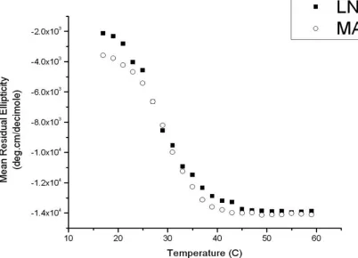
For the same solution conditions (pH 9, 125 mM boric acid, 10 mM NaCl), the CD data
(mean residue ellipticity as a function of temperature) (Figure 2.2) reveal similar folding
transition temperatures (Tf ∼ 30 °C) from random coil to the β-sheet secondary
conformation for both MAX1 and LNK1 (full wavelength spectra for MAX1 and LNK1
from which Figure 2.2 was constructed are shown in the Supporting Figure 2.3). The
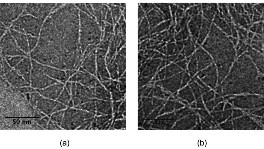
local nanostructures for both MAX1 and LNK1 fibrils are similar, in particular the fibril
thickness as observed by TEM (Figure 2.4); both peptides assemble into fibrils with
uniform width of approximately 3 nm. This similarity in the MAX1 and LNK1 fibril
morphology and the width of the fibril is consistent with each peptide assembling into a
fibril whose cross-section involves two stacked β hairpins that contact one another via a
28
Figure 2.2. Circular dichroism data (mean residue ellipticity in deg·cm/decimole at 218 nm v/s temperature °C) showing approximately the same folding transition temperature (∼30 °C) from random coil to β-sheet secondary conformation for both MAX1 and
29
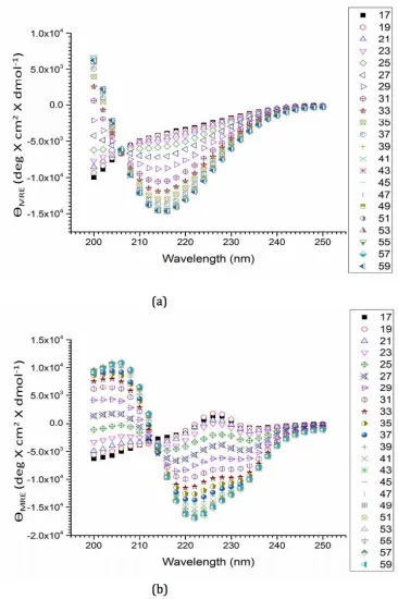
Figure 2.3. Mean residual ellipticity values as a function of incident wavelength (200-250 nm) at different temperatures (°C), indicated in the column to the right for (a) MAX1
(b) LNK1 both at 150 µM concentration at solution conditions pH 9 (125 mM boric acid,
30
Figure 2.4. Transmission electron micrographs of (a) MAX1 and (b) LNK1. Samples were prepared at pH 7, 50 mM bis-tris propane, 150 mM NaCl buffer, and stained with 1% (w/v) uranyl acetate in deionized water. Adapted with permission from
Biomacromolecules (2014).
2.5.2 Rheological behavior of hydrogel assembly
Solid MAX1 hydrogels exhibit a unique property of undergoing shear thinning
and flow under an applied shear stress (outside of the material linear viscoelastic regime)
but immediately recovering into solid gels on cessation of shear. An earlier study by Yan
et al.(152) exploring the hydrogel behavior during and after flow indicated the fracture of
the gel networks into domains much larger than the length scale of individual fibrils in
order to flow. The network morphology within the gel domains during flow was
structurally identical to the parent network at rest; the peptide fibrils displayed the same
cross-section, the same physical cross-linking points of fibrillar entanglement and
branching, and the same porosity. On cessation of shear, the large gel domains
31
rehealing behavior of MAX1 would not exist if the network disassembled into individual
fibrils during flow since there would be no immediate mechanism for the fibrils to
recross-link and percolate into a bulk network. Our hypothesis has been that it is the
frequent and fast fibrillar branching during MAX1 assembly that is key to this
shear-thinning but immediate network reformation behavior. If the network were composed of
fibrils with only physical entanglements for cross-links, the shear flow would disentangle
the fibrils, thus completely disrupting the network after shear. However, the branching
causes the network to fracture into large domains of intact network structure in response
to shear that does not allow the simple disentanglement of peptide fibril physical
cross-links during shear flow.
If the fibril branching in MAX1 is responsible for the observed shear-thinning and
immediate gel reformation behavior, then ridding the system of most fibril branching
should significantly affect the hydrogel flow properties. For MAX1, the putative fibril
interior interface between peptides contains solely valine residues. Such a “flat,”
featureless, hydrophobic interface is expected to tolerate fluctuations in the relative
orientations of peptides within the fibrillar assembly and potentially lead to fibril
branching. As mentioned earlier, the design of steric specificity in the hydrophobic core
of the LNK1 fibrils was an attempt to produce lock and key type interactions and
preclude fibrillar branching. Thus, a very different shear response is expected when
LNK1 hydrogel networks, presumably held together with primarily physical
entanglements as cross-links, are subject to the exact same shear treatment as the MAX1
32
To explore the rheological response of MAX1 and LNK1 networks to shear and
flow, self-assembled hydrogels from LNK1 or MAX1 were produced at a concentration
of 0.5% (w/ v) at pH 9 (125 mM boric acid, 10 mM NaCl). Under these conditions, both
hydrogels show similar preshear behavior with G′∼ 250 Pa ≫G″∼ 20 Pa for the MAX1
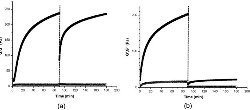
hydrogel and G′ ∼ 200 Pa and G″ ∼ 20 Pa for the LNK1 hydrogel, where G′ and G'' are
the storage and loss moduli, respectively. Figure 2.5a shows the shear-thinning and
recovery character of MAX1 hydrogels in which a MAX1 gel was subjected to
steady-state shear rate of 1000/s for 120 s. Upon cessation of shear, the hydrogel immediately
showed solid gel properties (G′∼ 75 Pa ⟫ G″) and quickly recovered to almost the same
value of storage modulus of the preshear, original MAX1 network (G′ ∼ 250 Pa) after
several hours. In stark contrast, when an LNK1 network, formed with the same solution
conditions as the MAX1 hydrogel network, was subject to the identical shear treatment, it
immediately displayed very weak hydrogel network properties (G′ ∼ 5 Pa > G″) and
failed to recover to even 10% of its original modulus value after several hours (Figure
2.5b). The LNK1 design was meant to prevent branching of the peptide fibrils during
assembly. The response to shear was consistent with this absence of branching, and the
shear treatment destroyed most physical entanglements between LNK1 fibrils that were
unable to reform in any significant way on cessation of shear. This lack of rehealing upon
cessation of shear is a clearly different shear response by the LNK1 hydrogel network,
33
Figure 2.5 Oscillatory time sweep measurements before and after application of steady-state shear (1000/s for 120 s, indicated by dotted line) on 0.5% (w/v) (a) MAX1 and (b) LNK1 networks under the same solution conditions (pH 9 125 mM boric acid 10 mM NaCl). Solid squares indicate G′ (Pa) and open circles G″ (Pa). Adapted with permission
from Biomacromolecules (2014).
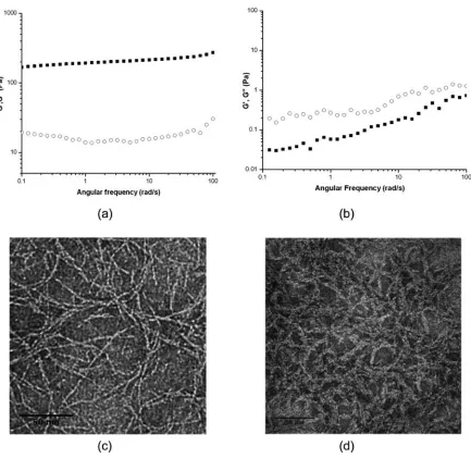
After a simple steady shear treatment of LNK1 inside the rheometer (Figure 2.5b),
the oscillatory shear data indicate a significant reduction in the hydrogel storage modulus
G′, consistent with a strong reduction of network-like properties of LNK1. In order to
more closely mimic conditions of potential clinical usage, both LNK1 and MAX1
hydrogels were subject to the syringe injection shear treatment as described in the
Materials and Methods section. The oscillatory frequency sweep data in Figure 2.6b
reveal a complete elimination of hydrogel network properties of LNK1 networks post
injection shear treatment. The LNK1 sample shows a greater value of the loss modulus,
G″, as compared to the storage modulus, G′, with G″ > G′ at all frequencies. This is a
clear signature of a material that is not a percolated hydrogel network but, rather, is a
particulate suspension or molecular solution. In stark contrast, MAX1 materials retain