Published online October 30, 2014 (http://www.sciencepublishinggroup.com/j/jgo) doi: 10.11648/j.jgo.20140206.12
The physiological changes in pregnancy and their
distribution according to trimester
Ayse Neslin Akkoca
1, *, Zeynep Tugba Ozdemir
2, Raziye Kurt
3, Bilge Bulbul Sen
1, Erhan Yengil
1,
Celalettin Karatepe
4, Oya Soylu Karapınar
3, Cahit Ozer
11Mustafa Kemal University Faculty of Medicine and Research Hospital, Department of Family Medicine, Hatay, Turkey 2Bozok University Faculty of Medicine and Research Hospital, Department of Internal Medicine, Yozgat, Turkey 3Mustafa Kemal University Faculty of Medicine and Research Hospital, Department of Gynecology, Hatay, Turkey
4Mustafa Kemal University Faculty of Medicine and Research Hospital, Department of Cardiovascular Surgery , Hatay, Turkey
Email address:
[email protected] (A. N. Akkoca)
To cite this article:
Ayse Neslin Akkoca, Zeynep Tugba Ozdemir, Raziye Kurt, Bilge Bulbul Sen, Erhan Yengil, Celalettin Karatepe, Oya Soylu Karapınar, Cahit Ozer. The Physiological Changes in Pregnancy and their Distribution According to Trimester. Journal of Gynecology and Obstetrics. Vol. 2, No. 6, 2014, pp. 86-90. doi: 10.11648/j.jgo.20140206.12
Abstract:
Aim: In the present study we aimed to investigate specific skin changes of pregnancy with respect to the trimesters. Materials and Methods: Pregnant women either outpatients or inpatients applied to obstetrics and gynecology department, internal medicine department and family medicine department were involved in this study. Results: 400 pregnant women either outpatients or inpatients were involved in this study. 21 pregnant were excluded from the study cause they did not come regularly to follow-ups. Out of 400 pregnant women, 116 (29%) patients were nullipara and 284 (71%) patients were multipara. Patients ages were between 17-49 years and mean age was 25. The most observed physiological skin changes in order were hyperpigmentation in 311 patients (71%), hypertrichosis in 124 patients (31%), hypothricosis in 15 patients (3.7%), hirsutism in 21 patients (5,2%), hair thickening in 76 patients (19%), hair loss in 92 patients (23%), nail lesions in 18 patients (4,5%), stria distensae in 166 patients (41%), palmar erythema in 127 patients (31%), spider angioma in 52 patients (13%), edema in 132 patients (33%), purpura in 3 patients (0.8%),varices in 50 patients (12,5%), hemorrhoid in 60 patients (15%), gingival hyperemia in 90 patients (22,5%), gingivitis in 50 patients (12,5%). Hyperpigmentation were present in 80% of third trimester and 70% of second trimester pregnant and there was a significant difference (p<0, 05). Hair changes (hypertrichosis, hypotrichosis, hirsutism, hair loss and thickening ) were present in 60% of the second trimester women and %70 of the third trimester of women and the difference was significant (p<0,05). Only hair thickening was present in the 30% of the third trimester pregnant which was significantly different (p<0,05). Stria distensae was observed in 55% of the first trimester pregnant and in 45% of the third trimester pregnant which was significantly different (p<0, 05). Conclusion: As a result, many skin changes, physiologic or none, were detected during the pregnancy. We propose that these changes might be related to age, parity and gestational week of pregnant women.Keywords:
Pregnant, Trimester, Skin Changes, Hirsutism, Hyperpigmentation, Hypertrichosis1. Introduction
Pregnancy is one of the most important periods in human life with hormonal, immunologic, vascular, metabolic and psychological changes. During this period, which is our largest organ (an average of 1.5 square meters) skin shows some speciality and this changes makes pregnancy period even more important. These changes create problems in terms of health in women as well as some of the causes of psychological problems.
The skin changes at gestation are; physiological skin
changes during pregnancy, and pregnancy-specific dermatoses and non-specific dermatoses of pregnancy (1,2). These changes can be seen in almost all pregnant women and in certain processes and varying degrees of severity and in skin, mucous membranes, glands, the vascular system and connective tissue .
fetoplacental unit. Duration of pregnancy, protein hormones such as human chorionic gonadotropin (hCG), human somatomammoprote's, human chorionic thyrotropin released and also progesterone and estrogen are released from the placenta at different levels . These hormones, primary or secondary, is thought to be responsible for many physiological changes in the skin developed during pregnancy.
Increased pigmentation in pregnancy; increased melanocyte stimulating hormone (MSH) are associated with serum levels of estrogen and progesterone and can be seen in up to 90% of pregnancies. They settle much more in areas where naturally melanocyte density is greater. This thickeners usually occur in the first three months . The pigmented areola, nipple, navel circumference, genital, perianal, and axillary areas are having darker appearance. In the midline of the abdominal wall "linea alba" turns to "linea nigra" . Darkening of areola around is called "secondary areola". This pigmentation is reduced after birth but not completely come to the situation before pregnancy.
Varying degrees of hirsutism almost seen in all pregnant women mostly on face, abdomen, pubis, and less on the limbs and back. Placental androgen production may result to hirsutism. Generally back in 6 months after the birth or in some pregnant women in the last 3 months. The bushy hair and bright, plump appearance comes out during pregnancy because of a prolonged anagen phase. After the birth numerous of hairs shows the transition to telogen phase and can cause to diffuse hair loss. 1-5 months after the birth this hair loss usually lasts . Reinstatement is done in about a year, rarely stay sparse. There can be seen a withdrawal of the frontoparietal in pregnants such as androgenic alopecia like male pattern and a withdrawal after birth may not return to the original state. Also, in some women, diffuse thinning of hair can be seen in the oncoming month of pregnancy
The fragility of the nail body, transverse grooving , the distal onycholysis and subungual hyperkeratosis may develop in pregnant women. Moreover, this nail growth rate increased at this period. The pathogenesis of nail changes is not known.
The most frequent change of connective tissue is stria distansea seen at abdomen, thigh, buttocks and breast. Approximately ocur in 90% of pregnant women, especially at 6-7 months of pregnancy. Stria distansea comes out dark pink in color in early stages and occur in the form of shiny atrophic strips. Oncoming months pale and cream colored striae take an atrophic appearance. This situation makes striae less visible but never disappear completely. The real reason is not clear of stria distansea. İt is thought that hormonal factors and the expansion of underlying tissue streches connective tissue and this is the cause of the stria distansea. At the same time adrenocorticosteroids and estrogen ruptures the collagen matrix of the dermis and this causes the weakening of elastic fibers.
The vascular changes in pregnancies qualitatively similar to those of hyperthyroidism or cirrhosis . The increase in
estrogen levels and increased vascularity are responsible from vascular changes. Hyperemia is physiological during pregnancy unless there is an increase in vascular proliferation. Temperature changes due to vasomotor instability and the redness, discoloration and spotting can be seen. Spider angiomas and palmar erythema are often together and they arise 2/3 of white pregnant women. Spider angiomas seen in face, body, upper extremities and where the superior vena cava releases. Palmar erythema seen in the form of redness at thenar and hypothenar areas. Sometimes, common redness or spotted erythema can be seen. They arise between 2-5. months of pregnancy and they decline in for 3 months after birth.
1/3 of pregnant there are varicose veins which occur in the legs due to the increase in venous pressure as a result of uterine growth and its pressure to the femoral and pelvic veins that cause vasodilation. For the same reason, vulvar varicosities and hemorrhoids are often seen.
Edema in the lower limbs can be seen in recent months at least 2/3 of pregnant women (3-5). About 2-2.5 liters of extra fluid is thought to be collected during pregnancy at the range of interstitial. 80% of normal pregnancies has moderate edema (6-10). Prolonged sitting or standing, the increase of capillary permability, varicose veins, venous prevention in the lower limbs, sodium and water retention due to venous pressure increased at legs and hot weather during pregnancy are all factors of edema (10). Due to edema of the lower extremities there can be pain, bloating sensation, night cramps, insomnia and fatigue common in pregnant women (11).
Gums, edema and hyperemia at different intensities is observed in almost all of the pregnants and often accompanied by gingivitis. This table can be together with vestibular vascularization. Especially in those with bad oral hygiene and can be painful and can show ulcerative cruise . Small, heavy bleeding due to trauma may be observed and develops in the last 3 months of pregnancy and postpartum dropps.
In this study unlike other studies in the literature ;we aimed to analyze the physiological changes of the skin in pregnant women according to the number of pregnancy and gestational age.
2. Materials and Methods
400 pregnant women either outpatients or inpatients applied to obstetrics and gynecology, internal medicine and family medicine department between the years of 2011-2013 were involved in this study.
purpura, varicose veins, hemorrhoids, gingival hyperemia and gingivitis.
3. Statistıcal Analysis
SPSS packet program was used for statistical analysis. T-test was used for Independent samples examination, chi-square test for non-numeric comparison of variables. P <0.05 was considered statistically significant.
4. Results
In our study, 400 pregnant patients were evaluated seen in Family Medicine, Internal Medicine, Obstetrics and Gynecology Clinic. Patients ages ranged from 17 to 49, the average age was 25. 116 (29%) pregnant women were nulliparous and 284 (71%) were multiparous. 12 (3%) of pregnant women were at first-trimester, 118 (29.5%) of second-trimester, 270 (67.5%) of in the third trimester. Distribution according to age groups and trimester are shown in Table-1
Table 1. Distribution of pregnant women according to age groups and trimester.
age 15-24 25-34 35 and over
trimester Number of pregnant Number of pregnant Number of pregnant chi-square P
1 1 (0,8%) 10(3,5%) 1(0,8%)
2 35 (26%) 73(31%) 10(35%)
3 90 (70,5%) 160(65,6%) 20(65%) 4,8 0,330
Evaluated 400 pregnant women, 380 (95%) of had at least one of the physiological skin changes and in 20 patients (5%) did not reveal any skin changes.
Table 2. The prevalence of the physiological skin changes of pregnant women according to trimester.
physiological skin changes 1stTRİMESTER Number of pregnant
2ndTRİMESTER Number of pregnant
3rdTRİMESTER
Number of pregnant chi-square P
NO 4(23%) 8(8,1%) 8(2,5%)
YES 8(78%) 111(90%) 261(98%) 19 0,000***
Evaluation of physiological changes in the skin are common in all three trimesters, although detected 98% of pregnant women in the 3rd trimester and 90% of pregnant women in the 2nd trimester and this was was statistically significant (p <0.05).
Table 3. The distribution of the physiological skin changes of pregnant women according to trimester.
1stTRİMESTER Number of pregnant
2ndTRİMESTER Number of pregnant
3rdTRİMESTER
Number of pregnant chi-square P
Pigmentation changes 5(55%) 84(70%) 223(84%) 14 0,001
Hairchanges 2(11%) 71(63%) 179(66%) 12 0,004
Nail complaint 2(11%) 3(2,5%) 10(8,5%)
Connective tissue changes 7(55%) 28(23%) 130(45%) 21 0,000
Vascular changes 8(78%) 70(60%) 180(68%) 3,4 0,187
Mucosal changes 6(44%) 38(35%) 90(35%) 94 0,8
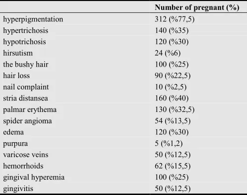
Table 4. The prevalence of the physiological skin changes.
Number of pregnant (%)
hyperpigmentation 312 (%77,5)
hypertrichosis 140 (%35)
hypotrichosis 120 (%30)
hirsutism 24 (%6)
the bushy hair 100 (%25)
hair loss 90 (%22,5)
nail complaint 10 (%2,5)
stria distansea 160 (%40)
palmar erythema 130 (%32,5)
spider angioma 54 (%13,5)
edema 120 (%30)
purpura 5 (%1,2)
varicose veins 50 (%12,5)
hemorrhoids 62 (%15,5)
gingival hyperemia 100 (%25)
gingivitis 50 (%12,5)
5. Discussion
gingival hyperemia (25%), the bushy hair (22%), hemorrhoids (15.5%), gingivitis (12.5%), spider angioma (13.5%), nail complaints (2.5%) and purpura (1%, 2).
Most studies report that more than 90% of pregnant women has hyperpigmentation. In our study, 77% of pregnant women showed an increase in the pigmentation.
There are basically three phases analyzed in hair cycle: anagen, catagen and telogen. In studies of estrogen, its effect was found at the anagen and telogen phase(15). Late in pregnancy telogen ratio has increased from 35% to about 50%. In the postpartum period, telogen effluvium returns to normal (16). Mostly acceptable, with high estrogen levels during pregnancy, trichogram follicular cycle is responsible for the prolongation of the anagen phase. Anagen hair present 81% of pregnant women in the first trimester, while 84% of non-pregnant women. For the second and third trimester this ratio is 90% and 94% respectively and the presence of anagen hair, is higher than that first trimester count. Furthermore, compared to non-pregnant ,it have been reported that pregnant women has increasing rate of thick hair and hair growth is slowed (17). 22% of our pregnant women described stopping at hair loss and bushy hair. The bushy hair was especially in the 3rd trimester and showed statistically significant superiority (p <0.05).
varying degrees of hirsutism can ocur especially in the pubic region, and on the back and the extremities at almost all pregnant women. Hirsutism regresses within six months postpartum. Other changes in pregnant women is also alopecia. May not return to normal after birth (17- 19.20). Other factors associated with postpartum hair loss are stress, blood loss, increased prolactin levels during lactation (17 to 19.20). In the study of Pençe et. al. hirsutism and androgenic alopecia incidence was found 2.7% and 1.95%, respectively, also reported in the study of Dertlioğlu et al. (21) as 20.7% and 20.2%. However, have not identified a significant relationship between hirsutism and androgenic alopecia with patient's age , number of pregnancies and with gestational age . In our study, these rates were 6% and 25%, respectively. The most frequent of these changes was hypertrichosis occurring in at least one area at 35% of women. In 25% of pregnant women hair loss was continued or described hair loss starting during pregnancy .
Nail changes during pregnancy are transverse ridging, nail brittleness, leuconychia, distal onycholysis and the hangnail and their etiology is unknown (22,23). İn the study of Dertlioğlu et al. (21) nail changes observed in 38.5% of pregnant women and the most common symptom was leuconychia (8.9%). Unlike other signs, nail changes were associated only with the patients age but were not associated with the gestational age and pregnancy number. 10 (2.5%) of our cases had nail changes that occur during pregnancy. Nail breakage and transverse ridging was very common.
The most common connective tissue change is stria distansea developing on abdomen, hips, buttocks and breast. Sitrie gravidarum occurs especially in the 6th-7th months of pregnancy approximately 90% of pregnant women (14.23). Stria distansea appears as atrophic bands after birth.
Sometimes it may be accompanied by mild pruritus. This situation makes striae less visible but never completely disappears (24,25). In the literature, Muzaffer et al. (2) reported incidence of stria in pregnancy as 77%, Kumari et al (26) as 79%. In our study, 40% of our pregnant women had stria in the body of at least one localization. We think that the ratio of the difference might have been associated with ethnic origin.
Vascular changes observed in pregnancy develops due to distention, instability and proliferation of the veins and regresses postpartum (14,28,30). Vascular changes such as spider angiomas seen in 67% of pregnant women between 2nd and 5th months and palmar erythema has been detected in 66% of the pregnant women in the first trimester (26). In a study of Dertlioğlu (21) et al. spider angioma and palmar erythema was found 35.6% and 30.4%, respectively; spider angioma was statistically correlated with age and gestational age of the patient, palmar erythema only statistically correlated with gestational age. In our study, 32% of pregnant women had palmar erythema. In 3.5% of pregnant women, weve found at least one spider angioma started during pregnancy. İt is known that more than 40% of pregnant women has varicose veins (2.26). In our study, 13% of had varicose veins (especially in the legs), while 15% had hemorrhoids.
During pregnancy due to increased capillary permeability and fragility, especially the second half of pregnancy purpura lesions may develop (2,13). We only found purpura in 1.2% of pregnant women.
İn the half of the women the edema is observed on face, eyelids, hands and feet . Edema is usually evident early in the morning and lost during the day. İt is important to distinguish this edema from cardiac, renal or pre-eclamptic edema (27,28,30). Vasomotor disturbances, causing symptoms such as "flushing", pallor, hot or cold flushes and cutis marmorata in the legs, and is common during pregnancy (29). In our study, 30% of pregnant women had edema.
More than 80% of women during pregnancy, hyperemia and edema occurs in the gingiva. This effect may be due to hormonal changes or can develop due to gingival poor hygiene, malnutrition and local irritant factors (13,14,26,30). In some publications the gingival changes have been reported as 16.4% (2). İn the study of Dertlioğlu et al. (21) the gingival changes identified at 43% of women but there was not statistically significant relationship between gestational age . In our study, 12.5% of pregnant women had gingivitis, gingival hyperemia have detected in 25% of.
References
[1] Pence B, Kundakçı N, Avşar F. Gebelerde deri değişiklikleri ve dermatozların incelenmesi (Examination of skin changes and dermatoses in pregnant women). T Klin Dermatoloji 1994; 4: 81-86
[2] Muzaffar F, Hussain I, Haroon TS. Physiologic skin changes during pregnancy: A study of 140 cases. Int J Dermatol 1998;37: 429-431.
[3] Kocatepe K. 9 Ay 10 gün hamilelik rehberi(9 months 10 days pregnancy guide). Papatya Yayıncılık Eğitim. İstanbul; 2006; 203-17.
[4] Reeder SJ, Martin LL, Koniak-Griffin D. Maternity nursing,family newborn, and women’s health care, 18th, Lippincott,Philadelphia: New York, 1997; 367-93, 430.
[5] Taşkın L. Doğum ve kadın sağlığı hemşireliği (Birth and women's health nursing), 7. Baskı. Sistem Ofset Matbaacılık: Ankara; 2005. 85-100,173,197-202.
[6] Beksaç MS, Demir N, Koç A, Yüksel A. Obstetrik maternalfetal ve perinatoloji (Maternalfetal obstetrics and perinatology). MN Medikal ve Nobel Basım Yayın Ticaret ve Sanayi Ltd. Şti; 2001; 676-787.
[7] Çiçek N, Mungan T. Klinikte obstetrik ve jinekoloji(Clinical obstetrics and gynecology). Güneş Tıp Kitabevleri: Ankara; 2007; 79-89.
[8] Davison JM. Edema in pregnancy. Kidney Int Suppl 1997;59:90-6.
[9] Pillitteri A. Maternal and child health nursing, 2th. J.B. Lippincott Company; 1992;173-84,238,65.
[10] Reynolds D. Severe gestational edema. J Midwifery Women’s Health 2003;48 :146-8.
[11] Young GL, Jewell D. Interventions for varicosities and leg oedema in pregnancy. Cochrane Database of Systematic Reviews 1998; 2. Art. No:Cd001066.
[12] McLean MA, Wilson R, Thompson JA, Krishnamurthy S,Walker JJ. Immunological changes in early pregnancy. Eur JObstet Gynaecol Reprod Biol 1992; 43: 67-72.
[13] Stern K, Davidsohn I. Effect of estrogen and cortisone on immune hemoantibodies in mice of inbred strains. J Immunol1955; 74: 479-484.
[14] Gebelikte Gözlenen Deri Değişiklikleri ve Gebelik Dermatozlarının İncelenmesi(Examination of skin changes and dermatoses during pregnancy) Fırat Tıp Dergisi 2011; 16 (4): 170-174
[15] Paus R, Cotasrelis G. The biology of hair follicles. N Eng J Med 1999;341: 491-497.
[16] Milikan L. Hirsutism, postpartum telogen effluvium, and male pattern alopecia. J Cosmetic Dermatol 2006; 5: 81-86.
[17] Fiedler VC, Hafeez A. Diffuse alopecia: Telogen hair loss. Disorders of Hair Growth’de. Ed. Olsen EA. McGraw-Hill NewYork1994; 241-253.
[18] Özdemir M, Özdemir S. Gebelikten etkilenen deri hastalıkları (Pregnancy affected skin diseases). Dermatose 2006; 5: 163-168.
[19] Craven AJ ve ark. Prolactin delays hair growth in mice.J Endocrinol 2006; 191: 415-425.
[20] Foitzik K, Krause K, Nixon AJ, Ford CA, Ohnemus U, Pearson AJ,Paus R. Prolactin and its receptor are expressed in murine hair follicle epithelium, show hair cycle-dependent expression, and induce catagen.Am J Pathol. 2003;162: 611-21.
[21] Dertlioğlu S, Çiçek D, Uçak H, Çelik H,Halisdemir N, , Gebelikte Gözlenen Deri Değişiklikleri ve Gebelik Dermatozlarının İncelenmesi, Fırat Tıp Dergisi 2011; 16 (4): 170-174
[22] Özdemir M, Özdemir S. Gebelikte görülen fizyolojik deri değişiklikleri (Physiological skin changes in pregnancy). Dermatose 2006; 5: 22-25.
[23] Coşkun B, Öztürk P, Çiçek D. Gebelik dermatozları (Dermatoses of pregnancy).Dermatose 2004; 3: 72-76.
[24] Lawley TJ, Yancey KB. Skin Changes and Diseases in Pregnancy.In: Freedberg IM, Eisen AZ, Wolff K, Austen KF, Goldsmith LA,Katz SI. Et al eds. Fitzpatrick’s Dermatology in General Medicine.6th ed. New York, McGraw-Hill, 2003 p.1361-6.
[25] Thomas RG, Liston WA. Clinical association of stria gravidarum.J Obstet Gynaecol 2004; 24: 270-271.
[26] Kumari R, Jaisankar TJ, Mohan D, Thappa A. Clinical studyof skin changes in pregnancy. Indian J Dermatol Venereol Leprol 2007; 73: 141
[27] Kroumpouzos G, Cohen LM. Dermatoses of pregnancy.J Am Acad Dermatol 2001;45:1-22.
[28] Graham-Brown RAC. The Ages of Man and their Dermatoses.In: BurnsT, Breathnach S, Cox N, Griffith C. eds. Rook’s Textbook of Dermatology. 7th ed. Oxford: Blackwell Science;2004. p. 70.11.
[29] Winton GB, Lewis CW. Dermatoses of pregnancy. J Am Acad Dermatol 1982;6:977-98.