ORIGINAL ARTICLE
IJPHY
ABSTRACT
Background: The International Headache Society (IHS), 2013 defined Cervicogenic Headache (CGH) as a secondary headache, which implies that headache is caused by a disorder of the cervical spine and its components bony, disc and soft tissue elements. CGH can be a perplexing pain disorder that is refractory to treatment if it is perceived. Patients with CGH exhibited decreases in the quality of life comparable to migraine-patients and patients with tension-type headache, with even lower scores for physical functioning. The objective of the study is to see the effectiveness of PRT versus ischemic compression on pressure pain threshold, range of motion, and headache disability in CGH patients.
Methods: Total of 60 patients of CGH was taken based on inclusion and exclusion criteria, who were divided into three groups, i.e., PRT GROUP A, Ischemic Compression GROUP B, and CONTROL group GROUP C. Group A received PRT, Group B received Ischemic Compression and Group C received conventional treatment 3 sessions per week for 4 weeks.
Results: Significant reduction in Headache disability followed by improved physical functioning measured by Head-ache disability index, improvement in Pressure pain threshold and measured by Pressure algometer and Range of motion measured by Universal goniometer in the group who received Positional release technique along with conven-tional treatment. (p < 0.05) Therefore, it is suggested that the Posiconven-tional release technique reduces Headache disability, Improves Pressure pain threshold and range of motion in college-going students with Cervicogenic headache.
Conclusion: PRT is an effective approach to improve the pressure pain threshold, Headache disability, and Range of motion, thus improving the patient's physical functioning.
Keywords: Cervicogenic headache (CGH), Positional release technique (PRT), Neck pain, Headache, Trigger point, Ischemic compression.
Received 20th June 2019, accepted 05th August 2019, published 09th August 2019
www.ijphy.org
10.15621/ijphy/2019/v6i4/185417
CORRESPONDING AUTHOR
Int J Physiother. Vol 6(4), 140-148, August (2019) ISSN: 2348 - 8336
EFFECT OF POSITIONAL RELEASE TECHNIQUE VERSUS ISCHEMIC
COMPRESSION ON PRESSURE PAIN THRESHOLD, RANGE OF
MO-TION, AND HEADACHE DISABILITY IN CERVICOGENIC HEADACHE
PATIENTS AMONG COLLEGE GOING, STUDENTS. A RANDOMIZED
CONTROLLED TRIAL
*1Premlata ²Priyanka Rishi ³Gurpreet Singh
*1Premlata
Physiotherapist, Narayana Super-Specialty Hospital, Gurugram, Haryana 122505, India. email: [email protected]
²Assistant Professor at SGT University, Gurugram, Haryana 122505, India. email: [email protected] ³Assistant Professor at SGT University,
SGT University, Gurugram, Haryana 122505, India. email: [email protected]
INTRODUCTION
Headache classification subcommittee of international headache society, 2013 has classified Cervicogenic head-ache as a secondary headhead-ache, which implies a disorder of the cervical spine causes headache and its segment bony, disc and soft tissue elements [1]. The IHS has differentiat-ed that there are 14 different subtypes and subcategories of headaches. These headaches have been named either pri-mary headaches, coming from the vascular or muscular re-gion, or secondary, which can result from another source, for example, inflammation or head and neck injuries [2]. Headaches are emerging from a musculoskeletal disorder of the cervical spine named Cervicogenic headaches (CGH) [3]. “Cervicogenic” headaches, first portrayed by Sjaastad, are described as unilateral frontotemporal headaches with clinical symptomatology like a migraine. Clinical presenta-tion of Cervicogenic headaches are: predominantly unilat-eral always on the same side; precipitation either by neck movement or pressure on certain tender spots in the neck; the variable presence of ipsilateral shoulder or arm pain; and pain, stiffness and decreased the range of movement of the neck. Onset might be related to a traumatic incident [4].
CGH is usually associated with sub-occipital neck pain. The IHS classification described as being the pain unilat-eral and bilatunilat-eral [5]. The characteristic feature of Cervi-cogenic headache is the “so-called trigger point, which ex-hibits as a circumscribed hypersensitive skin and muscle spot with a decreased pain threshold” [6]. While Sjaastad and his colleagues have observed that the manifestations of Cervicogenic headache might be encouraged by firm manual pressure on “certain tender spots in the neck,” they originally described them as being over the C2 nerve root, over the greater occipital nerve and the transverse proce-dure of C4-C5 [7].
Cervicogenic headache can be perplexing pain disorder that is refractory to treatment if it is perceived. The condi-tion’s pathophysiology and source of pain have been debat-ed, yet the pain is likely alluded from at least one or more muscular, neurogenic, bony, articular, or vascular struc-tures in the neck [8]. Patients with cervicogenic headache showed decreases in the quality of life comparable to mi-graine patients and patients with tension-type headache, with even lower scores for physical functioning [9]. The re-ported pervasiveness of CGH varies from 13.8% to 17.8% of the headache population in various epidemiological studies [10]. The predominance of CGH in the all general population is estimated to be somewhere in the range of 0.4% and 2.5%, with a female dominance (2:1) [11]. How-ever, Sjaastads and Bakketeig [12] reported the prevalence of 4.1% with no female preponderance, yet in pain man-agement clinics, the incidence is as high as 20% of chronic patient headache [13].
According to Silberstein, cervicogenic headache is char-acterized as pain either in the cranial region, neck, upper trapezius, or sternocleidomastoid (SCM) region, which ra-diates as per their particular pattern [14]. It has been
sug-gested that the suboccipital muscles are a causative factor in both cervicogenic neck pain and headache and also, may become atrophic further complicating the pain syndrome [15,16]. Chronic postural pressure has been proposed to cause hypertonicity of the suboccipital musculature, prompting tension being transmitted to the pain-sensitive Dura resulting in chronic headaches. Patients with CGHs often have the tightness of the SCM, upper trapezius, le-vator, scalenes, sub-occipitals, pectoralis minor, and major muscles [17].
Physical therapy is commonly used for the management of people with CGH [18]. Previous systemic reviews reported preliminary evidence for the application of upper cervical spine manipulation or mobilization for the management of CGH [19].Chaibi A et al., 2012 in a systemic review of manual therapies proposes that spinal manipulative thera-py may be a viable treatment in the management of CGH patients [20]. A study conducted in Australia revealed that upper cervical spine mobilization and manipulation was the most utilized intervention by physical therapists [21]. There are different treatment procedures that healthcare practitioners can use in the treatment of CGH, which in-corporates invasive and non-invasive techniques; the inva-sive treatment technique includes injections, dry needling, and surgery [22] The non-invasive treatment comprises of Ultrasound [23] TENS, Massage, Exercises, manipulation, [24] or mobilization [25] Treatment choices for trigger points incorporate ischemic compression [26] and Posi-tional Release Therapy (PRT) [27].
PRT is a method in which muscles are placed in a posi-tion of greatest comfort, and this causes normalizaposi-tion of muscle hypertonicity and fascial tension. Also, it decreas-es joint hypomobility, increasdecreas-es circulation, followed by a reduction in swelling, decreased pain, and increase mus-cle strength [27]. Therefore, the purpose behind this study was to assess the effectiveness of Positional release tech-nique versus ischemic compression alongside convention-al treatment in the management of CGH in college-going students and to check whether positional release technique and ischemic compression are viable for reducing trigger points in cervical muscles causing Cervicogenic headache. METHODOLOGY
This study was carried out at Physiotherapy OPD of SGT hospital, Gurugram. The Ethical Research Committee ap-proved this study at Faculty of Physiotherapy SGT Univer-sity (SGTU/FOP/2018/37), Gurugram. The whole proce-dure was explained to the subjects, and informed consent was taken from all the subjects included in the study. The therapist informed the subjects that the part to be treated should be exposed during the treatment according to the treatment need, and consent was taken prior. All the sub-jects selected for the study had undergone an assessment
which was done by the physician, and then subjects were
disability measured by Headache disability index (HDI).
All measurements were repeated after the last day of 2nd
and 4th week.
Sixty subjects were incorporated into the study who have fulfilled the criteria of Age – 18 to 30 years, Both male and female [28] and they had to be present with the diagnosis of CGH according to the criteria of Sjaastad and Fredrik-sen [29],1) Unilateral pain starting in the neck and radi-ating to the frontotemporal region, 2) Pain aggravated by neck movement, 3) Restricted cervical range of motion, 4) Joint tenderness in at least one of the upper cervical spine (C0-C3). Headaches frequency of at least once per week and more than three months.
Trigger point diagnosis by Simons et al [30]: Presence of a palpable taut band in a skeletal muscle, Presence of a hy-persensitive spot within the taut band, Palpable or local visible twitch on snapping palpation, Reproduction of re-ferred pain elicited pain by palpation of the sensitive spot, Headache disability index- moderate disability subjects were taken in the study, Pressure pain threshold- mini-mum 3.2kg/cm2 pressure pain threshold should be present across all cervical segments and tenderness grade 3 sub-jects were included in the study.
Participants were excluded if they had any malignancy, spi-nal infection, vestibular dysfunction, vertebral tumor, and fractures if they had a severe head or neck trauma within last 12 months, any neck or intracranial surgery, radiating pain to upper extremities and cervical disc condition and arthritis of the cervical spine. Participants were excluded if they had other primary headaches (i.e., migraine and ten-sion-type headache) bilateral headaches — any contraindi-cation to manual therapy [28].
Subjects were divided into three groups by block random sampling method, i.e., GROUP A (Positional release tech-nique group), GROUP B (Ischemic compression group), and GROUP C (Conventional treatment group). Treat-ment was given to the subjects for three times a week for four weeks, for 45 minutes of duration [31].
Figure1. Flow chart of selection criteria of the subjects
GROUP B N= 20 GROUP C N= 20 GROUP A N= 20
ULTRASONIC THERAPY + HOT PACK ISCHEMIC
COMPRESSION THERAPY + CONVENTIONAL TREATMENT POSITIONAL RELEASE
TECHNIQUE + CONVENTIONAL TREATMENT
N= 90 headache subjects
15 excluded
10 subjects were of tension-type headache and migraine. 5 subjects were of cervical spondylosis
N= 75
15 subjects excluded because they were not matching the inclusion criteria.
Randomization was done and subjects divided into three groups. N= 60
Figure2. Flow chart of treatment protocol given to sub-jects
Measurements of all variables were taken at baseline, last day of the 2nd week and last day of the
4th week. The analysis was done using the appropriate statistical tool to interpret the results.
Positional release technique was given to subjects + Ultrasonic therapy + Hot pack after the end of the treatment session for relaxation Protocol performed: 3 sessions per week for 4 weeks 45 minutes duration
Ultrasonic therapy + Hot pack were given to subjects after the end of the session for relaxation Protocol performed: 3 sessions per week for 4 weeks 45 minutes duration Ischemic compression
was given to subjects + Ultrasonic therapy + Hot pack after the end of the session for relaxation Protocol performed: 3 sessions per week for 4 weeks 45 minutes duration
60 subjects were divided into three groups.
20 subjects were included in GROUP A
20 subjects were included in GROUP B
20 subjects were included in GROUP C
PROCEDURE
Group A (positional release technique [27]and conven-tional treatment)
Subjects in Group A got PRT [22] along with conven-tional treatment, which includes ultrasonic therapy for 5 minutes for each muscle (SCM, UPPER TRAPEZIUS, and RCPMN). The intensity was depended according to the location of the muscle, either superficial or deep (1Hz for deep muscles & 3Hz for superficial muscles). 3MHz at 1.0 watts/cm2 intensity was for Upper trapezius, 3MHz at 1.0 watts/cm2 intensity was used for Sternocleidomastoid muscle, and 1MHz at 1.0 watts/cm2 intensity was used for Rectus capitis posterior minor muscle then PRT was given at the most hyperirritable spot in the muscle belly. Three muscles were taken for the treatment i.e.
Upper trapezius: Positional release technique (PRT) was given at the point of a tension of the upper trapezius, which was diagnosed by the therapist. The subjects were in a supine position, the therapist kept the upper trapezius muscle in the greatest comfort position, the subjects head was flexed laterally toward the trigger point, and her shoul-der was abducted 90 degrees. In that position, the thera-pist monitored the trigger point with her index finger and maintained pressure from the thumb on that position until release was felt. This was repeated for 3-4 times with 20 second relaxation time with every repetition.
Sternocleidomastoid: PRT was given at the point of the tension of the sternocleidomastoid muscle, which was di-agnosed by the therapist. The subjects were in a supine po-sition; the therapist kept the sternocleidomastoid muscle in a position of greatest comfort and therapist palpated to find a tender spot with a pincer grasp. Therapist monitored the tender point with her index finger and maintained pressure on that tender point by turning the neck on the same side until release was felt. This was repeated for 3-4 times with 20 second relaxation time with every repetition.
was in a supine position; the therapist kept the rectus capi-tis posterior minor muscle in a position of greatest comfort and therapist palpated to find a tender spot with a pincer grasp. Therapist monitored the tender point with her index finger and maintained pressure on that tender point by ex-tending the neck until release was felt. This was repeated for 3-4 times, with 20 seconds of relaxation time with every repetition. A hot pack was given for 10 minutes to subjects after treatment for relaxation.
Group B (ischemic compression [26] and conventional treatment)
Subjects in Group B got Ischemic compression along with conventional treatment, which includes ultrasonic therapy for 5 minutes for each muscle (SCM, UPPER TRAPEZIUS, and RCPMN). The intensity was depended according to the location of the muscle, either superficial or deep (1Hz for deep muscles & 3Hz for superficial muscles). 3MHz at 1.0 watts/cm2 intensity was for Upper trapezius, 3MHz at 1.0 watts/cm2 intensity was used for Sternocleidomastoid muscle, and 1MHz at 1.0 watts/cm2 intensity was used for Rectus capitis posterior minor muscle then Ischemic com-pression was given at the most hyperirritable spot in the muscle belly. Threemuscles were taken for the treatment i.e.
Upper trapezius: For upper trapezius muscle subjects was in a supine position on the couch; therapist kept the upper trapezius muscle in lengthening position the therapist was utilized a pincers grasp, placing the thumb and index finger over the active trigger point. The pressure was maintained until a release of the tissue barrier was felt. The pressure will be applied intermittently initially and then continu-ously for 90 seconds according to subjects tolerability. This was repeated 3-4 times with 20 second relaxation period.
Sternocleidomastoid: For SCM muscle, the therapist has utilized a pincers grasp, placed her thumb and index fin-ger over the active trigfin-ger point of the sternocleidomastoid muscle. The pressure was maintained until a release of the tissue barrier was felt. The pressure was applied intermit-tently initially and then continuously for 90 seconds ac-cording to subjects tolerability. This was repeated 3-4 times with 20 second relaxation period.
Rectus capitis posterior minor:For Rectus Capitis Poste-rior Minor (RCPMN) subjects were in a prone position, and the therapist has utilized a pincers grasp, placed her thumb and index finger over the active trigger point of RCPMN muscle. The pressure was maintained until a re-lease of the tissue barrier was felt. The pressure was applied intermittently initially and then continuously for 90 sec-onds according to subjects tolerability. This was repeated 3-4 times with 20 second relaxation period. A hot pack was given to subjects for 10 minutes after treatment for relax-ation.
`GROUP C (ultrasonic therapy and hot pack)
Subjects in Group C got ultrasonic therapy, which included Ultrasonic therapy for 5 minutes for each muscle (SCM, UPPER TRAPEZIUS, and RCPMN). The intensity was
depended according to the location of the muscle, either superficial or deep (1Hz for deep muscles & 3Hz for su-perficial muscles). 3MHz at 1.0 watts/cm2 intensity was for Upper trapezius, 3MHz at 1.0 watts/cm2 intensity was used for Sternocleidomastoid muscle, and 1MHz at 1.0 watts/ cm2 intensity was used for Rectus capitis posterior minor muscle. A hot pack was given for 10 minutes to the subjects after treatment for relaxation.
DATA ANALYSIS
The data were analyzed by using the software package SPSS 24 for window version. Mean, and standard deviation of all the variables were calculated. The level of significance was set at p<0.05. Paired T-test was used to compare within the group differences in the analysis of the data collected for Pressure pain threshold, Range of motion, and Headache disability at baseline, 2nd, and 4th week of intervention. RESULTS
Mean comparison of Age, Weight, Height, and BMI was done. The study was done on 60 subjects who were equally divided into three groups, with 20 subjects in each group. Between-group analysis of these baseline characteristics showed that there was no significant difference in mean age, mean height, mean weight, and mean BMI of the sub-jects in all three groups(p>0.05). (Refer figure 1).
Figure 1: Shows the mean comparison of Age, Weight, Height, and BMI between Group A, Group B, and
Group C.
NS: Nonsignificant
The between-group analysis of Pressure pain threshold of all three muscles i.e., Upper trapezius, Sternocleidomastoid muscle and Rectus capitis posterior minor muscle (Right and Left side), Range of motion of all four ranges i.e., Flex-ion, ExtensFlex-ion, Lateral flexion and Rotation(Right and Left side) and Headache disability was done using Independent T-test which shows that there was no significant difference seen in the mean value of Group A, Group B and Group
C at baseline but at 2nd week the mean value of all three
groups has improved significantly and at 4th week Group A
Table 1: Pre and post-treatment values ofHeadache Dis-ability
HEADACHE DISABILITY Mean ± SD p-value
Pre values Group A
Group B 56.80± 13.0347.10± 9.85 .012
Group B
Group C 47.10± 9.8547.30 ±9.78 .95
Group A
Group C 56.80± 13.0347.30 ±9.78 .013
Post values Group A
Group B 10.80 ± 4.8335.20± 8.52 .005
Group B
Group C 39.20 ± 9.5935.20± 8.52 .010
Group A
Group C 10.80 ± 4.8339.20 ± 9.59 .001
*p-value< .05 or .001 (significant or highly significant re-spectively)
*p-value> .05 (non-significant)
Note: Data was presented as mean + SD. Results of the analysis were done by independent T-test, which shows that group A showed the Highly significant difference, which is an experimental group where participants re-ceived Positional release technique along with convention-al treatment. P-vconvention-alue (P<0.01) was seen at the 4th week, which showed that there was a highly significant difference in Headache disability at the end of 4th week.
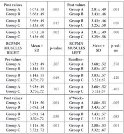
Table 2: Pre and Post-treatment values of Pressure pain threshold in Upper trapezius, Sternocleidomastoid, and Rectus capitis posterior minor muscle (Right and left
side). UPPER TRAPEZIUS RIGHT Mean ± SD p-value UPPER TRAPE-ZIUS LEFT Mean ±
SD p-val-ue
Pre values Group A
Group B 3.58± 8.013.21± 8.01 .131
Pre values Group A
Group B 3.47± .453.33± .48 .022
Group B
Group C 3.21± 8.013.21± .62 .017 Group BGroup C 3.33± .482.8± .64 .064
Group A
Group C 3.58± 8.013.21± .62 .312 Group AGroup C 3.47± .452.8± .64 .954
Post values Group A
Group B 4.27± 6.043.21±8.01 .002
Post values Group A
Group B 3.53 ± .673.87± .46 .003
Group B
Group C 3.21±8.013.82± .73 .018 Group BGroup C 3.53 ± .673.52 ± .84 .020
Group A
Group C 4.27± 6.043.82± .73 .001 Group AGroup C 3.52 ± .843.87± .46 .001
STERNO- CLEIDO-MASTOID MUSCLE RIGHT Mean ± SD p-value STERNO- CLEIDO-MASTOID MUSCLE LEFT Mean ±
SD p-val-ue
Pre values Group A
Group B 3.89± .564.08± .50 .274
Pre values Group A
Group B 3.61± .493.83± .46 .154
Group B
Group C 4.08± .503.63 ±.60 .015 Group BGroup C 3.83± .463.46 ±.60 .035
Group A
Group C 3.89± .563.63 ±.60 .163 Group AGroup C 3.61± .493.46 ±.60 .390
Post values Group A
Group B 3.07± .583.66± .49 .005
Post values Group A
Group B 2.81± .493.43± .46 .001
Group B
Group C 3.66± .493.43± .60 .012 Group BGroup C 3.43± .463.25± .58 .020
Group A
Group C 3.07± .583.43± .60 .002 Group AGroup C 2.81± .493.25± .58 .000
RCPMN MUSCLES RIGHT Mean ± SD p-value RCPMN MUSCLES LEFT Mean ±
SD p-val-ue
Pre values Group A
Group B 3.93± .494.14± .55 .207
Baseline– Group A
Group B 3.68± .523.83± .57 .376
Group B
Group C 4.14± .553.73±.72 .049 Group BGroup C 3.83± .573.52±.67 .120
Group A
Group C 3.93± .493.73±.72 .307 Group AGroup C 3.68± .523.52±.67 .405
Post values Group A
Group B 3.11± .503.69± .54 .004
4thWeek–
Group A
Group B 2.88± .533.43± .57 .005
Group B
Group C 3.69± .543.52±.72 .030 Group BGroup C 3.43± .573.32±.67 .085
Group A
Group C 3.11± .503.52± .72 .001 Group AGroup C 2.88± .533.32± .67 .001
*p-value< .05 or .001 (significant or highly significant re-spectively)
*p-value> .05 (non-significant)
Note: Data was presented as mean + SD. Results of the anal-ysis were done by independent T-test, which shows that group A showed the Highly significant difference, which is an experimental group where participants received Po-sitional release technique along with conventional treat-ment. P-value for upper trapezius muscle right and left side was (P<0.001) and (p<0.001), for sternocleidomastoid muscle right and left side it was (p<0.002) and (p<0.000) and for rectus capitis posterior minor muscle right and left side it was (p<0.001) and (p<0.001) was seen at 4th week which showed that there was a highly significant difference in pressure pain threshold readings at the end of 4th week.
Table 3: Pre and Post-treatment values Cervical Flexion and Extension Range of motion.
FLEXION Mean ± SD p-val-ue EXTEN-SION Mean ± SD p-val-ue
Pre values Group A
Group B 43.00± 8.0144.75± 4.44 .398
Pre values Group A
Group B 47.75± 7.8642.75± 4.44 .39
Group B
Group C 44.75± 4.4448.00±5.23 .041 Group BGroup C 47.75± 7.8641.25±4.83 .041
Group A
Group C 43.00± 8.0148.00±5.23 .025 Group AGroup C 47.75± 7.8641.25±4.83 .003
Post values Group A
Group B 53.75 ±6.0450.00± 4.29 .006
Post values Group A
Group B 57.75 ±7.8647.75± 4.99 .005
Group B
Group C 51.70 ± 4.7750.00± 4.29 .025 Group BGroup C 45.10 ± 4.9447.75± 4.99 .030 Group A
Group C 51.70 ± 4.7753.75 ±6.04 .003 Group AGroup C 45.10 ± 4.9457.75 ±7.86 .002 *p-value< .05 or .001 (significant or highly significant re-spectively)
Table 4: Pre and post-treatment values of Lateral flexion and Rotation (Right and Left side) Range of motion.
LATERAL FLEXION
RIGHT Mean ± SD p-val-ue
LATERAL FLEXION
LEFT Mean ± SD
p-val-ue Prevalues
Group A
Group B 35.50± 6.8632.25± 3.90 .072
Pre values Group A
Group B 37.75± 6.3837.50± 5.50 .895
Group B
Group C 32.25± 3.9035.00±4.29 .038 Group BGroup C 37.50± 5.3039.00±5.76 .405
Group A
Group C 35.50± 6.8635.00±4.29 .784 Group AGroup C 37.75± 6.3839.00±5.76 .519
Post values Group A
Group B 42.25 ±4.4437.25± 3.80 .005
Post values Group A
Group B 43.50 ±2.8641.25± 3.93 .004
Group B
Group C 38.35 ± 4.7437.25± 3.80 .423 Group BGroup C 43.00 ± 5.7641.25± 3.93 .035 Group A
Group C 38.35 ± 4.7442.25 ±4.44 .003 Group AGroup C 43.00 ± 5.7643.50 ±2.86 .002 ROTATION
RIGHT Mean ± SD p-val-ue
ROTA-TION
LEFT Mean ± SD
p-val-ue Pre values
Group A
Group B 71.00± 9.6870.00± 9.46 .743
Pre values Group A
Group B 76.00±10.5977.25± 9.10 .691
Group B
Group C 70.00± 9.4670.50±9.30 .867 Group BGroup C 78.25±10.0477.25± 9.10 .743
Group A
Group C 71.00± 9.6870.50±9.30 .657 Group AGroup C 76.00±10.5978.25±10.04 .495
Post values Group A
Group B 86.75 ±7.3074.75± 8.96 .005
Post values Group A
Group B 88.25 ±6.7481.25± 7.76 .004
Group B
Group C 74.50 ± 9.3074.75± 8.96 .050 Group BGroup C 81.25 ± 8.9081.25± 7.76 0.30 Group A
Group C 74.50 ± 9.3086.75 ±7.30 .002 Group AGroup C 81.25 ± 8.9088.25 ±6.74 .001 *p-value< .05 or .001 (significant or highly significant re-spectively)
*p-value> .05 (non-significant
Note: Data was presented as mean + SD. Results of the anal-ysis were done by independent T-test, which shows that group A showed the Highly significant difference, which is an experimental groupwhere participants received Posi-tional release technique along with convenPosi-tional treatment. P-value for flexion range of motion was (P<0.003) and for extension it was (p<0.002), for lateral flexion right and left side it was (p<0.003) and (p<0.002) and for rotation right and left side it was (p<0.002) and (p<0.001) was seen at 4th week which showed that there was a highly significant dif-ference in cervical range of motions at the end of 4th week. DISCUSSION
This trial provided evidence that Positional release tech-nique versus ischemic compression therapy, when com-pared with conventional treatment regime, was effective for cervicogenic headache patients, even though there was no statistical evidence of an additive effect when the two treatments were utilized at the same time. Beneficial effects were found in headache frequency and intensity as well as in pressure pain threshold, cervical range of motion, and headache disability for both restorative techniques utilized alone and in combination with conventional treatment. The subject in this study had similar baseline values for all
dependent variable, suggesting that all groups had a homo-geneous distribution of subjects.
One of the inclusion criteria of this study was that all par-ticipants had to be between 18 and 30 years of age. The age range was restricted to eliminate chances of degenerative changes [33]. Active myofascial trigger points are also most commonly found in people younger than 50 years of age, as these are the most active years [34]. Thus the mean age of all three groups fell within the seage limits. All the subjects met the inclusion criteria for the study falling between the age of 18 and 30 years, with the mean age of the 60 partici-pants being 23. 20 years. No statistically significant differ-ence was found with regards to age; thus, all three groups are comparable in terms of age.
In this study within-group analysis of group A, Head-ache disability index showed significant improvement in headache disability which was 80.99%, in group B it was
25.26%, and in group C it was 17.12% by the end of 4th
week. (Refer table 1) Therefore, group A shows a highly significant difference by reducing headache disability and improving physical functioning in college-going students with Cervicogenic headache by 80.99%. The evidence for proving our result is that a recent pilot study by Premlata et al., 2019, have also shown a reduction in pain and head-ache disability in cervicogenic headhead-ache patients and have significant improvement in headache disability by 80.29% [35]. A. Kumaresan et al., 2012., proposed in their study that PRT, with the treatment time, selected produced sig-nificant pain relief. Pain relief could have occurred due to the decrease in the intrafusal and extrafusal fiber disparity and reset of the inappropriate proprioceptive activity [36]. The treatment of PRT relaxes the muscle spindle mecha-nism of the counterstained tissue, decreasing abberent gamma and alpha neuronal activity, thereby breaking the sustained contraction followed by a reduction in pain and improvement in patient’s physical functioning [37].
the greatest comfort position. The resultant relaxation of tissue leads to an improvement in vascular circulation and removal of the chemical mediators of inflammation. Thus, PRT may eliminate peripheral & central sensitization.This technique may also reduce the central sensitization directly by the damping influence on the facilitated segment in the spinal cord [38].
In this study within-group analysis of group A, there was significant improvement in cervical range of motion (re-fer table 3 and 4) that is flexion, extension, lateral flexion and rotation which was measured by Goniometer showed 24.88% improvement in cervical flexion, 20.96% in cer-vical extension, 18.87% and 15.38% on the right and left side lateral flexion and 22.11% and 16.05% on the right and left side rotation. In group B it was 23.04% cervical flexion, 11.70% cervical extension, lateral flexion right and left side 15.52% and 9.86% and for the rotation, the right and left side was 6.71% and 2.65%. In group C significant improvement was occurred for Flexion showed 5.72% and extension 9.46%, for lateral flexion right and left side it was 8.57% and 10.25%, for Rotation right and left side it was
10.67%, and the end saw 3.85% improvement of 4th week.
Thus, group A showed Highly significant improvement by gaining a range of motion by the end of the 4thweek. Bode’s Pardo et al., 2013 [28], in their study manual treatment for cerviogenic headache and active trigger point thera-py in the sternocleidomastoid muscle experienced a more significant increase in the active cervical range of motion in subjects who received trigger point therapy than those received simulated trigger point therapy. D’Ambrogio et al.,1997, demonstrated that PRT is a technique in which muscles are placed in a position of greatest comfort, and this causes normalization of muscle hypertonicity & fas-cial tension, a reduction of joint hypomobility which in turn improved Range of motion, increased circulation & reduced swelling, decreased pain and enhanced muscular strength [27].
Wong et al., 2004, [39]in their randomized controlled study evaluated the reliability and validity of a tender point pal-pation scale (TPPS) and the effect of Strain Counter Strain (SCS) on painful tender points. The experimental design employed a convenience sample of 49 volunteers with bilat-eral hip tender points, randomly assigned to three groups, each receiving SCS, exercise (EX), or SCS and EX. Pain. By the end of the study, all groups demonstrated signifi-cant pain decreases in both muscle groups showed with the Visual analog scale (VAS) and (TPPS). However, the SCS groups tended toward more significant pain reductions than the exercise group for hip abductors and adductors. Lewis et al., 2001 [40]in their pilot study showed that four cases of low back pain were treated with SCS as the sole treatment. Outcome measures were derived from the Mc-Gill Pain Questionnaire and the Oswestry Low Back Pain Disability Questionnaire. All patients registered reductions in pain and disability following SCS intervention. No ex-perimental evidence for the effectiveness of SCS was of-fered; however, outcomes suggested that a controlled study
was warranted to examine the efficacy of SCS for the treat-ment of low back pain.
There are so many evidence to support this study where the reduction of headache symptoms after the manage-ment of trigger points by different procedures was seen but the effectiveness of PRT in the improvement of patients with cervicogenic headache remains unclear [39]. This study focuses on a patient with Cervicogenic headache and trigger points in their cervical muscles (Upper trapezius, Sternocleidomastoid, and Rectus capitis posterior minor muscles) where Positional Release Technique treated them. The main goal of Positional release technique is to reduce pain by reducing swelling, decreasing headache disability followed by improving physical functioning. We discovered that patients with CGH received Positional release thera-py versus ischemic compression along with conventional treatment experienced a decrease in pressure pain sensi-tivity and improvement in pressure pain threshold, gain in cervical range of motion is experienced and enhance-ment in physical functioning by decreasing the headache disability. PRT can be an ideal treatment for treating CGH patients. The study was limited to small sample size. It can be done on large sample size.The samples were limited to an age group between 18-30 years. It can be generalized to other age groups. So, the elderly population can also be taken into consideration for the study with other variables.
CONCLUSION
This study showed that all the variables had improved sig-nificantly in the Positional release technique group so it was concluded that positional release technique could be an effective application to improve Pressure pain thresh-old, gaining Range of motion and reducing Headache dis-ability by improving patients physical functioning.
Acknowledgment: The study was self-funded
CONFLICT OF INTEREST:No REFERENCES
[1] Olesen J, Bes A, Kunkel R, Lance JW, Nappi G, Pfaffen-rath V, et al., The international classification of head-ache disorders, (beta version). Cephalalgia. 2013 Jul 1;33(9):629-808.
[2] Headache Classification Subcommittee of the Interna-tional Headache Society. The internaInterna-tional classifica-tion of headache disorders. Cephalalgia. 2004;24(1):9-160.
[3] Jull G, Trott P, Potter H, Zito G, Niere K, Shirley D, Emberson J, Marschner I, Richardson C. A random-ized controlled trial of exercise and manipulative therapy for cervicogenic headache. Spine. 2002 Sep 1;27(17):1835-43.
[4] Jaeger B. Are “cervicogenic” headaches due to myofas-cial pain and cervical spine dysfunction?. Cephalalgia. 1989 Sep;9(3):157-64.
[5] Treleaven, J., G. Jull, and L. Atkinsin, cervical muscu-loskeletal dysfunction in post-concussional headache. Cephalalgia, 1994. 14(4): p. 273-9; discussion 257.
Cervicogen-ic headache-The clinCervicogen-ical pCervicogen-icture, radiologCervicogen-ical findings and hypotheses on its pathophysiology. Headache 1987;27:495-99
[7] Sjaastad O, Saunte C, Hovdal H, Breivik H, Grønbæk E. “Cervicogenic” headache. An hypothesis. Cephalal-gia 1983;3:249-56.
[8] Biondi DM. Cervicogenic headache: a review of di-agnostic and treatment strategies. The Journal of the American Osteopathic Association. 2005 Apr 1;105(4_ suppl):16S-22S.
[9] Van Suijlekom HA, Lame I, Stomp-van den Berg SG, Kessels AG, Weber WE. Quality of life of patients with cervicogenic headache: a comparison with control subjects and patients with migraine or tension-type headache. Headache. 2003;43:1034-1041.
[10] Vavrek D, Haas M, Peterson D. Physical examination
and self-reported pain outcomes from a randomized trial on chronic cervicogenic headache. Journal of manipulative and physiological therapeutics. 2010 Jun 1;33(5):338-48.
[11] Bodes-Pardo G, Pecos-Martín D,
Gallego-Izquier-do T, Salom-Moreno J, Fernández-de-las-Peñas C, et al., Manual treatment for cervicogenic headache and active trigger point in the sternocleidomastoid mus-cle: a pilot randomized clinical trial. Journal of ma-nipulative and physiological therapeutics. 2013 Sep 1;36(7):403-11.
[12] Sjaastad O, Bakketeig L. Prevalence of cervicogenic
headache: Vaga study of headache epidemiology. Acta Neurol Scand 2008; 117(3): 173-80.
[13] Haldeman S, Daagenais S. cervicogenic headaches: a
criticial review. Spine J. 2001;1(1):31-46.
[14] Silberstein SD, Lipton RB, Goadsby PJ, Headache in
clinical practice. 1998.
[15] McPartland JM, Brodeur RR. Rectus captis posterior
minor: a small but important suboccipital muscle. J Bodywork Movement Ther 1999;3:30-5.
[16] Hack GD, Koritzer RT, Robinson WL, Hallgren RC,
Greenman PE. Anatomic relation between the rectus capitis posterior minor muscle and the duramater. Spine 1995;20:2484-6.
[17] Treleaven, J., G. Jull, and L. Atkinsin, cervical
muscu-loskeletal dysfunction in post-concussional headache. Cephalalgia, 1994. 14(4): p. 273-9; discussion 257.
[18] Pöllmann W, Keidel M, Pfaffenrath V. Headache and
the cervical spine: a critical review. Cephalalgia. 1997 Dec;17(8):801-16.
[19] McDermaid CS, Hagino C, Vernon H. Systematic
re-view of randomized clinical trials of complementary/ alternative therapies in the treatment of tension-type and cervicogenic headache. Complementary thera-pies in Medicine. 1999 Sep 1;7(3):142-55.
[20] Chaibi A, Russell MB. Manual therapies for
cervico-genic headache: a systematic review. The journal of headache and pain. 2012 Jul;13(5):351.
[21] Jull G. use of high and low velocity cervical
manipu-lative therapy procedures by Australian manipumanipu-lative
physiotherapists. Austr J Physiother 2002;48:189-93.
[22] Jensen R, Stovner LJ. Epidemiology and
comorbid-ity of headache. The Lancet Neurology. 2008 Apr 1;7(4):354-61.
[23] Rickards LD. The effectiveness of non-invasive
treat-ments for active myofascial trigger point pain: a sys-tematic review of the literature. International journal of osteopathic medicine. 2006 Dec 1;9(4):120-36.
[24] Bogduk N, Corrigan B, Kelly P, Schneider G, Farr R.
Cervical headache. Med J Aus. 1985;3:206-7.
[25] Jull G. Headaches of cervical origin. In: Grant R,
editor, Physical therapy of the cervical and thoracic spine. New yorkyork: Churchill Livingston Inc: 1988. P. 195-217.
[26] Travel JG, Simons DG. Myofascial pain and
dysfunc-tion: the trigger point manual: Lippincott Williams & Wilkins; 1992.
[27] D’Ambrogio KJ, Roth GB. Positional release therapy:
Assessment & treatment of musculoskeletal dysfunc-tions: Mosby Incorporated; 1997.
[28] Bodes-Pardo G, Pecos-Martín D,
Gallego-Izquier-do T, Salom-Moreno J, Fernández-de-las-Peñas C, et al., Manual treatment for cervicogenic headache and active trigger point in the sternocleidomastoid mus-cle: a pilot randomized clinical trial. Journal of ma-nipulative and physiological therapeutics. 2013 Sep 1;36(7):403-11.
[29] Sjaastad O, Fredriksen TA. Cervicogenic headache:
criteria, classification and epidemiology. Clin Exp Rheumatol 2000; 18:S3-6.
[30] Simons DG, Travell J, Simons LS. Myofascial pain and
dysfunction. The trigger point manual. Vol. 1. 2 ed. Baltimore: Williams & Wilkins; 1999.
[31] Kumar GY, Sneha P, Sivajyothi N. Effectiveness of
Muscle energy technique, Ischaemic compression and Strain counterstrain on Upper Trapezius Trigger Points: A comparative study. International Journal of Physical Education, Sports and Health. 2015;1(3):22-6.
[32] Draper DO, Mahaffey C, Kaiser D, Eggett D, Jarmin J.
Thermal ultrasound decreases tissue stiffness of trig-ger points in upper trapezius muscles. Physiotherapy theory and practice. 2010 Jan 1;26(3):167-72.
[33] Britnell SJ, Cole JV, Isherwood L, Stan MM, Britnell
N, Burgi S, et al., Postural health in women: the role of physiotherapy. Journal of obstetrics and gynaecology Canada. 2005 May 1;27(5):493-500.
[34] Vecchiet L. Muscle pain and aging. Journal of
muscu-loskeletal Pain. 2002 Jan 1;10(1-2):5-22.
[35] Premlata el al, To evaluate the role Positional release
technique on Pain and Headache disability in cervi-cogenic headache patients in middle aged population. A Pilot study. International journal of medical science and diagnostic research. 2019 Jan; Vol 3; Issue 1: 68-74.
[36] Kumaresan A, Deepthi G, Anandh V, Prathap S.
Of Trapezitis. International Journal of Pharmaceuti-cal Science and Health Care. 2012;1(2):71-81.
[37] Speicher T, Draper DO. Top 10 positional-release
therapy techniques to break the chain of pain, part 2. Athletic Therapy Today. 2006 Nov;11(6):56-8.
[38] Marzieh Mohamadi Tension-Type - Headache treated
by Positional Release Therapy a case report. Manual Therapy. 2012; 17: 456-458.
[39] Wong CK, Schauer C. Reliability, validity and
effec-tiveness of strain counterstrain techniques. Jour-nal of Manual & Manipulative Therapy. 2004 Apr 1;12(2):107-12.
[40] Lewis C, Flynn TW. The use of strain-counterstrain