Iran J Public Health, Vol. 48, No.11, Nov 2019, pp.2074-2078
Case Report
A Novel Frameshift Mutation in Abnormal Spindle-Like
Microcephaly (ASPM) Gene in an Iranian Patient with Primary
Microcephaly: A Case Report
Afsaneh BAZGIR, Mehdi AGHA GHOLIZADEH, Faezeh SARVAR, *Zahra PAKZAD
Department of Medical Genetics, Fardis Central Lab, Alborz, Iran *Corresponding Author: Email: [email protected]
(Received 10 Mar 2019; accepted 11 Jul 2019)
Introduction
Autosomal recessive primary microcephaly (MCPH) is a genetically heterogeneous condition that is distinguished by reduced head circumfer-ence and mental retardation at birth (1). Depend-ing on different population and consanguineous populations, the birth incidence of primary mi-crocephaly varies from 1.3 to 150/100000 (2). To date, 18 genes (MCPH1-MCPH18) have been known to be mutated in MCPH patients (3). The most common gene known in primary micro-cephaly are mutations in the abnormal spindle-like microcephaly (ASPM) gene at the MCPH5 locus. These mutations include deletions, duplica-tions, single base-pair changes and intronic
vari-ants that resulting in frameshift and protein-truncating. (4). ASPM gene has the important role in centriole biogenesis, normal function of mitotic spindle and neocortical development (5). The diagnosis of MCPH is challenging due to the genetic heterogeneity (3). Recently, the utilization of whole exome sequencing (WES) has improved to investigate the genetic etiology of heterogene-ous disorders (6).
In this study, we report an 8 years old female with MCPH with a novel homozygous frameshift mutation in exon 9 of the ASPM gene using WES. This variant has not been previously re-ported in another Iranian family and might be a
Abstract
Autosomal recessive primary microcephaly (MCPH) is a rare genetic disorder, leading to the defect of neurogenic brain development. Individuals with MCPH reveal reduced head circumference and intellectual disability. Several MCPH loci have been identified from several populations. Genetic heterogeneity of this disorder represents mo-lecular testing challenge. An 8 yr old female, born from consanguineous parents, was attended to Fardis Central Lab, Alborz, Iran. Based on the reduced circumference and intellectual disability, MCPH was diagnosed. Whole exome sequencing of the patient identified a novel homozygous frameshift mutation (c.2738dupT, p.Cys914fs) in
exon 9 Abnormal Spindle-like Microcephaly )ASPM( gene. By Sanger sequencing, segregation analysis showed
that both parents were heterozygous carriers for this variant. The novel frameshift mutation likely truncates the protein, resulting in loss of normal function ASPM in homozygous mutation carriers. The study might add a new pathogenic variant in mutations of the ASPM gene as a causative variant in patients with MCPH and might be helpful in genetic counseling of consanguineous families.
new possible candidate to describe the patient’s phenotype (7, 8).
Case presentation
An 8-yr-old female presented to our laboratory (Fardis Central Laboratory, Alborz) at March 2018 with microcephaly, intellectual disability, developmental delay, craniofacial abnormalities, epilepsy, muscle weakness in left arm, attention-deficit/hyperactivity disorder (ADHD) and poly-neuropathy. While motor milestones were nor-mal, her cognitive and language skills were de-layed. Developmental delay milestone was at 12
to 15 months old by delaying in reaching lan-guage and thinking skills. Craniofacial include microcephaly due to craniosynostosis.
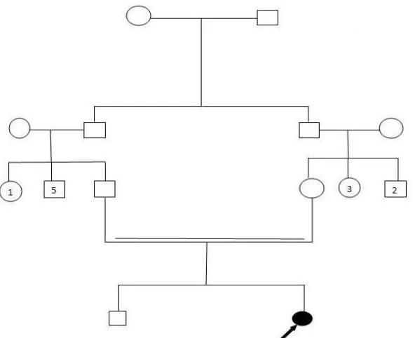
On physical exam at 8 years of age, the patient’s weight was 18 kg, height 113 cm and head cir-cumference 40 cm which was 9 SD below the population age – and sex-related mean. In the case of patients’ family history, the parents were first cousins originating from the Northwest of Iran with normal head circumference and have normal intelligence. There was no history of any genetic disorders including neurological disease in the family (Fig. 1).
Fig. 1: Pedigree and sequencing chromatograph of the studied family. Filled circle represents an affected individual.
Double lines show consanguineous
Consent form was obtained from the parents ear-lier to beginning of the examination.
Genomic DNA was extracted from peripheral blood from the proposita and her parents using DNA extraction kit (Roche, USA). Human whole exome enrichment was performed in proposita using Agilent SureSelect V6 Target Enrichment Kit and the library was sequenced on Illumina Hiseq 4000 platform with an average depth of 110.7X for target regions (Macrogen, South
Ko-rea).The resulting VCF (Variant Call Format) file contains approximately 107500 variants. The Variants were called using with GATK and fil-tered to focus analysis on those variants within the microcephaly genes. All exon and flanking 10bp were detected and analyzed.
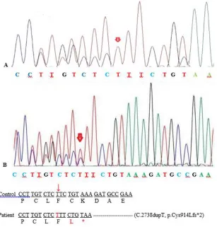
Sanger sequencing, segregation analysis showed that both parents are heterozygous carriers for this variant. (Fig. 2). This variant has not been previously reported and it was absent in ExAC and 1000G databases. Based on the ACMG guideline, this variant can be classified as a likely
pathogenic variant. In silico computational analy-sis (Mutation Taster, and Combined Annotation Dependent Depletion (CADD)) predicted that the novel frameshift mutation (p.Cys914Lysfs*2)
in ASPM can cause premature stop codon, result-ing to truncate the protein synthesis.
Fig. 2: Partial DNA sequence of ASPM gene. The sequence chromatograms showed the homozygous mutation
c.2738dupT in the patient (A) and the heterozygous mutation in the parents (B)
For confirmation of the mutation detection, Sanger DNA sequencing was performed. Primers were designed through primer3 software for am-plification of exon 9 of ASPM gene. Primers used were as follows:
5'TGTGCTTGCTACCCTACACT 3' (Forward) and 5'ACGAGGGATGGTTAGAATTACTG3' (Reverse). Direct sequencing was carried out us-ing 3130 automated DNA sequencer (Applied Biosystems) and the sequences were analyzed by sequencing analysis software, version 5.3.1.
Discussion
dif-fers in different populations. It is reported 13.3% in an Iranian population (7). The ASPM gene lo-cated on chromosome 1q31.3 at MCPH5 locus and contains 28 exons and 3,477 amino acids (NM_018136). The ASPM protein has an im-portant role for the normal functioning of the mitotic spindle in embryonic neuroblasts (9). Many of mutations consist of nonsense, frame-shift, deletion, single base pair change, a break-point translocation and splice affecting mutations identified in throughout the ASMP gene and most of them result in truncating of protein (9-11). ASPM mutations among an Iranian family with MCPH most likely leading to functional im-pairment of the gene product (7). Mutations of ASPM can influence of orientation of the mitotic spindle, resulting in the decrease of the brain size and affect the asymmetrical to symmetrical cell division ratios, leading to decrease the neuronal cells (12).
In this study, we identified a novel homozygous frame-shift mutation (c.2738dupT) in exon 9 of the ASPM gene in an Iranian patient with MCPH through WES. This variant has not been previ-ously reported and in silico analysis indicated that this variant could lead to a premature stop codon in 2 codons downstream, resulting in early pro-tein truncation (p.Cys914Lysfs*2). Most of the
identified mutations in ASPM gene are predicted to produce a truncated protein. No correlation has been observed between the position of the mutation in the gene and degree mental retarda-tion or clinical outcome but in a study showed that patients with p.R1327* mutation had worst growth pattern, IQ and occurrence seizure (13). This may indicate that nonsense- medicated de-cay is the common mechanism of ASPM muta-tions. Consistent with previous reports of ASPM mutations, the detected variant is also predicted to induce nonsense-mediated mRNA decay (NMD) and result in loss of normal function of ASPM in homozygous mutation carriers. A few mutations in the downstream of this variant have been reported in different population as patho-genic, providing support for this variant as a pos-sible candidate to describe the patient’s pheno-type (7, 14, 15).
Conclusion
To our knowledge, the c.2738dupT mutation in the ASPM gene was first reported and this vari-ant is probably to cause the primary microcephaly reported for this patient. The study might add a new pathogenic variant in mutations of the ASPM gene as causative variant in patients with MCPH and might be helpful in genetic counsel-ing of consanguineous families. However, further studies in other cases should be conducted to clarifying the pathogenic effect of the variant.
Ethical considerations
Ethical issues (Including plagiarism, informed consent, data fabrication, double publication, etc.) have been completely observed by the au-thors.
Acknowledgements
We thank the patient and her parents for their cooperation in the study.
Conflict of interest
The authors have no conflicts of interest to de-clare.
References
1. Woods CG, Bond J, Enard W (2005). Autosomal recessive primary microcephaly (MCPH): a review of clinical, molecular, and evolutionary findings. Am J Hum Genet, 76:717-728.
2. Kaindl AM, Passemard S, Kumar P, et al (2010). Many roads lead to primary autosomal recessive microcephaly. Prog Neurobiol, 90:363-383.
3. Jayaraman D, Bae B-I, Walsh CA (2018). The Genetics of Primary Microcephaly. Annu Rev Genomics Hum Genet, 19:177-200.
heterogeneity and mutation continuum. Orphanet J Rare Dis, 6:39.
5. Jayaraman D, Kodani A, Gonzalez DM, et al (2016). Microcephaly proteins Wdr62 and Aspm define a mother centriole complex regulating centriole biogenesis, apical complex, and cell fate. Neuron, 92:813-828. 6. Koboldt DC, Steinberg KM, Larson DE, Wilson
RK, Mardis ER (2013). The next-generation sequencing revolution and its impact on genomics. Cell, 155:27-38.
7. Darvish H, Esmaeeli-Nieh S, Monajemi G, et al (2010). A clinical and molecular genetic study of 112 Iranian families with primary microcephaly. J Med Genet, 47(12):823-8. 8. Akbariazar E, Ebrahimpour M, Akbari S, et al
(2013). A novel deletion mutation in ASPM gene in an Iranian family with autosomal recessive primary microcephaly. Iran J Child Neurol, 7:23-30.
9. Bond J, Roberts E, Mochida GH, et al (2002). ASPM is a major determinant of cerebral cortical size. Nat Genet, 32:316-20.
10. Bond J, Scott S, Hampshire DJ, et al (2003). Protein-truncating mutations in ASPM cause
variable reduction in brain size. Am J Hum Genet, 73:1170-1177.
11. Pichon B, Vankerckhove S, Bourrouillou G, Duprez L, Abramowicz MJ (2004). A translocation breakpoint disrupts the ASPM gene in a patient with primary microcephaly. Eur J Hum Genet, 12:419-21.
12. Thornton GK, Woods CG (2009). Primary microcephaly: do all roads lead to Rome? Trends Genet, 25:501-510.
13. Abdel‐Hamid MS, Ismail MF, Darwish HA, Effat LK, Zaki MS, Abdel‐Salam GM (2016). Molecular and phenotypic spectrum of ASPM‐related primary microcephaly: Identification of eight novel mutations. Am J Med Genet A, 170:2133-2140.
14. Nicholas A, Woods G, Swanson E, et al (2009). The molecular landscape of ASPM mutations in primary microcephaly. J Med Genet, 46(4):249-53.