INTRODUCTION
An imbalance in the autonomic nervous system (ANS) contributes to cardiovascular pathology and increased cardio-vascular mortality [1]. Heart rate recovery (HRR), measured after an exercise stress test (EST), represents a simple and useful tool for the assessment of cardiac autonomic activity, particularly parasympathetic tone. During exercise, increased sympathetic and decreased parasympathetic activity lead to increased HR. Immediately after the exercise, parasympa-thetic reactivation and subsequent sympaparasympa-thetic withdrawal occur, resulting in HR decrease (return to baseline) [2,3]. An
abnormal HRR directly after exercise cessation has shown to be predictive of cardiovascular events and all-cause mortality in healthy and diseased individuals [4-6]. Moreover, slower baseline HRR was associated with risk factors for atheroscle-rosis, such as higher blood pressure and low-density lipopro-tein cholesterol (LDL-C) levels [7]. However, other studies showed conflicting results regarding the diagnostic/predictive ability of HRR for the presence and severity of coronary artery disease (CAD), and the underlying mechanisms are yet to be characterized [2,8-11].
The SYNTAX (SYNergy between percutaneous coronary intervention with TAXus and cardiac surgery) score is an angiographic tool to determine the complexity of CAD [12]. The prognostic value of the SYNTAX score in predicting car-diovascular events, mortality, and morbidity in patients with CAD has been previously demonstrated [13,14] and the tem shows several advantages compared to other scoring sys-tems. For instance, the SYNTAX score can be used to assess
Decreased heart rate recovery may predict a high
SYNTAX score in patients with stable coronary artery
disease
Sadık Volkan Emren1*, Rahman Bilal Gediz2, Oktay Şenöz3, Uğur Karagöz1, Ersin Çağrı Şimşek4, Fatih Levent5,
Emre Özdemir1, Mustafa Ozan Gürsoy1, Cem Nazli1
1Department of Cardiology, Katip Celebi University School of Medicine, Izmir, Turkey, 2Department of Cardiology, Manisa State Hospital,
Manisa, Turkey, 3Department of Cardiology, Çiğli State Hospital, Izmir, Turkey, 4Department of Cardiology, Tepecik Training and Education
Hospital, Izmir, Turkey, 5Department of Cardiology, Tınaztepe Hospital, Izmir, Turkey
ABSTRACT
An impaired heart rate recovery (HRR) has been associated with increased risk of cardiovascular events, cardiovascular, and all-cause mortality. However, the diagnostic ability of HRR for the presence and severity of coronary artery disease (CAD) has not been clearly elucidated. Our aim was to investigate the relationship between HRR and the SYNTAX (SYNergy between percutaneous coronary intervention with TAXus and cardiac surgery) score in patients with stable CAD (SCAD). A total of 406 patients with an abnormal treadmill exercise test and ≥50% coronary stenosis on coronary angiography were included. The HRR was calculated by subtracting the HR in the first minute of the recovery period from the maximum HR during exercise. The SYNTAX score ≥23 was accepted as high. Correlation of HRR with SYNTAX score and independent predictors of high SYNTAX score were determined. A high SYNTAX score was present in 172 (42%) patients. Mean HRR was lower in patients with a high SYNTAX score (9.8 ± 4.5 vs. 21.3 ± 9, p < 0.001). The SYNTAX score was negatively correlated with HRR (r: -0.580, p < 0.001). In multivariate logistic regression analysis, peripheral arterial disease (OR: 13.3; 95% CI: 3.120–34.520; p < 0.001), decreased HRR (OR: 0.780; 95% CI: 0.674–0.902; p = 0.001), peak systolic blood pressure (OR: 1.054; 95% CI: 1.023–1.087; p = 0.001), and peak HR (OR: 0.950; 95% CI: 0.923–0.977; p < 0.001) were found to be independent predictors of a high SYNTAX score. Our results showed that HRR is significantly correlated with the SYNTAX score, and a decreased HRR is an independent predictor of a high SYNTAX score in patients with SCAD.
KEY WORDS: Heart rate recovery; SYNTAX score; coronary artery disease; CAD; HRR
DOI: http://dx.doi.org/10.17305/bjbms.2019.3725 Bosn J Basic Med Sci. 2019;19(1):109-115. © 2019 ABMSFBIH
*Corresponding author: Sadık Volkan Emren, Department of Cardiology, Katip Celebi University Atatürk Training Research and Education Hospital, Basın sitesi 35360, Izmir, Turkey. Phone: +9005052644578.
E-mail: [email protected]
the atherosclerotic burden but also to determine the number, location, complexity, and functional impact of obstructive lesions. The current guidelines on coronary revascularization recommend the use of the SYNTAX score to guide decision making between percutaneous coronary intervention (PCI) and coronary artery bypass grafting (CABG) in patients with CAD [15,16]. To the best of our knowledge, there is a lack of research on the relationship between HRR and the SYNTAX score in patients with CAD.
The aim of this study was to investigate whether HRR predicts a high SYNTAX score in patients with stable CAD (SCAD).
MATERIALS AND METHODS
This is a retrospective analysis of a multicenter observa-tional study conducted between May 2017 and April 2018 in five coronary units in Turkey (Katip Celebi University School of Medicine, Çiğli State Hospital, Tepecik Training and Education Hospital and Tınaztepe Hospital in Izmir, and Manisa State Hospital in Manisa). Patients with SCAD who had an abnormal treadmill exercise test and ≥50% stenosis in coronary arteries, detected by coronary angiography, were included in the study. The patients underwent coronary angi-ography within two weeks of EST. All patients achieved 85% of age-predicted maximal HR.
The exclusion criteria were as follows: submaximal EST, history of prior acute coronary syndrome or coronary revas-cularization, arrhythmia, conduction block, heart failure, moderate or severe valvular disease, thyroid diseases, ane-mia, pacemaker implantation, or the use of antiarrhythmics, digoxin, nitrates, and drugs affecting the HR such as beta blockers, calcium channel blockers and ivabradine.
Exercise stress testing
After a fasting period of 6–12 hours, the patients under-went EST. None of the patients used rate control drugs before the test. The test was performed according to the Bruce protocol, in which the speed and gradient were increased at 3-minute intervals. Baseline blood pressure was recorded and a 12-lead electrocardiogram was performed before EST, and then at each exercise stage, peak exer-cise, and at 1-minute intervals during the recovery period. After peak exercise, patients maintained the upright posi-tion during an active cool-down for at least 2 minutes at a speed of 2.4 km/h and an inclination of 2.5%. The test was terminated in case of ≥1 mm ST segment elevation with-out Q wave, ≥2 mm horizontal or downsloping ST segment depression, a decrease in systolic blood pressure ≥10 mmHg, moderate to severe angina, malignant cardiac arrhythmia,
systolic blood pressure ≥250 mmHg or diastolic blood pres-sure ≥155 mmHg.
The functional capacity of patients during EST was esti-mated in metabolic equivalents [METs] (1 MET = 3.5 mL/kg per minute of oxygen consumption, which refers to the rest-ing state). Other variables such as restrest-ing systolic and diastolic blood pressure, peak systolic and diastolic blood pressure, resting heart rate (RHR), peak HR and Duke Treadmill score (DTS): exercise time - [(5 × max ST deviation) - (4 × tread-mill angina index)], were also obtained. The HRR was calcu-lated by subtracting the HR in the first minute of the recovery period from the maximum HR during exercise.
Calculation of SYNTAX score
Standard coronary angiography was performed through the radial or femoral artery. At least 4 and 2 angiographic images were obtained for the left and right coronary systems, respectively. The coronary angiograms were evaluated by two independent experienced interventional cardiologists who were blinded to the clinical data and EST results of the patients. Coronary lesions with a lumen diameter >1.5 mm and at least 50% diameter stenosis were further analyzed. The SYNTAX score was calculated using the online SYNTAX score calculator 2.10 (www.syntaxscore.com). Patients were divided into two groups based on high (≥23) and low (<23) SYNTAX scores.
Clinical variables including the presence of diabetes melli-tus, smoking, hyperlipidemia, chronic renal failure, peripheral arterial disease (PAD), hypertension and obstructive pulmo-nary disease were recorded. The baseline characteristics (age, gender, weight, and height) and laboratory parameters (uric acid level and leukocyte count) were also obtained.
Statistical analysis
RESULTS
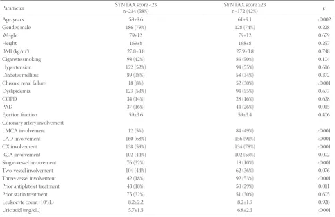
A total of 406 patients met the criteria and were included in the study. The mean age of the patients was 59 ± 8 years and the majority were male (79%). Clinical and demographic data of patients with high and low SYNTAX scores are shown in Table 1. The mean age, uric acid level, and the number of patients with family history of CAD, chronic renal failure, PAD and abnormal HRR values were higher in the group with a high SYNTAX score (p < 0.05). The number of major epicar-dial coronary arteries involved was also higher in patients with a high SYNTAX score (p < 0.005).
The EST findings are summarized in Table 2. All patients achieved at least 8 METs and 85% of the age-predicted maximal HR during EST. The mean HRR was lower in patients with a high compared to those with a low SYNTAX score (9.8 ± 4.5 vs. 21.3 ± 9, p < 0.001). Resting and peak diastolic and systolic blood pressures were higher in patients with a high SYNTAX score. Peak HR was lower in patients with a low SYNTAX score and RHR was higher in patients with a high SYNTAX score. The DTS and MET values were similar in both groups.
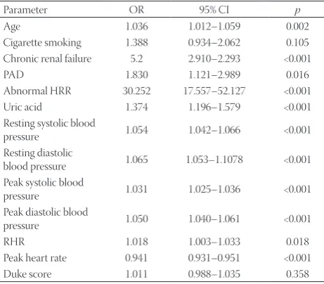
Univariate and multivariate predictors of the SYNTAX score are presented in Table 3 and 4, respectively. In the multivariate logistic regression analysis, PAD (OR: 13.3; 95% CI: 3.120–34.520; p < 0.001), decreased HRR (OR: 0.780; 95% CI: 0.674–0.902; p = 0.001), peak systolic blood pressure
(OR: 1.054; 95% CI: 1.023–1.087; p = 0.001), and peak HR (OR: 0.950; 95% CI: 0.923–0.977; p < 0.001) were found to be independent predictors of the SYNTAX score.
A negative correlation of the HRR with the SYNTAX score (r: -0.580, p < 0.001) is shown in Figure 1.
DISCUSSION
HRR is a measure of ANS function. Attenuated HRR has been associated with increased risk of cardiovascular events and all-cause mortality [4-6]. Although slower HRR was associated with risk factors for atherosclerosis [7], con-flicting results were reported on the diagnostic ability of HRR for the presence and severity of CAD [2,8-11]. In this study, we investigated whether HRR predicts a high SYNTAX score in patients with SCAD. We demonstrated a negative correlation between low HRR and high SYNTAX score, i.e., CAD patients with a high SYNTAX score had lower HRR. Furthermore, our multivariate logistic regression anal-ysis showed that HRR as well as peak systolic blood pressure and peak HR were independent predictors of the SYNTAX score in patients with SCAD.
The SYNTAX score is a validated tool for the assessment of complexity and severity of CAD. It assists in determining the optimal revascularization strategy for patients with CAD and serves as a prognostic indicator of cardiovascular morbidity
TABLE 1. Baseline clinical and laboratory characteristics of patients with SCAD
Parameter SYNTAX score <23 n=234 (58%) SYNTAX score ≥23 n=172 (42%) p
Age, years 58±8.6 61±9.1 <0.002
Gender, male 186 (79%) 128 (74%) 0.228
Weight 79±12 79±12 0.679
Height 169±8 168±8 0.257
BMI (kg/m2) 27.8±3.8 27.9±3.8 0.748
Cigarette smoking 98 (42%) 86 (50%) 0.104
Hypertension 122 (52%) 94 (55%) 0.616
Diabetes mellitus 89 (38%) 58 (34%) 0.372
Chronic renal failure 18 (8%) 52 (30%) <0.001
Dyslipidemia 123 (53%) 94 (55%) 0.677
COPD 34 (14%) 28 (16%) 0.628
PAD 37 (16%) 44 (26%) 0.015
Ejection fraction 59±3.6 59±3.4 0.406
Coronary artery involvement
LMCA involvement 12 (5%) 84 (49%) <0.001
LAD involvement 160 (68%) 156 (91%) <0.001
CX involvement 138 (59%) 134 (78%) <0.001
RCA involvement 102 (44%) 102 (59%) 0.002
Single-vessel involvement 76 (32%) 18 (10%) <0.001
Two-vessel involvement 104 (44%) 62 (36%) 0.076
Three-vessel involvement 42 (18%) 92 (53%) <0.001 Prior antiplatelet treatment 43 (18%) 50 (29%) 0.011
Prior statin treatment 75 (32%) 51 (30%) 0.605
Leukocyte count (109/L) 8.2±2.2 8.2±1.9 0.928
Uric acid (mg/dL) 5.7±1.3 6.8±2.3 <0.001
and mortality [17]. To date, various clinical and demographic variables have been associated with the SYNTAX score, including traditional cardiovascular risk factors such as advanced age, male gender, smoking history, and renal insuf-ficiency [18]. Moreover, serum uric acid was shown to be an independent predictor of the complexity of CAD as evaluated by the clinical SYNTAX score (CSS) [19].
EST parameters have also been related to the severity of CAD. In the current study, both resting and peak blood pres-sure were higher in patients with a high SYNTAX score. These patients may have had uncontrolled hypertension, where higher baseline systolic and diastolic blood pressure led to an exaggerated systolic and diastolic blood pressure response during EST, as demonstrated previously [20]. The DTS has been negatively correlated with the SYNTAX score [21,22], although the correlation coefficients varied greatly between different studies. For example, in 65 patients with a positive exercise test and who underwent coronary angiography, Acar et al. [21] found a strong negative correlation between the DTS and SYNTAX score (r = -0.91, p < 0.001). On the other hand, in a cohort of 258 patients with EST and coronary angiogra-phy, Dzenkeviciute et al. [22] demonstrated a weak correla-tion (r = -0.173; p = 0.007) between the two scores. Günaydın et al. [23] reported that the DTS was an independent predic-tor of a high SYNTAX score in 267 patients who underwent coronary angiography after a positive EST [23]. In our study, patients with a high SYNTAX score had a more negative DTS,
TABLE 2. Exercise stress test results in patients with SCAD in relation to the SYNTAX score
Parameter SYNTAX score <23n=234 (58%) SYNTAX score ≥23n=172 (42%) p
Resting systolic blood pressure 136±19 163±25 <0.001 Resting diastolic blood pressure 82±3 108±19 <0.001 Peak systolic blood pressure 175±30 244±54 <0.001 Peak diastolic blood pressure 91±21 140±32 <0.001
RHR 84±14 88±14 0.017
Peak heart rate 146±22 99±27 <0.001
METs 11.5±1.6 11.7±2.5 0.249
Duke score -4.5 (-9.75–(-1)) -6 (-12–(-1)) 0.001
HRR 21.3±9 9.8±4.5 <0.001
Results are presented as mean±SD or median (IQR). SCAD: Stable coronary artery disease; SYNTAX: SYNergy between percutaneous coronary interven-tion with TAXus and cardiac surgery; RHR: Resting heart rate; HRR: Heart rate recovery; IQR: Interquartile range; MET: Metabolic equivalent
TABLE 3. Predictors of high SYNTAX score in patients with SCAD
according to univariate logistic regression analysis
Parameter OR 95% CI p
Age 1.036 1.012–1.059 0.002 Cigarette smoking 1.388 0.934–2.062 0.105 Chronic renal failure 5.2 2.910–2.293 <0.001 PAD 1.830 1.121–2.989 0.016 Abnormal HRR 30.252 17.557–52.127 <0.001 Uric acid 1.374 1.196–1.579 <0.001 Resting systolic blood
pressure 1.054 1.042–1.066 <0.001 Resting diastolic
blood pressure 1.065 1.053–1.1078 <0.001 Peak systolic blood
pressure 1.031 1.025–1.036 <0.001 Peak diastolic blood
pressure 1.050 1.040–1.061 <0.001 RHR 1.018 1.003–1.033 0.018 Peak heart rate 0.941 0.931–0.951 <0.001 Duke score 1.011 0.988–1.035 0.358
SCAD: Stable coronary artery disease; SYNTAX: SYNergy between percutaneous coronary intervention with TAXus and cardiac surgery; PAD: Peripheral arterial disease; RHR: Resting heart rate; HRR: Heart rate recovery; CI: Confidence interval; OR: Odds ratio
TABLE 4. Predictors of high SYNTAX score in patients with SCAD
according to multivariate logistic regression analysis
Parameter OR 95% CI p
PAD 13.3 3.120–34.520 0.001 HRR 0.780 0.674–0.902 0.001 Peak systolic blood pressure 1.054 1.023–1.087 0.001
Peak heart rate 0.950 0.923–0.977 <0.001 SCAD: Stable coronary artery disease; SYNTAX: SYNergy between percutaneous coronary intervention with TAXus and cardiac sur-gery; PAD: Peripheral arterial disease; HRR: Heart rate recovery; CI: Confidence interval; OR: Odds ratio
FIGURE 1. A negative correlation of heart rate recovery (HRR)
but the DTS was not a predictor of a high SYNTAX score according to the multivariate logistic regression analysis. There are several differences between our and the study of Günaydın et al. [23] which may explain the discrepancy in results. For example, in contrast to Günaydın study, our study popula-tion was larger (406 vs. 267 patients in Günaydın study), we included patients at higher cardiovascular risk, such as those with PAD and chronic renal failure, and we excluded patients with normal coronary arteries and noncritical stenosis (≤50%). Moreover, we analyzed a larger number of variables, includ-ing clinical variables, ANS and EST parameters, to determine independent predictors of a high SYNTAX score. These fac-tors may have increased the accuracy of regression analysis for prediction of SYNTAX score in our study.
RHR is another measure of ANS function, and it was demonstrated that increased RHR, as well as decreased HR increment (maximum HR - RHR before exercise) and decreased HR decrement (maximum HR - RHR after exer-cise), are associated with the severity of CAD [24]. In another study including patients with SCAD, RHR was an indepen-dent predictor of SYNTAX score [25]. However, contrary to our study, Yılmaz et al. [25] did not exclude patients who were using HR-controlling medications such as beta blockers and calcium channel blockers. In the current study, the RHR was higher and peak HR was lower in patients with a high SYNTAX score, still only the peak HR was able to predict the SYNTAX score.
HRR is defined as a decrement in HR predominantly due to the reactivation of parasympathetic system after exercise cessation [2]. Georgoulias et al. suggested HRR 1 minute after peak exercise to be a reliable index of the severity of myocar-dial ischemia [26]. Moreover, an attenuated HRR has been related to CAD risk factors such as hypertension, diabetes mellitus and metabolic syndrome [27,28]. However, while some studies reported an association between abnormal HRR and the presence of CAD [8,9,11], other studies did not find a similar relationship [2,10]. These discrepancies may be explained by differences in study designs. First, exercise and recovery protocols varied between the studies. To allow a reli-able comparison with previous studies, here we applied the most commonly used standard Bruce protocol with an active cool-down. Second, different tools for the assessment of the severity of CAD were utilized in those studies, such as the Gensini score and Duke CAD prognostic index. In the current study, we preferred to use the SYNTAX score to assess disease severity, as recommended by the current coronary revascular-ization guidelines [15].
To the best of our knowledge, the relationship between HRR and SYNTAX score has not been previously shown in patients with CAD. In this study, the difference in the mean HRR between high and low SYNTAX score groups was
11.5 beats. For 1 beat per minute increase in HRR, the hazard ratio for high SYNTAX score was 0.780 (95% CI: 0.674–0.902,
p = 0.001). Although the study protocols and cut-off points for HRR differed between studies, the majority have demon-strated an increased risk of cardiovascular events in patients with an abnormal HRR. Indeed, even minor changes in HRR may affect cardiovascular outcomes. A meta-analysis includ-ing prospective cohort studies showed that for every 10 beats per minute decrements in HRR, the hazard ratio for cardio-vascular events and all-cause mortality was 1.13 (95% CI 1.05– 1.21) and 1.09 (95% CI 1.01–1.19), respectively in the general population. Moreover, their dose-response analysis suggested that the higher the HRR, the better the outcome [29].
The underlying mechanisms linking CAD presence/sever-ity and abnormal HRR are not clear, although several explana-tions have been proposed [30-34]. Delayed postexercise HRR is mainly attributed to impaired vagal reactivation following exercise. Moreover, in patients who had myocardial infarc-tion, the role of enhanced sympathetic activity at maximum exercise in the inhibition of parasympathetic reactivation after exercise was suggested, resulting in attenuated HRR [33]. Chronic sympathetic hyperactivity increases hemodynamic stress and cardiac workload which may lead to endothe-lial dysfunction and ultimately atherosclerosis. Conversely, increased parasympathetic activity has a protective effect by reducing HR and blood pressure [30]. Attenuated HRR may also increase myocardial oxygen consumption and have a deleterious effect on the myocardium [31]. Peçanha et al. [32] suggested a model for HRR in which the fast phase (a rapid decline in HR in the first minute of recovery promoted by vagal reactivation) is mainly determined by the deactivation of central command and mechanoreflex, while the slow phase (a more gradual decline in HR after the first minute due to an interaction of vagal reactivation and sympathetic withdrawal) is mediated predominantly by the metaboreflex and ther-moregulatory deactivations [32]. The results of some studies suggested that HR response to central command activation in patients with CVD is increased at rest [34]. Similarly, mech-anoreflex, metaboreflex and thermoregulation mechanisms may be impaired in CVD [32].
the validity and utility of HRR in predicting SYNTAX score in patients with CAD.
CONCLUSION
HRR is negatively correlated with the SYNTAX score in patients with SCAD. Decreased HRR may be used to predict high SYNTAX score in CAD patients.
DECLARATION OF INTERESTS
The authors declare no conflict of interests.
REFERENCES
[1] Ellestad MH. Chronotropic incompetence. The implications of heart rate response to exercise (compensatory parasympathetic hyperactivity?) Circulation 1996;93(8):1485-7.
https://doi.org/10.1161/01.CIR.93.8.1485.
[2] Shetler K, Marcus R, Froelicher VF, Vora S, Kalisetti D, Prakash M, et al. Heart rate recovery: Validation and methodologic issues. J Am Coll Cardiol 2001;38(7):1980-7.
https://doi.org/10.1016/S0735-1097(01)01652-7.
[3] Emren V, Kocabas U. Is heart rate recovery index a predictive factor for cardioinhibitory syncope? Kardiol Pol 2018;76(2):347-52. https://doi.org/10.5603/KP.a2017.0228.
[4] Chaitman BR. Abnormal heart rate responses to exercise pre-dict increased long-term mortality regardless of coronary disease extent: The question is why? J Am Coll Cardiol 2003;42(5):839-41. [5] Morshedi-Meibodi A, Larson MG, Levy D, O’Donnell CJ, Vasan RS.
Heart rate recovery after treadmill exercise testing and risk of car-diovascular disease events (The Framingham Heart Study). Am J Cardiol 2002;90(8):848-52.
https://doi.org/10.1016/S0002-9149(02)02706-6.
[6] Nishime EO, Cole CR, Blackstone EH, Pashkow FJ, Lauer MS. Heart rate recovery and treadmill exercise score as predictors of mortality in patients referred for exercise ECG. JAMA 2000;284(11):1392-8. https://doi.org/10.1001/jama.284.11.1392.
[7] Kizilbash MA, Carnethon MR, Chan C, Jacobs DR, Sidney S, Liu K. The temporal relationship between heart rate recovery imme-diately after exercise and the metabolic syndrome: The CARDIA study. Eur Heart J 2006;27(13):1592-6.
https://doi.org/10.1093/eurheartj/ehl043.
[8] Akyüz A, Alpsoy S, Akkoyun DC, Degirmenci H, Güler N. Heart rate recovery may predict the presence of coronary artery disease. Anadolu Kardiyol Derg 2014;14(4):351-6.
https://doi.org/10.5152/akd.2014.4824.
[9] Ghaffari SB, Kazemi B, Aliakbarzadeh P. Abnormal heart rate recov-ery after exercise predicts coronary artrecov-ery disease severity. Cardiol J 2011;18(1):47-54.
[10] Vivekananthan DP, Blackstone EH, Pothier CE, Lauer MS. Heart rate recovery after exercise is a predictor of mortality, independent of the angiographic severity of coronary disease. ACC Current Journal Review 2004;13(1):30-1.
https://doi.org/10.1016/j.accreview.2003.10.035.
[11] Lipinski MJ, Vetrovec GW, Froelicher VF. Importance of the first two minutes of heart rate recovery after exercise treadmill testing in predicting mortality and the presence of coronary artery disease in men. Am J Cardiol 2004;93(4):445-9.
https://doi.org/10.1016/j.amjcard.2003.10.039.
[12] Yadav M, Palmerini T, Caixeta A, Madhavan MV, Sanidas E, Kirtane AJ, et al. Prediction of coronary risk by SYNTAX and derived scores: Synergy between percutaneous coronary intervention with taxus and cardiac surgery. J Am Coll Cardiol 2013;62(14):1219-30. https://doi.org/10.1016/j.jacc.2013.06.047.
[13] Kato S, Saito N, Kirigaya H, Gyotoku D, Iinuma N, Kusakawa Y, et al. Incremental prognostic value of the SYNTAX score to late gadolin-ium-enhanced magnetic resonance images for patients with stable coronary artery disease. Heart Vessels 2016;31(6):871-80.
https://doi.org/10.1007/s00380-015-0685-x.
[14] Yang CH, Hsieh MJ, Chen CC, Chang SH, Wang CY, Lee CH, et al. SYNTAX score: An independent predictor of long-term cardiac mortality in patients with acute ST-elevation myocardial infarction. Coron Artery Dis 2012;23(7):445-9.
https://doi.org/10.1097/MCA.0b013e3283587835.
[15] Kolh P, Windecker S, Alfonso F, Collet JP, Cremer J, Falk V, et al. 2014 ESC/EACTS Guidelines on myocardial revascularization: The Task Force on Myocardial Revascularization of the European Society of Cardiology (ESC) and the European Association for Cardio-Thoracic Surgery (EACTS). Developed with the special contribu-tion of the European Associacontribu-tion of Percutaneous Cardiovascular Interventions (EAPCI). Eur J Cardiothorac Surg 2014;46(4):517-92. https://doi.org/10.1093/ejcts/ezu366.
[16] Patel MR, Calhoon JH, Dehmer GJ, Grantham JA, Maddox TM, Maron DJ, et al. ACC/AATS/AHA/ASE/ASNC/SCAI/SCCT/STS 2017 appropriate use criteria for coronary revascularization in patients with stable ischemic heart disease: A report of the American College of Cardiology Appropriate Use Criteria Task Force, American Association for Thoracic Surgery, American Heart Association, American Society of Echocardiography, American Society of Nuclear Cardiology, Society for Cardiovascular Angiography and Interventions, Society of Cardiovascular Computed Tomography, and Society of Thoracic Surgeons. J Am Coll Cardiol. 2017;69(17):2212-41. https://doi.org/10.1016/j.jacc.2017.02.001.
[17] Serruys PW, Morice MC, Kappetein AP, Colombo A, Holmes DR, Mack MJ, et al. Percutaneous coronary intervention versus cor-onary-artery bypass grafting for severe coronary artery disease. N Engl J Med 2009;360(10):961-72.
https://doi.org/10.1056/NEJMoa0804626.
[18] Montero-Cabezas JM, Karalis I, Wolterbeek R, Kraaijeveld AO, Hoefer IE, Pasterkamp G, et al. Classical determinants of coro-nary artery disease as predictors of complexity of corocoro-nary lesions, assessed with the SYNTAX score. Neth Heart J 2017;25(9):490-7. https://doi.org/10.1007/s12471-017-1005-0.
[19] Xiong Z, Zhu C, Qian X, Zhu J, Wu Z, Chen L. Predictors of clin-ical SYNTAX score in coronary artery disease: Serum uric acid, smoking, and Framingham risk stratification. J Invasive Cardiol 2011;23(12):501-4.
[20] Abiodun OO, Balogun MO, Adebayo RA, Akintomide AO. Blood pressure control and exaggerated blood pressure response in Nigerians with essential hypertension. Clin Med Insights Cardiol 2014;8:53-6.
https://doi.org/10.4137/CMC.S15961.
[21] Acar Z, Korkmaz L, Agac MT, Erkan H, Dursun I, Kalaycioglu E, et al. Relationship between Duke Treadmill Score and coronary artery lesion complexity. Clin Invest Med 2012;35(6):E365-9. https://doi.org/10.25011/cim.v35i6.19208.
[22] Dzenkeviciute V, Sapoka V, Kasiulevicius V, Rinkuniene E, Steponeniene R, Einikyte R, et al. Value of Duke treadmill score in predicting coronary artery lesion and the need for revascularisa-tion. Kardiol Pol 2017;75(5):439-44.
https://doi.org/10.5603/KP.a2017.0032.
[23] Günaydın ZY, Bektas O, Gurel YE, Karagoz A, Kaya A, Kiris T, et al. The value of the Duke treadmill score in predicting the presence and severity of coronary artery disease. Kardiol Pol 2016;74(2):127-34. https://doi.org/10.5603/KP.a2015.0143.
[24] Cay S, Ozturk S, Biyikoglu F, Yildiz A, Cimen T, Uygur B, et al. Association of heart rate profile during exercise with the sever-ity of coronary artery disease. J Cardiovasc Med (Hagerstown) 2009;10(5):394-400.
https://doi.org/10.2459/JCM.0b013e328329c715.
https://doi.org/10.1177/0003319716649881.
[26] Georgoulias P, Orfanakis A, Demakopoulos N, Xaplanteris P, Mortzos G, Vardas P, et al. Abnormal heart rate recovery imme-diately after treadmill testing: Correlation with clinical, exercise testing, and myocardial perfusion parameters. J Nucl Cardiol 2003;10(5):498-505.
https://doi.org/10.1016/S1071-3581(03)00530-0.
[27] Erdogan D, Gonul E, Icli A, Yucel H, Arslan A, Akcay S, et al. Effects of normal blood pressure, prehypertension, and hypertension on autonomic nervous system function. Int J Cardiol 2011;151(1):50-3. https://doi.org/10.1016/j.ijcard.2010.04.079.
[28] Seshadri N, Acharya N, Lauer MS. Association of diabetes mellitus with abnormal heart rate recovery in patients without known coro-nary artery disease. Am J Cardiol 2003;91(1):108-11.
https://doi.org/10.1016/S0002-9149(02)03014-X.
[29] Qiu S, Cai X, Sun Z, Li L, Zuegel M, Steinacker JM, et al. Heart rate recovery and risk of cardiovascular events and all-cause mortality: A meta-analysis of prospective cohort studies. J Am Heart Assoc 2017;6(5). pii: e005505.
https://doi.org/10.1161/JAHA.117.005505.
[30] Curtis BM, O’Keefe JH Jr. Autonomic tone as a cardiovascular risk factor: The dangers of chronic fight or flight. Mayo Clin Proc
2002;77(1):45-54.
https://doi.org/10.4065/77.1.45.
[31] Imai K, Sato H, Hori M, Kusuoka H, Ozaki H, Yokoyama H, et al. Vagally mediated heart rate recovery after exercise is accelerated in athletes but blunted in patients with chronic heart failure. J Am Coll Cardiol 1994;24(6):1529-35.
https://doi.org/10.1016/0735-1097(94)90150-3.
[32] Peçanha T, Silva-Júnior ND, Forjaz CL. Heart rate recovery: Autonomic determinants, methods of assessment and association with mortality and cardiovascular diseases. Clin Physiol Funct Imaging 2014;34(5):327-39.
https://doi.org/10.1111/cpf.12102.
[33] Ushijima A, Fukuma N, Kato Y, Aisu N, Mizuno K. Sympathetic excitation during exercise as a cause of attenuated heart rate recov-ery in patients with myocardial infarction. J Nippon Med Sch 2009;76(2):76-83.
https://doi.org/10.1272/jnms.76.76.
[34] Strike PC, Magid K, Brydon L, Edwards S, McEwan JR, Steptoe A. Exaggerated platelet and hemodynamic reactivity to mental stress in men with coronary artery disease. Psychosom Med 2004;66(4):492-500.