Copyright © 2004, American Society for Microbiology. All Rights Reserved.
Identification of
Cryptococcus neoformans
Temperature-Regulated
Genes with a Genomic-DNA Microarray†
Peter R. Kraus,
1Marie-Jose´e Boily,
1Steven S. Giles,
2Jason E. Stajich,
1Andria Allen,
1Gary M. Cox,
2Fred S. Dietrich,
1John R. Perfect,
2and Joseph Heitman
1,2,3,4*
Departments of Molecular Genetics and Microbiology,1Medicine,2and Pharmacology and Cancer Biology3
and Howard Hughes Medical Institute,4Duke University Medical Center, Durham, North Carolina
Received 5 May 2004/Accepted 30 June 2004
The ability to survive and proliferate at 37°C is an essential virulence attribute of pathogenic microorgan-isms. A partial-genome microarray was used to profile gene expression in the human-pathogenic fungus Cryptococcus neoformansduring growth at 37°C. Genes with orthologs involved in stress responses were induced
during growth at 37°C, suggesting that a conserved transcriptional program is used byC.neoformansto alter
gene expression during stressful conditions. A gene encoding the transcription factor homolog Mga2 was induced at 37°C and found to be important for high-temperature growth. Genes encoding fatty acid biosyn-thetic enzymes were identified as potential targets of Mga2, suggesting that membrane remodeling is an
important component of adaptation to high growth temperatures.mga2⌬mutants were extremely sensitive to
the ergosterol synthesis inhibitor fluconazole, indicating a coordination of the synthesis of membrane com-ponent precursors. Unexpectedly, genes involved in amino acid and pyrimidine biosynthesis were repressed at 37°C, but components of these pathways were found to be required for high-temperature growth. Our findings demonstrate the utility of even partial-genome microarrays for delineating regulatory cascades that contribute to microbial pathogenesis.
Pathogenic microorganisms must be able to survive at the physiological temperature of the host in order to proliferate and cause disease. A hallmark of human-pathogenic fungi is their ability to grow at the human body temperature of 37°C. The ability to grow at temperatures as high as 42°C is an established virulence factor for pathogenicSaccharomyces
cer-evisiae strains (42), and growth at 37°C is associated with a
mycelium- to yeast-form switch that is essential for the viru-lence of the human pathogens Blastomyces dermatitidis,
His-toplasma capsulatum, Coccidioides immitis, Paracoccidioides
brasiliensis, and Sporothrix schenckii (8, 41). Thermally
regu-lated dimorphism occurs in Cryptococcus neoformans during mating and haploid fruiting and in self-fertile diploid strains but is not a requirement for pathogenesis (4, 47). Although a temperature-dependent morphogenetic switch is not essential for the virulence ofC.neoformans, the ability to grow at 37°C is considered an established virulence attribute (9). At least 38
Cryptococcusspecies other thanC.neoformanshave been
de-scribed, andC.neoformansis the only species that can consis-tently grow at mammalian body temperatures and is the only common human pathogen (9).
C. neoformansis a basidomycete that is the most common
cause of fungal meningitis.C.neoformansinfects both immu-nocompetent and immunocompromised hosts, but disease is generally expressed when the host has impaired immunity. Virulence factors forC.neoformansthat have been genetically defined include (i) the ability to synthesize the antioxidant
pigment melanin, (ii) the production of an antiphagocytic poly-saccharide capsule, (iii) urease and phospholipase production, and (iv) the ability to survive and proliferate at 37°C (13, 14, 37, 45). Proteins that have been identified in C. neoformans as being essential or important for growth at high temperatures include Ras1, the p21-activated kinase (PAK) kinase Ste20, the phospholipid-binding protein Cts1, the vacuolar ATPase Vph1, the thiol peroxidase Tsa1, the cell integrity mitogen-activated protein (MAP) kinase Mpk1, and the Ca2⫹
/calmod-ulin-dependent phosphatase calcineurin (3, 21, 22, 34, 43, 45, 57). Deletion or disruption of the genes encoding each of these proteins results in either attenuation or complete loss of viru-lence in mammalian models of cryptococcosis.
The increasing availability of entire genome sequences has resulted in the proliferation of large-scale techniques for study-ing gene function. For fungal systems where complete genome sequences are not yet available, these techniques have been adapted to provide valuable information about gene expres-sion and function. A major strength of these approaches is that they can provide information that may be difficult to obtain by traditional techniques of genetic analysis; they can also estab-lish a foundation for further study when complete genome sequences become available. cDNA library subtraction and serial analysis of gene expression (SAGE) have been used to identifyC.neoformansgenes that are preferentially transcribed under conditions of high-temperature growth and in animal models of infection (2, 46, 49, 50, 52). Furthermore, DNA microarrays were adapted for studying phase-regulated gene expression inH.capsulatumprior to completion of the genome sequence (30).
In this study, we used a similar approach and constructed a shotgun genomic-DNA microarray to assess C. neoformans
transcription during growth at the human physiological
tem-* Corresponding author. Mailing address: Department of Molecular Genetics and Microbiology, 322 CARL Building, Box 3546, Research Dr., Duke University Medical Center, Durham, NC 27710. Phone: (919) 684-2824. Fax: (919) 684-5458. E-mail: [email protected].
† Supplemental material for this article may be found at http://ec .asm.org/.
1249
on September 8, 2020 by guest
http://ec.asm.org/
perature of 37°C. Implementation of this 6,274-element array resulted in the identification of 239 genes that displayed dif-ferential expression during growth at 37 and 25°C. One differ-entially regulated gene,MGA2, encodes a transcription factor homolog that is required for normal growth in vitro at a range of growth temperatures. Microarray experiments were per-formed to identify candidate target genes of theMGA2 tran-scription factor which encode orthologs of components of the fatty acid biosynthesis machinery.mga2⌬mutants are hyper-sensitive to the ergosterol synthesis inhibitor fluconazole, sug-gesting a coordination between the metabolism of fatty acid membrane components and the metabolism of sterol mem-brane components. In addition, we report the unexpected find-ing that the transcription of genes in amino acid and pyrimi-dine biosynthetic pathways decreases at 37°C, yet mutations in components of these pathways result in a growth defect at high temperatures. Our studies highlight the potential of genomic approaches for revealing molecular principles of microbial pathogenesis.
MATERIALS AND METHODS
Strains and media.The strains used were as follows: H99 (MAT␣), JF99 (MATaura5), PK23 (MATaura5[URA5]), PK28 (MAT␣mga2⌬::nat), and AI34 (MAT␣clc1⌬::nat). The growth medium used was 2% yeast extract–1% pep-tone–2% dextrose (YPD). For solid media, 2% agar was added. Nourseothricin was added to solid media at 100g/ml, and fluconazole was added at 10 mg/ml. An allele to delete theMGA2gene and replace it with the nourseothricin resistance cassette by homologous recombination was generated as previously described (16, 23). The construct was transformed into strain H99 by biolistic transformation, and nourseothricin-resistant transformants were screened for deletion of theMGA2open reading frame (ORF) by PCR and Southern analysis of genomic DNA. TheMGA2gene was reintroduced by transforming a plasmid containing the entireMGA2gene plus 1 kb each of 5⬘and 3⬘flanking DNAs linked to a cassette that confers resistance to G418 (23). Transformants were selected on G418 and tested for the ability to grow in the presence of 10g of fluconazole/ml.
Array construction, sequencing, and annotation.The array consists of two components: cDNA clones from strains JEC21 and H99 amplified by PCR with gene-specific primers (130 elements) and shotgun H99 genomic clones (6,144 elements). The H99 genomic library was constructed as follows: 3 to 5g of genomic DNA was sheared with a Hydroshear device (Gene Machines) to generate 1.5- to 3-kb DNA fragments, which were cloned as described previously (39), picked and placed in Luria-Bertani-Hogness medium, and stored at⫺80°C. For sequencing, clones were grown in 96-well plates containing Terrific broth medium in a Higro orbital shaker (Gene Machines), and DNA was isolated by using a RevPrep robot (Gene Machines). Sequencing reactions were performed by using an MJ Research thermal cycler with standard BigDye chemistry (Ap-plied Biosystems). Samples were analyzed on a PE3700 96-capillary sequencer, and data were analyzed and assembled by using the Pare/Phrased sequence package.
PCRs to amplify genomic library inserts were performed by using 64 96-well plates containing bacterial cultures, each of which harbored an independent library transformant. A total of 2l of bacterial stock was used for PCR ampli-fication of inserts with vector-specific primers. PCR products were analyzed on 1% agarose gels, precipitated, washed, and printed on polylysine-coated glass slides at the Duke Center for Genome Technology.
Array annotation was performed by comparing array clone sequences with The Institute for Genomic Research (TIGR) JEC21 genome database by using BLASTx (21 January 2004 version, C. neoformansGenome Project). Array clones were assigned JEC21 locus names only when a single BLAST match with an expect value (E value) of between 10⫺50and 0 was found. The median E value
for all array element sequences compared to JEC21 sequences was 10⫺104;
however, a large majority of the highly scoring segment pairs produced an E value of 0. Complete annotations, including E values and accession numbers of orthologs, were completed only for elements of interest based on hybridization data. Gene names were assigned to some array clones based on the names of orthologs from other fungal species.
Culture conditions and RNA preparation.H99 cultures for the 37°C time course were inoculated into YPD medium and grown at 25°C for 16 h with shaking. The cultures were diluted to an optical density at 600 nm (OD600) of 0.2
and returned to 25°C until the culture density reached an OD600of 0.8
(approx-imately 4 h). A portion of the cultures was harvested by centrifugation at this time, which was considered the 0-h time point. Cells were diluted in prewarmed YPD medium to an OD600of 0.1 and incubated for 12 h. Portions of the cultures
were harvested every 3 h during the time course. For steady-state experiments, the cultures were inoculated into YPD medium and grown for 16 h at 25 or 37°C, diluted to an OD600of 0.2 with fresh YPD medium, and incubated at 25 or 37°C
until an OD600of 0.8 was reached. For the comparison of wild-type andmga2⌬
mutant cells, cultures from isogenic strains H99 (wild type) and PK28 (mga2⌬::nat) were inoculated into YPD medium, grown for 16 h at 30°C, diluted to an OD600of 0.2 with fresh YPD medium, and incubated at 30°C until an
OD600of 0.8 was reached. Cells from all experiments were harvested by
centrif-ugation, frozen in ethanol-dry ice, and lyophilized for RNA preparation. RNA was prepared by using TRIzol reagent (Invitrogen, Carlsbad, Calif.) according to the manufacturer’s instructions.
Microarray hybridization.Fluorescence-labeled cDNA was generated by in-corporating amino-allyl-dUTP during reverse transcription of 10g of total RNA. Cy3 or Cy5 dye (Amersham, Piscataway, N.J.) was coupled to the amino-allyl group as described previously (18). For the 37°C time course, equal amounts of samples from all time points were pooled to generate a reference sample, which was labeled with Cy3. A sample from each time point was individually labeled with Cy5 and competitively hybridized against the reference sample. For steady-state experiments, samples from cells grown at 25°C were labeled with Cy3 and samples from cells grown at 37°C were labeled with Cy5. Dyes were switched for the reverse fluor control. For the comparison of wild-type and mga2⌬mutant cells, three hybridizations were performed by using wild-type cDNA labeled with Cy5 andmga2⌬cDNA labeled with Cy3. In parallel, reverse fluor control hybridization was performed for each of these hybridizations.
Data analysis.Arrays were scanned on a GenePix 4000B scanner (Axon Instruments, Foster City, Calif.) and analyzed by using GenePix Pro (version 4.0), Cluster, TreeView, and CryptoArray, a Microsoft Excel macro for normal-izing and formatting data (19). Data were normalized for each array element by dividing the background-corrected median pixel intensity of each spot by the sum of the median intensities of all spots on the array (6). This normalization was performed individually for data generated at each wavelength. Gene expression ratios were determined by dividing the normalized intensity in the red channel (635 nm) by the intensity in the green channel (532 nm) for each element. CryptoArray also parses the data to exclude ribosomal DNA and mitochondrial DNA elements and provides the data in the appropriate format for Cluster. For the time course experiments, ratio measurements for each time point were divided by the corresponding ratio measurements for the 0-h time point to cancel the pooled reference sample.
Northern analysis.Samples of 10g of total RNA were separated on 1% agarose gels containing 1.85% formaldehyde and transferred to Nytran SuPer-Charge membranes (Schleicher & Schuell, Keene, N.H.). The probes used were generated by PCR amplification of cDNA or genomic DNA with gene-specific primers. For genes from the genomic-DNA portion of the array (MGA2,SLG1, CLC1, andRDS1), ORF-specific probes were designed and used for Northern analysis. For knownC.neoformansgenes included on the microarray (ILV5, SMG1,FHB1, andAOX1), the same PCR products that were printed on the array were used as probes for Northern analysis. Probe labeling and hybridiza-tion were performed as described previously (29).
RESULTS
Array construction. To assessC. neoformans gene
expres-sion on a large scale, we constructed a 6,274-element shotgun genomic-DNA microarray by using PCR to amplify clones from a library of genomic-DNA fragments. Genomic libraries that were constructed for theC.neoformansserotype A strain H99 sequencing project were used as a platform for PCR amplification of genomic DNA. These libraries contain genom-ic-DNA inserts ranging from 1.6 to 3.2 kb in length. Inserts of this size are likely to contain coding regions and therefore are suitable for use as microarray probe elements. A total of 6,144 genomic clones were amplified and comprise approximately 12 Mb ofC. neoformansgenomic DNA. Two different methods
on September 8, 2020 by guest
http://ec.asm.org/
were used to estimate that this portion of the array represents approximately 0.5⫻coverage of the 19-MbC.neoformans ge-nome (38, 59). Both ribosomal DNA and mitochondrial DNA clones are present in the genomic library and therefore are also present on the microarray (139 and 325 elements, respec-tively). For designation of genes within each clone, the anno-tation information provided by the sequencing project for closely related C. neoformans strain JEC21 was an essential resource. We performed BLASTx searches of each array ele-ment against the JEC21 genome database, and a majority of array elements yielded only one significant BLAST match. Based on the sizes of the inserts in the genomic library, it is possible that multiple ORFs are present on a given array ele-ment. Elements predicted to contain more than one ORF by the BLASTx comparison to the JEC21 genome database were excluded from further analysis. In addition to the genomic-DNA probes, 130 PCR products, each containing a single known C. neoformans gene, were also included on the array (see Table S1 in the supplemental material).
Assessing temperature-regulated transcription. We used
two approaches to identify genes that display differential ex-pression during growth at 25 and 37°C. First, we shifted C.
neoformanscultures from 25 to 37°C and assessed gene
expres-sion at multiple time points. Time course experiments were performed twice, and representative results are shown in the cluster analysis in Fig. 1. Second, we compared steady-state gene expression in cultures of cells actively growing in loga-rithmic phase at exclusively 25 or 37°C. These experiments were performed in triplicate, and a reverse fluor control was used in the second trial. By using both temperature shift and steady-state experiments, we reasoned that genes required for both the initiation and the maintenance of growth at 37°C would be revealed in the microarray data analysis. For each experiment, samples were collected and total RNA was pre-pared. cDNAs were generated from the total RNA samples and differentially labeled with Cy3 (pooled reference sample for the time course experiment and 25°C samples for the steady-state experiment) or Cy5 (individual time point samples for the time course experiment and 37°C samples for the steady-state experiment). These cDNAs were subjected to competitive hybridization on the genomic-DNA microarray. The data for the time course and steady-state experiments are provided in Table S2 in the supplemental material.
Initial annotation of the elements on the microarray was performed by using BLASTx searches of the C. neoformans
JEC21 genome annotation database. For the 239 elements that showed differential expression in at least one experiment, a more complete analysis of the clone identity was performed by using BLASTx searches of the nonredundant database at the National Center for Biotechnology Information. For most cases, our annotation of these clones was similar to that of the corresponding locus in theC.neoformansJEC21 genome an-notation database. Genes with no published name were as-signed one if other fungal orthologs had the same designation or were assigned a JEC21 locus designation if no consensus gene name exists. Several clones identified in our experiments did not have significant BLAST matches to the JEC21 genome database or any other database, and these clones were not included in the list of differentially expressed genes.
Genes induced at 37°C.Table 1 summarizes the genes that
were induced during growth at 37°C compared to 25°C. The induction values reported represent the values at which the greatest difference between the 37°C culture and the 25°C culture was observed in either the time course or the steady-state experiment. Genes with orthologs that have known roles in responding to stress were induced at 37°C, including an ortholog of a WSC domain protein gene (SLG1), a chitin synthase gene, a trehalose synthase gene, a trehalose-associ-ated protein kinase gene (RIM15), and genes involved in pro-moting resistance to reactive oxygen species (catalases and oxidases). InS. cerevisiae, these and other related genes are induced by heat shock and other types of cellular stress as part of a general stress response (25).
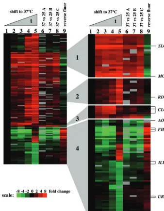
We performed a cluster analysis to group genes according to similarities in expression profiles (Fig. 1) (19). Genes whose expression changed at least threefold in at least one experi-ment were analyzed by hierarchical clustering. Cluster 1 con-tains genes that were induced in both the time course and the steady-state experiments and includes the WSC domain or-tholog SLG1 and theMGA2 gene, encoding a transcription activator homolog. WSC domain proteins are a family of cell surface proteins that are important for adaptation to heat stress and may act to sense stress-induced perturbations of the cell surface and to signal via the protein kinase C-activated cell integrity MAP kinase pathway (27, 55). Cluster 2 contains genes that were induced at 37°C in the time course experiment, with peak induction occurring at late time points after the shift to 37°C (9 and 12 h). Genes in this cluster include an ortholog of theS.pombestress response generds1⫹. The expression of
rds1⫹is induced in response to heat stress inS.pombe, but no
function has as yet been reported for this gene (40). Putative orthologs ofrds1⫹are present in theNeurospora crassa,
Mag-naporthe grisea, Aspergillus nidulans, Fusarium graminearum,
and Ustilago maydis genomes (24; Center for Genome
Re-search, www.broad.mit.edu). Cluster 3 includes multiple clones containing theCLC1gene, which displays strong induction at 37°C at early time points but less pronounced differences in gene expression at later time points and in the steady-state experiment. CLC1 encodes a voltage-gated chloride channel important for melanin synthesis, and no role for CLC1 in high-temperature growth has been reported (31, 61). Northern analysis confirmed that theMGA2, SLG1,CLC1,RDS1, and
SMG1genes were more highly expressed at 37°C than at 25°C in either the time course experiment or the steady-state exper-iment (Fig. 2).
Genes repressed at 37°C.Table 1 summarizes the genes that
were repressed during growth at 37°C compared to 25°C. Strik-ingly, genes that participate in amino acid biosynthesis ( SPE3-LYS9,ILV2, andILV5) and pyrimidine biosynthesis (CTP syn-thase;URA2) displayed significantly reduced expression during growth at 37°C compared to 25°C. We also observed a strong reduction in the expression ofFHB1, encoding a flavohemo-globin important for resistance to nitrosative stress (17). Fur-thermore, three different ribosomal protein genes were re-pressed at 37°C compared to 25°C. TheAOX1gene, encoding an alternative oxidase, was previously identified by a cDNA library subtraction technique as a gene that is preferentially transcribed at 37°C (2). However, we found that the expression of theAOX1gene was reduced both at early time points after
on September 8, 2020 by guest
http://ec.asm.org/
the shift from 25 to 37°C and at 37°C compared to 25°C in the steady-state experiment (Fig. 1, cluster 4). The expression of
AOX1was greater after 12 h of growth at 37°C than at the 0-h time point, indicating that growth phase or culture density influences the expression of AOX1. Northern analysis con-firmed thatFHB1, AOX1, andILV5were expressed at lower levels during growth at 37°C than during growth at 25°C in the time course experiment or the steady-state experiment (Fig. 2).
Correlation of gene expression profile and mutant
pheno-type.One goal of this study was to identify genes important for
survival at 37°C based on expression profiles after a shift to 37°C or in a steady-state experiment. We identified a gene,
MGA2, encoding a transcription factor homolog, whose ex-pression was markedly induced after the shift from 25 to 37°C.
InS. cerevisiae, Mga2 and its paralog, Spt23, are involved in
regulating transcription in response to cold shock and hypoxia (32, 44). Both transcription factors activate the expression of
OLE1, encoding a fatty acid desaturase, suggesting that cell and/or organelle membranes may require remodeling for re-sponses to growth temperature changes (60).
FIG. 1. Gene expression profiles in temperature shift and steady-state microarray experiments. Elements whose expression changed at least threefold in at least one time point after the shift to 37°C or in the steady-state experiment were analyzed by hierarchical clustering. Each lane represents a microarray experiment, and each row represents the expression of an element on the array. For the temperature shift experiment, cDNAs from the 0-, 3-, 6-, 9-, and 12-h time points were labeled with Cy5 (red), and a reference sample consisting of equal amounts of samples from all time points was labeled with Cy3 (green). A sample from each time point was competitively hybridized against the reference sample on the microarray. The time course experiment was normalized against the 0-h time point as described in Materials and Methods. In the 37 versus 25°C experiments, the 37°C samples were labeled with Cy5 and the 25°C samples were labeled with Cy3. The reverse fluor experiment was carried out with samples labeled “B,” and the dye labeling was reversed. For all experiments, red indicates increased gene expression and green indicates decreased gene expression. Missing data are represented in gray. Genes with more than two data points missing were removed from the cluster analysis. Clusters of interest have been enlarged and are discussed in the text.
on September 8, 2020 by guest
http://ec.asm.org/
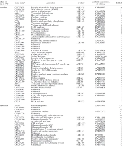
TABLE 1. Temperature-regulated genes
Effect on
gene at 37°C Gene namea Annotation E valueb Genbank accession no.of ortholog Fold changec
Induction CNC05450 Putative short-chain dehydrogenase 6.6E⫺38 CAB86467 16
CNA06780 WSC domain protein (SLG1) 1.2E⫺63 12
CNL00800 Amino acid permease 3.1E⫺102 CAA65074 11
MGA2 Transcriptional activator 2.9E⫺20 CAB92703 11
CNC06420 Hypothetical protein 1.0E⫺73 CAC36937 8.2
CND03740 Catalase, putative 8.0E⫺136 AAG45152 7.9
CNE01110 Predicted protein 2.7E⫺80 CAD37156 7.5
PPS1 Putative dual-specificity phosphatase 2.4E⫺42 CAD21139 7
THR4 Threonine synthase AAK83373 7
CLC1 Voltage-gated chloride channel AAO73005 6.3
CNA02540 S1/P1 nuclease 2.2E⫺29 BAB96801 6
GLT1 Glutamate synthase 0E⫹00 CAA61505 5.8
CNH04480 Trehalose synthesis 1.3E⫺90 BAA31349 5.3
DUR3 Urea transporter 1.1E⫺85 AAB65069 5.1
CNI03330 O-Methyl transferase 1.2E⫺36 AAK44417 4.9
LYS2 Aminoadipate semialdehyde dehydrogenase 6.4E⫺12 CAA85072 4.9
CND06130 Unknown 4.5
SMG1 Putative aryl alcohol oxidase AAM83033 4.5
CNI01590 Superoxide dismutase 1.2E⫺69 AAK82369 4.5
CNF03810 Unknown 4.4
CNA04360 Unknown 4.3
CNA07560 Unknown 4.2
CNA05600 Catalase A related 1.7E⫺170 AAK15808 4.2
RDS1 Stress response protein 6.9E–98 CAB52275 4.2
CNE00600 Carboxypeptidase F 5.9E–172 CAA56075 4.1
CHD02910 Malate synthase 7.0E⫺185 AABD01988 4.1
CNB05770 Putative ABC transporter 0E⫹00 CAD10327 4
CNM01770 Similar to benzodiazepine receptor 8.1E-17 CAA22182 4
CNL01900 Unknown 3.9
CNK00270 UDP-N-acetyl-glucosamine-1-P transferase 6.9E-98 CAA67366 3.9
CNB00750 Unknown 3.7
CNB02490 Putative short-chain dehydrogenase 5.8E-62 AAK89074 3.7
CNB04620 Similar to HSCARG protein 6.5E-31 AAH30039 3.7
CNE04710 Unknown 3.6
CNC00290 Putative multiple-drug resistance protein 1.3E-128 CAD29613 3.6
CNB00710 Unknown 3.5
CNF04870 Proline oxidase 2.6E-21 AAB88789 3.4
CNH02020 Conserved hypothetical protein 8.5E-19 CAD37054 3.4
CND01100 12-Oxophytodienoate reductase 2.6E-68 CAB43506 3.4
RIM15 Trehalose-associated protein kinase 9.6E-131 CAD27468 3.3
PMA1 Plasma membrane ATPase 1.2E-285 AAK94188 3.2
CNE00040 Putative transketolase 5E-38 AAD36826 3.1
CNF05280 Unknown 3.1
CNF00410 Unknown 3.1
CNA05300 Chitin synthase 6 2.1E-302 AAB84285 3.1
CNI00390 C2H2zinc finger protein 2.4E-14 CAB63541 3
CNFp2250 Unknown 3
CNH03900 Unknown 3
CHL1 DNA helicase 1.1E-122 AAB18749 3
Repression FHB1 Flavohemoglobin AAP41066 ⫺25
CNG00908 Unknown ⫺11
CNB05100 Unknown ⫺8.3
AOX1 Alternative oxidase AAM22475 ⫺7.1
CNA01720 Unknown ⫺5.3
ILV5 Acetohydroxyacid reductoisomerase ⫺4.5
CNC06370 Hypothetical WD-repeat protein 3.6E⫺101 CAB11489 ⫺4.5
CNH00300 Ribosomal protein P.0 5.0E⫺88 AAK11262 ⫺3.9
ILV2 Acetolactate synthase AAR29084 ⫺3.8
CNC00200 CTP synthase 1.4E⫺193 CAA15716 ⫺3.7
CND01770 Ribosomal protein S26E 2.9E⫺35 AAC95384 ⫺3.6
RPL22 Ribosomal protein L22 AAN75181 ⫺3.5
PKR1 Protein kinase A regulatory subunit AAG30146 ⫺3.4
CND04910 Putative dityrosine transporter 4.6E⫺41 CAD70885 ⫺3.4
CNM01290 Unknown ⫺3.4
CNE02070 HpcH/HpaI aldolase family 1.2E⫺38 PF03328 ⫺3.4
PAN6 Pantothenate biosynthesis AAN75165 ⫺3.3
CNK02850 Putative allantoate transporter 4.8E⫺58 CAA22656 ⫺3.3
URA2 Pyrimidine biosynthesis 0E⫹00 CAA91130 ⫺3.2
CNC04280 Pumilio family RNA binding protein 1.6E⫺69 CAB03616 ⫺3.2
CND04330 Cdc20/fizzy 9.0E⫺101 CAB57442 ⫺3.2
SPE3-LYS9 Chimeric spermidine synthase-saccharopine
dehydrogenase AAK83327 ⫺3.2
aNamed genes include known genes with specifically amplified PCR products included on the array or genomic clones of previously identifiedC. neoformansgenes (no E value is provided) and clones with significant homology to other fungal orthologs that have a gene name. Genes that do not fit these criteria were given the C. neoformans JEC21 annotation database locus name.
bE value comparingC. neoformansgene and its ortholog, using information from BLASTx searches of the nonredundant database at the National Center for Biotechnology Information and from theC. neoformansJEC21 annotation database (http://www.tigr.org/tdb/e2k1/cna1/cna1.shtml).
cPeak fold induction ratio from the time course and steady-state experiments. Genes displaying both induction and repression over the course of the experiments were categorized as induced or reduced based on cluster analysis (Fig. 1).
on September 8, 2020 by guest
http://ec.asm.org/
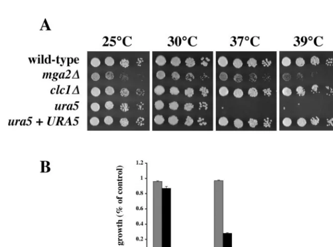
TheC.neoformans ortholog ofMGA2was deleted by ho-mologous recombination, and its role in promoting high-tem-perature growth was assessed. Compared to the isogenic wild-type strain H99, the mga2⌬ mutant strain displayed slow growth at 25 and 30°C (Fig. 3A). Themga2⌬growth defect was more pronounced at 37 and 39°C (Fig. 3), suggesting that this transcription factor homolog plays a significant role in promot-ing high-temperature growth. Because fatty acid metabolism and sterol biosynthesis are coordinated inS.cerevisiae(11, 36), we tested whether the mga2⌬mutation had an effect on C.
neoformans growth in the presence of the ergosterol
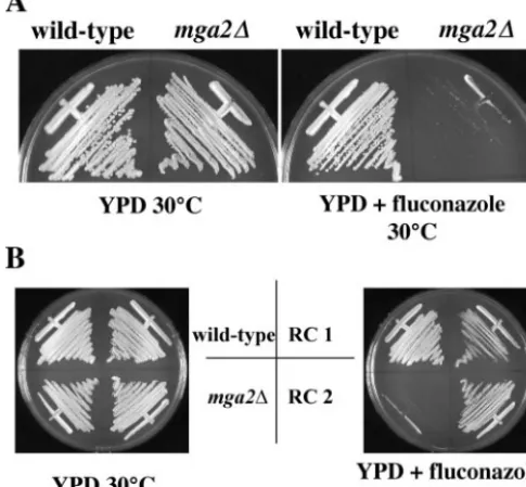
biosyn-thesis inhibitor fluconazole. Themga2⌬mutant strain was hy-persensitive to 10 g of fluconazole/ml, while the growth of wild-type strain H99 was unaffected (Fig. 4A). In a genetic cross with a wild-typeMATastrain, the phenotypes conferred by themga2⌬mutation segregated in a 1:1 ratio of wild type to mutant, and all mutant phenotypes cosegregated 100% with the nourseothricin resistance marker (data not shown). Rein-troduction of theMGA2gene into themga2⌬mutant restored its ability to grow in the presence of 10g of fluconazole/ml and complemented the growth defects (Fig. 4B and data not shown). The hypersensitivity of themga2⌬mutant to flucon-azole likely was due to defects in sterol metabolism, because themga2⌬mutant was also hypersensitive to fenpropimorph, another sterol synthesis inhibitor, but not to the protein syn-thesis inhibitor cycloheximide (data not shown). These findings suggest that the mechanisms coordinating fatty acid metabo-lism and sterol biosynthesis are conserved inC.neoformans.
The microarray results indicated that multiple genes in two biosynthetic pathways, pyrimidine biosynthesis and isoleucine
FIG. 2. Northern analysis of temperature-regulated expression. (A) Total RNA from 25°C samples and 37°C samples was probed with gene-specific probes. (B) Total RNA from 0-, 3-, 6-, 9-, and 12-h time points was probed with gene-specific probes.ACT1was included as a loading control in both experiments.
FIG. 3. Phenotypic analysis of mutant strains. (A) Fivefold serial dilutions of cultures of H99 (wild type), PK28 (mga2⌬), AI34 (clc1⌬), JF99 (ura5), and PK23 (ura5mutant JF99 withURA5 reintroduced) were inoculated into YPD medium and grown for 3 days at the indicated temperatures. (B) Cultures of H99 (wild type) (gray bars) and PK28 (mga2⌬) (black bars) were inoculated into YPD medium to an OD600of 0.05
and incubated at 25 or 37°C for 24 h. Growth was determined by comparing the OD600of each experimental culture to that of a control culture
of the same strain grown at 30°C for 24 h. Error bars represent the standard deviation of the mean from three independent experiments.
on September 8, 2020 by guest
http://ec.asm.org/
or valine biosynthesis, were repressed after a shift in growth temperature from 25 to 37°C (Table 1). Interestingly, disrup-tion of ILV2(encoding acetolactate synthase) confers a 37°C growth defect (33). Therefore, we examined whether a muta-tion in the pyrimidine biosynthetic pathway also resulted in an inability to grow at 37°C. Strain JF99 was isolated as a spon-taneous 5-fluoroorotic acid (5-FOA)-resistant mutant of wild-type strain KN99a, which is isogenic with strain H99. 5-FOA is converted into the toxic compound 5-fluorouracil when the pyrimidine synthetic pathway is intact, and growth on 5-FOA occurs only when pyrimidine synthesis is defective. Strain JF99 displayed a marked growth defect compared to strain H99 when incubated at 37°C, and this growth defect at 37°C was complemented by reintroduction of theURA5gene (Fig. 3A). These findings demonstrate that the growth defect is due to a mutation in URA5 and not to secondary mutations. Similar results were obtained with six other independent 5-FOA-resis-tant isolates (data not shown).
TheCLC1gene encodes a voltage-gated chloride channel implicated in melanin biosynthesis and ion homeostasis (31, 61). Based on the microarray results, CLC1was strongly in-duced after a shift from 25 to 37°C (Fig. 1). This induction was confirmed by Northern analysis (Fig. 2B). Disruption of the
CLC1gene had no effect on the ability of C. neoformans to grow at 37°C (Fig. 3A).
Identifying putative targets of Mga2.An important
applica-tion of microarray technology is the identificaapplica-tion of novel
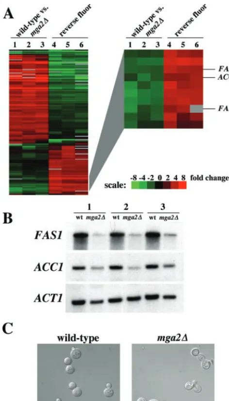
transcription factors and the definition of their targets. Here we sought to identify targets of the Mga2 transcription factor homolog. Microarray analysis of wild-type gene expression compared to mga2⌬ mutant gene expression was performed (Table 2; see Table S3 in the supplemental material). Notably, the orthologs of the S. cerevisiae FAS1and ACC1genes, en-coding the beta subunit of fatty acid synthase and acetyl coen-zyme A decarboxylase, respectively, displayed significantly re-duced expression in the mga2⌬ mutant. Cluster analysis of three independent wild-type and mga2⌬ mutant microarray experiments and the corresponding reverse fluor control ex-periments revealed similar expression patterns for theC.
neo-formans FAS1 and ACC1 genes (Fig. 5A). The reduction in
FAS1andACC1expression was confirmed by Northern anal-ysis (Fig. 5B). These results further support the hypothesis that Mga2 regulates fatty acid biosynthesis inC.neoformans.
Among the genes that were induced in themga2⌬mutant strain compared to the wild-type strain were several whose products are implicated in regulating polarized cell growth. The expression of two homologs of small GTPase effectors was significantly higher in the mga2⌬ mutant strain than in the wild-type strain (Table 2). GTPase modules play important roles in directing polarized growth in S. cerevisiaeand other fungi (10, 26, 58). In addition, the expression ofPAK1, encod-ing a PAK kinase important for filamentous growth durencod-ingC.
neoformans mating, was higher in the mga2⌬ mutant strain
than in the wild-type strain (57). Taken together, these results indicate that cells lacking Mga2 are defective in the regulation of polarized cell growth. Cultures of wild-type and mga2⌬
mutant cells were grown in YPD medium at 37°C for 24 h and examined by microscopy (Fig. 5C). Approximately 50% of
mga2⌬mutant cells displayed defective morphology, including multiple buds and misshapen buds (n⫽200). Less than 1% of wild-type cells grown under the same conditions displayed de-fects in bud morphology. These findings support the hypothesis that Mga2 plays a role in the morphogenesis ofC.neoformans.
DISCUSSION
We developed and implemented a genomic-DNA microar-ray to identify genes that display differential expression during growth at 25 or 37°C in the human-pathogenic fungusC.
neo-formansby using an approach similar to that taken by Sil and
colleagues to study phase-regulated gene expression inH.
cap-sulatum(30). When this work began, sequencing projects were
ongoing for three related varieties of C. neoformans, but no finished genome sequence was available. In conjunction with sequence and annotation information provided by these se-quencing projects, our microarray data allow the identification of genes whose expression is regulated by growth temperature. In addition, microarray experiments were used to identify pos-sible targets of a previously uncharacterized transcription fac-tor homolog inC.neoformans. Our studies illustrate how even partial-genome microarrays can be used to identify genes with altered expression patterns and delineate regulatory networks. A goal of this study was to identify genes important for growth at 37°C, a crucial virulence attribute ofC.neoformans. Analysis of gene expression changes that occur after a shift from 25 to 37°C and the steady-state comparison of 25°C growth versus 37°C growth revealed important features about
FIG. 4. Deletion ofMGA2results in hypersensitivity to the ergos-terol biosynthesis inhibitor fluconazole. (A) Cells from wild-type (H99) andmga2⌬mutant (PK28) strains were inoculated into YPD medium and YPD medium containing 10g of fluconazole/ml and grown at 30°C for 3 days. (B) Reintroduction ofMGA2into themga2⌬mutant restores growth in the presence of fluconazole. Strain PK28 (mga2⌬::nat) was transformed with a plasmid containing theMGA2
gene and a cassette that confers resistance to G418. Transformants were selected on G418 at 30°C. Two independent transformants (RC1 and RC2) were tested for growth in the presence of 10g of
flucon-azole/ml.
on September 8, 2020 by guest
http://ec.asm.org/
how this fungus adapts to alterations in growth environment. Genes that are important for stress responses are induced upon a shift from 25 to 37°C. Among the induced genes are orthologs of trehalose synthase, catalase, superoxide dis-mutase, and a presumptive sensor of cell wall stress. It is important to note that for those genes that were induced only at later time points after the shift from 25 to 37°C, induction may be the result of long-term adaptation to growth at 37°C or an indirect consequence of increased cell density. An example of this phenomenon is the pattern of expression of the alter-native oxidaseAOX1, which is initially repressed after a shift to 37°C but then is strongly induced by 12 h after the shift.FHB1, encoding flavohemoglobin, displays a similar pattern of expres-sion asAOX1. BothFHB1andAOX1play roles in responding to nitrosative and oxidative stresses, respectively (2, 17). It is unclear why these genes would be initially repressed after
exposure to heat stress; however, the regulation of these genes or their products at posttranscriptional levels in response to environmental changes is possible.
Heat shock induced gene expression is a phenomenon ob-served in both S. cerevisiae and Candida albicans (20, 25). These two fungi differ in thatS.cerevisiaeundergoes a general stress response, involving an extensive gene expression pro-gram that is initiated regardless of the specific stress encoun-tered, whereasC.albicansdoes not. Our studies suggest that it is possible to test whether a general stress response exists inC.
neoformansby using the genomic microarray. One important
component of the response to high-temperature growth in
bothS.cerevisiaeandC.albicansis the induction of heat shock
proteins that act as molecular chaperones. Our array contains orthologs of the heat shock proteinsHSP78andHSP104, and these genes display an approximately twofold increase in
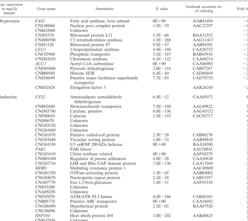
ex-TABLE 2. Gene expression in wild-type versusmga2⌬mutant strains
Gene expression inmga2⌬
mutant Gene name Annotation E value
Genbank accession no.
of ortholog Fold changea
Repression FAS1 Fatty acid synthase, beta subunit 0E⫹00 AAB41494 ⫺4.5
CNL00860 Nuclear pore complex protein 1.5E⫺70 AAC27297 ⫺4.3
CNK02060 Unknown ⫺3.5
CNJ03370 Ribosomal protein L11 3.3E⫺68 BAA31552 ⫺3.1
CNH00590 C1 tetrahydrofolate synthase 1.3E⫺288 AAG11417 ⫺3.1
CNI01120 Ribosomal protein S7 9.5E⫺47 AAB94301 ⫺3
LEU1 2-Isopropylmalate synthase 4.4E⫺148 CAA20723 ⫺3
CNC03960 Phosphate transporter 3.1E⫺187 BAB43910 ⫺2.9
CND02650 Chorismate synthase 6.1E⫺122 CAA68214 ⫺2.9
ACC1 Acetyl CoA carboxylase 0E⫹00 CAA86983 ⫺2.9
CND05600 Pyruvate dehydrogenase 2.0E⫺116 CAB97287 ⫺2.8
CNB00560 Histone H2B 6.4E⫺40 CAD60694 ⫺2.6
CNE04690 Putative major facilitator superfamily
transporter 7.7E⫺83 CAD70735 ⫺2.4
CNE02420 Elongation factor 3 AAK26245 ⫺2.2
Induction LYS2 Aminoadipate semialdehyde
dehydrogenase 6.4E⫺12 CAA85072 6.7
CNB02680 Monosaccharide transporter 7.5E⫺188 AAL89822 5.6
CND03740 Catalase, putative 8.0E⫺136 AAG45152 4.6
CNF00610 Calnexin 1.5E⫺143 CAC82717 4.2
CNJ00670 Unknown 4.1
CNG03520 Unknown 3.7
CNG04460 Unknown 3.7
CNG01070 Putative coiled-coil protein 2.3E⫺28 CAB88270 3.4
CNG03440 Vacuolar sorting protein 1.0E⫺33 AAB49810 3.4
CNG04330 U5 snRNP 200-kDa helicase 0E⫹00 BAA34508 3.3
PAK1 PAK kinase AAL58841 3.3
CNG01610 Chitin synthase related 0E⫹00 AAF04279 3
CNM01490 Regulator of purine utilization 4.8E⫺28 CAA58838 3
CNG02710 LIM and Rho GAP domain protein 7.6E⫺136 CAA17694 2.9
MDR1 Multidrug resistance protein 1 AAC49889 2.9
CNG01350 GTPase-activating protein 1.3E⫺65 AAB84002 2.9
CNG04670 Nucleoporin repeat protein 2.1E⫺28 CAB53357 2.9
CNA07770 Exo-1,3-beta-glucanase 1.4E⫺11 AAF65310 2.8
CNF03200 Unknown 2.8
CNA08250 Unknown 2.8
CNF03070 ATM/ATR PI-3 kinase 4.4E⫺166 CAB40165 2.8
CNB05770 Putative ABC transporter 0E⫹00 CAA54692 2.8
CNG06090 Hypothetical protein 2.1E⫺65 BAA07526 2.7
CNC06090 Unknown 2.7
HSP104 Heat shock protein 104 1.8E⫺202 AAK60625 2.7
CNK02930 Unknown 2.5
CNF04580 Conserved hypothetical protein 1.4E⫺289 CABB91226 2.2
aAverage fold change in expression in three independent trials of themga2⌬mutant compared to wild-type strain H99.
on September 8, 2020 by guest
http://ec.asm.org/
pression in the 3-h time point after the shift from 25°C growth to 37°C growth (data not shown). An ortholog of the SSA family of heat shock chaperones showed no significant change in expression. The failure to detect induction of these heat shock protein genes to similar levels as inS.cerevisiaeis likely
due to differences in experimental design, in that the first time point taken in our study was 3 h after the temperature shift, rather than 10 to 30 min, as in theS.cerevisiaeandC.albicans
studies.
Recently, Steen et al. analyzed temperature-regulated tran-scription in C. neoformansusing SAGE (49). In that report, gene expression at 25 and 37°C was studied for both the sero-type D strain B3501A and the serosero-type A strain H99. Although SAGE and microarray hybridization use different technologies to probe gene expression, it is reasonable to expect overlap in the microarray data and the SAGE data. We find similarities in the genes whose expression is higher at 37°C than at 25°C in our transcriptional profiling of H99 and the SAGE report analyzing the serotype D strain B3501A. Most notably, the genes encoding homologs of an aryl alcohol oxidase (SMG1) and a peripheral benzodiazepine receptor were more highly expressed at 37°C in both studies. Among genes that are re-pressed at 37°C, the ribosomal proteins P0 and S26 were found in our study and the SAGE analysis of both temperature-regulated transcription and gene expression during experimen-tal cryptococcal meningitis, suggesting that alterations in pro-tein synthesis are important for tolerating high-temperature growth and in vivo growth (49, 50).
That differences exist in our study and the SAGE studies is also not surprising given differences in experimental design. An advantage of microarray studies is the ability to analyze changes in gene expression over a time course, while the SAGE experiments described by Steen et al. (49) were per-formed at a single time point. Additionally, Steen et al. found significant differences in the patterns of expression for strains B3501A and H99 in their study. Transcriptional analysis of strain B3501A reveals that genes involved in lipid metabolism, such as genes encoding⌬9 fatty acid desaturase and fatty acid synthase, were more highly expressed at 25°C. We found that a transcription factor homolog that may target lipid metabo-lism genes is strongly induced at 37°C (see below). These differences could reflect unique properties of C. neoformans
serotype D strains such as B3501A relative to the more ther-motolerant serotype A strain H99. However, changes in mem-brane composition and fluidity are likely to occur during growth at either low- or high-temperature extremes (1, 48, 51). Microarray analysis identified a gene, MGA2, that shows significantly higher expression during growth at 37°C and that is also important for normal growth at high temperatures. However, this approach does not appear to be generally ap-plicable. InS.cerevisiae, genes that protect against DNA dam-age are not significantly induced in the presence of DNA-damaging agents (7). Of the previously identified genes important forC.neoformanshigh-temperature growth (CNA1,
CNB1, CTS1,STE20,MPK1, andRAS1), none displayed
sig-nificantly higher gene expression during growth at 37°C than during growth at 25°C.MPK1 encodes a MAP kinase in the protein kinase C-mediated cell integrity pathway and therefore might be expected to display an increase in expression upon a temperature shift; however, no such increase in expression was observed. Furthermore, CLC1 was induced strongly in the earliest time points after the shift to 37°C (Fig. 1); however, a
clc1⌬ mutant was not affected in its ability to tolerate high-temperature growth (Fig. 3A). Strikingly, we observed a sig-nificant reduction in expression of genes involved in isoleucine
FIG. 5. Gene expression in wild-type and mga2⌬mutant strains. (A) Three independent microarray experiments and the corresponding reverse fluor control experiments were performed with cDNAs from wild-type (Cy3-labeled) and mga2⌬ (Cy5-labeled) strains. Elements whose expression changed at least threefold in at least one experiment were analyzed by hierarchical clustering. Each lane represents one experiment, and each row represents gene expression. A cluster of interest containing theFAS1andACC1genes is enlarged. Green color in lanes 1 to 3 represents decreased gene expression. (B) Northern analysis of wild-type (wt) andmga2⌬mutant gene expression. Three independent samples of total RNA from wild-type (H99) andmga2⌬ mutant (PK28) strains were probed with FAS1- and ACC1-specific probes.ACT1was included as a loading control. (C) Morphogenesis of wild-type andmga2⌬mutant strains. Cultures of wild-type (H99) and
mga2⌬mutant (PK28) strains were inoculated into YPD medium and incubated at 37°C for 24 h. Differential interference contrast images were captured at a magnification of⫻1,000 with a Zeiss Axioskop 2 Plus microscope equipped with an AxioCam MRM digital camera.
on September 8, 2020 by guest
http://ec.asm.org/
or valine biosynthesis and pyrimidine biosynthesis after a shift to 37°C, yet mutations inactivating each of these pathways result in a high-temperature growth defect. This is an unex-pected result, and an explanation may be that the ilv2⌬ and
ura5⌬mutants accumulate a toxic intermediate at 37°C but not at 25°C or that the uptake of essential nutrients is significantly altered by the growth temperature, the presence of the muta-tions, or both. This expression pattern may not occur for all amino acid biosynthetic pathways, as genes encoding homologs of lysine and threonine biosynthetic enzymes are induced rather than repressed at 37°C (Table 1). These results suggest caution may be warranted in the use ofURA5as a selectable marker and in the interpretation of experiments using ura5
mutant strains. Interestingly, we found no evidence of temper-ature-sensitive growth defects in serotype D ura5 mutants, whereas serotype Aura5mutants are clearly temperature sen-sitive (A. Idnurm, P. Kraus, and J. Heitman, unpublished data). Although identification of genes that display differential expression may not in all cases reveal physiologically relevant patterns of gene expression, microarray analysis can be used effectively as a screen to prioritize candidates for further study. Hence, an essential component of studies with theC. neofor-mansgenomic microarray is incorporation of information re-garding phenotypic consequences of alterations in gene expres-sion.
Long-chain fatty acids are used by eukaryotic cells as an energy source as well as for building blocks of cell and or-ganelle membrane lipids.S.cerevisiaehas served as an excel-lent model system for studying fatty acid metabolism, and considerable information is known about how fatty acids are synthesized, transported, and degraded (54). An important component of the regulation of fatty acid metabolism in yeast cells is the activation of gene expression, with signals such as glucose and unsaturated fatty acids acting to induce the ex-pression of a panoply of target genes. The exex-pression of genes involved in saturated fatty acid biosynthesis, includingACC1
and FAS1, is coregulated (12). Our results suggest that these genes are also coregulated inC.neoformans, and a transcrip-tion factor homolog that may target these genes was identified.
C.neoformansMga2 is homologous to twoS.cerevisiae
tran-scription factors, Mga2 and Spt23. These factors activate ex-pression ofOLE1, encoding⌬9 desaturase, in response to low oxygen levels and low growth temperatures (32, 60). Other targets of these factors in yeast cells, if any, are unknown. Membrane composition is altered whenS.cerevisiaecells are exposed to temperature extremes, and activation of desatu-rases is thought to provide increased membrane fluidity (48, 51). InC.neoformans, increased production of long-chain fatty acids may be necessary for tolerating high-temperature growth and could explain the increased expression of a factor regulat-ing their production. It is important to note that expression of the potential targets of Mga2,FAS1andACC1, was not sig-nificantly higher at 37°C than at 25°C (Table 1; see Table S2 in the supplemental material). Mga2 and Spt23 are synthesized as precursors that are anchored to the endoplasmic reticulum membrane and then activated by ubiquitin- or proteasome-dependent processing (28). Hence, an increase in the expres-sion of the MGA2andSPT23 genes may not result in a con-comitant increase in activity of the transcription factors. S.
cerevisiae MGA2expression is induced by the unfolded protein
response, suggesting that additional pathways may be targeted by this transcription factor (53). Increased expression ofMGA2
inC.neoformansmay be a consequence of the unfolded
pro-tein response or endoplasmic reticulum-mediated propro-tein deg-radation, both of which are activated by heat stress inS.
cer-evisiae(25, 53). Nevertheless,C.neoformans MGA2plays an
important role in cell growth, particularly at high growth tem-peratures.
Fatty acid metabolism is coordinated with sterol synthesis in yeast cells, and antifungal drugs that inhibit ergosterol biosyn-thesis may also have secondary effects on the enzymes that catalyze fatty acid biosynthesis (11, 35, 36). TheC.neoformans mga2⌬ mutant is hypersensitive to the ergosterol synthesis inhibitor fluconazole, indicating a conserved link between fatty acid and sterol metabolism. Membrane remodeling via fatty acid biosynthesis, saturation-desaturation, and sterol metabo-lism is likely to be an important component of the regulatory mechanisms enabling responses to stress in C. neoformans. Interestingly, the sterol composition ofC.neoformanscan be altered after passage through a mammalian host (15). Addi-tionally, lipids and sterols are important components of polar-ized cell growth inS.cerevisiaeandS.pombe, and our findings suggest that this relationship is conserved inC.neoformans(5, 56). The effects of improper regulation of fatty acid and sterol metabolism inC.neoformansinfluence the ability to undergo normal morphogenesis, which is essential for growth, mating, and haploid fruiting (4). The wiring of regulatory pathways that respond to stress, coordinate fatty acid and sterol synthesis, and control cellular morphogenesis likely differs between S.
cerevisiaeandC.neoformansand may differ among the closely
related varieties of C. neoformans. Detailed examination of these pathways will provide additional insight into the biology underlying stress responses and potentially reveal new areas to pursue for antifungal therapy.
ACKNOWLEDGMENTS
We are indebted to Anita Sil and to Joseph DeRisi (University of California, San Francisco) for essential assistance in initiating this work and for communicating unpublished results; Mark DeLong for assistance with array annotation; Andrew Alspaugh and Jo Kingsbury for discussions; Holly Dressman, Ben Isett, and Laura-Leigh Rowlette (Duke Center for Genome Technology) for technical assistance with microarrays; and James Fraser, Alexander Idnurm, Andrew Alspaugh, and John McCusker for critical reading of the manuscript.
We acknowledge theC.neoformansGenome Project, Stanford Ge-nome Technology Center, funded by the NIAID/NIH under cooper-ative agreement AI47087, and TIGR, funded by the NIAID/NIH un-der cooperative agreement U01 AI48594, for providing gene sequence and annotation information prior to publication. These studies were supported in part by R01 grants AI39115, AI42159, and AI50438 from NIAID to J.H., grant A128388 to J.R.P., and program project grant P01-AI44975 from NIAID to the Duke University Mycology Research Unit. Gary M. Cox is a Burroughs Wellcome New Investigator in Molecular Pathogenic Mycology. Joseph Heitman is a Burroughs Wellcome Scholar in Molecular Pathogenic Mycology and an Associ-ate Investigator of the Howard Hughes Medical Institute.
REFERENCES
1. Aguilar, P. S., J. E. Cronan, Jr., and D. de Mendoza.1998. ABacillus subtilis gene induced by cold shock encodes a membrane phospholipid desaturase. J. Bacteriol.180:2194–2200.
2. Akhter, S., H. C. McDade, J. M. Gorlach, G. Heinrich, G. M. Cox, and J. R. Perfect.2003. Role of alternative oxidase gene in pathogenesis of Crypto-coccus neoformans. Infect. Immun.71:5794–5802.
3. Alspaugh, J. A., L. M. Cavallo, J. R. Perfect, and J. Heitman.2000.RAS1
on September 8, 2020 by guest
http://ec.asm.org/
regulates filamentation, mating and growth at high temperature of Crypto-coccus neoformans. Mol. Microbiol.36:352–365.
4. Alspaugh, J. A., R. C. Davidson, and J. Heitman.2000. Morphogenesis of Cryptococcus neoformans. Contrib. Microbiol.5:217–238.
5. Bagnat, M., and K. Simons.2002. Lipid rafts in protein sorting and cell polarity in budding yeastSaccharomyces cerevisiae. Biol. Chem.383:1475– 1480.
6. Baldi, P., and G. W. Hatfield.2002. DNA microarrays and gene expression: from experiments to data analysis and modeling. Cambridge University Press, Cambridge, United Kingdom.
7. Birrell, G. W., J. A. Brown, H. I. Wu, G. Giaever, A. M. Chu, R. W. Davis, and J. M. Brown.2002. Transcriptional response ofSaccharomyces cerevisiaeto DNA-damaging agents does not identify the genes that protect against these agents. Proc. Natl. Acad. Sci. USA99:8778–8783.
8. Cannon, R. D., W. E. Timberlake, N. A. Gow, D. Bailey, A. Brown, G. W. Gooday, B. Hube, M. Monod, C. Nombela, F. Navarro, et al.1994. Molecular biological and biochemical aspects of fungal dimorphism. J. Med. Vet. My-col.32(Suppl. 1):53–64.
9. Casadevall, A., and J. R. Perfect.1998.Cryptococcus neoformans. ASM Press, Washington, D.C.
10. Casamayor, A., and M. Snyder.2002. Bud-site selection and cell polarity in budding yeast. Curr. Opin. Microbiol.5:179–186.
11. Casey, W. M., G. A. Keesler, and L. W. Parks.1992. Regulation of parti-tioned sterol biosynthesis inSaccharomyces cerevisiae. J. Bacteriol.174:7283– 7288.
12. Chirala, S. S.1992. Coordinated regulation and inositol-mediated and fatty acid-mediated repression of fatty acid synthase genes inSaccharomyces cer-evisiae. Proc. Natl. Acad. Sci. USA89:10232–10236.
13. Cox, G. M., H. C. McDade, S. C. Chen, S. C. Tucker, M. Gottfredsson, L. C. Wright, T. C. Sorrell, S. D. Leidich, A. Casadevall, M. A. Ghannoum, and J. R. Perfect.2001. Extracellular phospholipase activity is a virulence factor forCryptococcus neoformans. Mol. Microbiol.39:166–175.
14. Cox, G. M., J. Mukherjee, G. T. Cole, A. Casadevall, and J. R. Perfect.2000. Urease as a virulence factor in experimental cryptococcosis. Infect. Immun.
68:443–448.
15. Currie, B., H. Sanati, A. S. Ibrahim, J. E. Edwards, Jr., A. Casadevall, and M. A. Ghannoum.1995. Sterol compositions and susceptibilities to ampho-tericin B of environmentalCryptococcus neoformansisolates are changed by murine passage. Antimicrob. Agents Chemother.39:1934–1937.
16. Davidson, R. C., J. R. Blankenship, P. R. Kraus, M. d. Berrios, C. M. Hull, C. D’Souza, P. Wang, and J. Heitman.2002. A PCR-based strategy to generate integrative targeting alleles with large regions of homology. Micro-biology148:2607–2615.
17. de Jesus-Berrios, M., L. Liu, J. C. Nussbaum, G. M. Cox, J. S. Stamler, and J. Heitman.2003. Enzymes that counteract nitrosative stress promote fungal virulence. Curr. Biol.13:1963–1968.
18. DeRisi, J. L., V. R. Iyer, and P. O. Brown.1997. Exploring the metabolic and genetic control of gene expression on a genomic scale. Science278:680–686. 19. Eisen, M. B., P. T. Spellman, P. O. Brown, and D. Botstein.1998. Cluster analysis and display of genome-wide expression patterns. Proc. Natl. Acad. Sci. USA95:14863–14868.
20. Enjalbert, B., A. Nantel, and M. Whiteway.2003. Stress-induced gene ex-pression inCandida albicans: absence of a general stress response. Mol. Biol. Cell14:1460–1467.
21. Erickson, T., L. Liu, A. Gueyikian, X. Zhu, J. Gibbons, and P. R. Williamson.
2001. Multiple virulence factors ofCryptococcus neoformansare dependent on VPH1. Mol. Microbiol.42:1121–1131.
22. Fox, D. S., G. M. Cox, and J. Heitman.2003. Phospholipid-binding protein Cts1 controls septation and functions coordinately with calcineurin in Cryp-tococcus neoformans. Eukaryot. Cell2:1025–1035.
23. Fraser, J. A., R. L. Subaran, C. B. Nichols, and J. Heitman.2003. Recapit-ulation of the sexual cycle of the primary fungal pathogenCryptococcus neoformansvarietygattii: implications for an outbreak on Vancouver Island. Eukaryot. Cell2:1036–1045.
24. Galagan, J. E., S. E. Calvo, K. A. Borkovich, E. U. Selker, N. D. Read, D. Jaffe, W. FitzHugh, L. J. Ma, S. Smirnov, S. Purcell, B. Rehman, T. Elkins, R. Engels, S. Wang, C. B. Nielsen, J. Butler, M. Endrizzi, D. Qui, P. Ianak-iev, D. Bell-Pedersen, M. A. Nelson, M. Werner-Washburne, C. P. Selitren-nikoff, J. A. Kinsey, E. L. Braun, A. Zelter, U. Schulte, G. O. Kothe, G. Jedd, W. Mewes, C. Staben, E. Marcotte, D. Greenberg, A. Roy, K. Foley, J. Naylor, N. Stange-Thomann, R. Barrett, S. Gnerre, M. Kamal, M. Kamvysselis, E. Mauceli, C. Bielke, S. Rudd, D. Frishman, S. Krystofova, C. Rasmussen, R. L. Metzenberg, D. D. Perkins, S. Kroken, C. Cogoni, G. Macino, D. Catcheside, W. Li, R. J. Pratt, S. A. Osmani, C. P. DeSouza, L. Glass, M. J. Orbach, J. A. Berglund, R. Voelker, O. Yarden, M. Plamann, S. Seiler, J. Dunlap, A. Radford, R. Aramayo, D. O. Natvig, L. A. Alex, G. Mannhaupt, D. J. Ebbole, M. Freitag, I. Paulsen, M. S. Sachs, E. S. Lander, C. Nusbaum, and B. Birren.2003. The genome sequence of the filamentous fungus Neu-rospora crassa. Nature422:859–868.
25. Gasch, A. P., P. T. Spellman, C. M. Kao, O. Carmel-Harel, M. B. Eisen, G. Storz, D. Botstein, and P. O. Brown.2000. Genomic expression programs in
the response of yeast cells to environmental changes. Mol. Biol. Cell11:
4241–4257.
26. Harris, S. D., and M. Momany.2004. Polarity in filamentous fungi: moving beyond the yeast paradigm. Fungal Genet. Biol.41:391–400.
27. Heinisch, J. J., A. Lorberg, H. P. Schmitz, and J. J. Jacoby.1999. The protein kinase C-mediated MAP kinase pathway involved in the maintenance of cellular integrity inSaccharomyces cerevisiae. Mol. Microbiol.32:671–680. 28. Hoppe, T., K. Matuschewski, M. Rape, S. Schlenker, H. D. Ulrich, and S.
Jentsch.2000. Activation of a membrane-bound transcription factor by reg-ulated ubiquitin/proteasome-dependent processing. Cell102:577–586. 29. Hull, C. M., R. C. Davidson, and J. Heitman.2002. Cell identity and sexual
development inCryptococcus neoformansare controlled by the mating-type-specific homeodomain protein Sxi1alpha. Genes Dev.16:3046–3060. 30. Hwang, L., D. Hocking-Murray, A. K. Bahrami, M. Andersson, J. Rine, and
A. Sil.2003. Identifying phase-specific genes in the fungal pathogen His-toplasma capsulatumusing a genomic shotgun microarray. Mol. Biol. Cell
14:2314–2326.
31. Idnurm, A., J. L. Reedy, J. C. Nussbaum, and J. Heitman.2004.Cryptococcus neoformansvirulence gene discovery through insertional mutagenesis. Eu-karyot. Cell3:420–429.
32. Jiang, Y., M. J. Vasconcelles, S. Wretzel, A. Light, C. E. Martin, and M. A. Goldberg.2001.MGA2is involved in the low-oxygen response element-dependent hypoxic induction of genes inSaccharomyces cerevisiae. Mol. Cell. Biol.21:6161–6169.
33. Kingsbury, J. M., Z. Yang, T. M. Ganous, G. M. Cox, and J. H. McCusker.
2004.Cryptococcus neoformansIlv2p confers resistance to sulfometuron methyl and is required for survival at 37°C and in vivo. Microbiology150:
1547–1558.
34. Kraus, P. R., D. S. Fox, G. M. Cox, and J. Heitman.2003. TheCryptococcus neoformansMAP kinase Mpk1 regulates cell integrity in response to anti-fungal drugs and loss of calcineurin function. Mol. Microbiol.48:1377–1387. 35. Kuchta, T., C. Leka, R. Kubinec, and N. J. Russell.1997. Inhibition of ergosterol biosynthesis is not accompanied by a change in fatty acid compo-sition inSaccharomyces cerevisiaetreated with the antifungal agent 6-amino-2-n-pentylthiobenzothiazole. FEMS Microbiol. Lett.150:43–47.
36. Kwast, K. E., L. C. Lai, N. Menda, D. T. James III, S. Aref, and P. V. Burke.
2002. Genomic analyses of anaerobically induced genes inSaccharomyces cerevisiae: functional roles of Rox1 and other factors in mediating the anoxic response. J. Bacteriol.184:250–265.
37. Kwon-Chung, K. J., and J. C. Rhodes.1986. Encapsulation and melanin formation as indicators of virulence in Cryptococcus neoformans. Infect. Immun.51:218–223.
38. Lander, E. S., and M. S. Waterman.1988. Genomic mapping by fingerprint-ing random clones: a mathematical analysis. Genomics2:231–239. 39. Lengeler, K. B., D. S. Fox, J. A. Fraser, A. Allen, K. Forrester, F. S. Dietrich,
and J. Heitman.2002. Mating-type locus ofCryptococcus neoformans: a step in the evolution of sex chromosomes. Eukaryot. Cell1:704–718.
40. Ludin, K. M., N. Hilti, and M. E. Schweingruber.1995.Schizosaccharomyces pombe rds1, an adenine-repressible gene regulated by glucose, ammonium, phosphate, carbon dioxide and temperature. Mol. Gen. Genet.248:439–445. 41. Maresca, B., and G. S. Kobayashi.2000. Dimorphism inHistoplasma
cap-sulatumandBlastomyces dermatitidis. Contrib. Microbiol.5:201–216. 42. McCusker, J. H., K. V. Clemons, D. A. Stevens, and R. W. Davis.1994.
Genetic characterization of pathogenicSaccharomyces cerevisiaeisolates. Genetics136:1261–1269.
43. Missall, T. A., M. E. Pusateri, and J. K. Lodge.2004. Thiol peroxidase is critical for virulence and resistance to nitric oxide and peroxide in the fungal pathogen,Cryptococcus neoformans. Mol. Microbiol.51:1447–1458. 44. Nakagawa, Y., N. Sakumoto, Y. Kaneko, and S. Harashima.2002. Mga2p is
a putative sensor for low temperature and oxygen to induceOLE1 transcrip-tion inSaccharomyces cerevisiae. Biochem. Biophys. Res. Commun.291:707– 713.
45. Odom, A., S. Muir, E. Lim, D. L. Toffaletti, J. Perfect, and J. Heitman.1997. Calcineurin is required for virulence ofCryptococcus neoformans. EMBO J.
16:2576–2589.
46. Rude, T. H., D. L. Toffaletti, G. M. Cox, and J. R. Perfect.2002. Relationship of the glyoxylate pathway to the pathogenesis ofCryptococcus neoformans. Infect. Immun.70:5684–5694.
47. Sia, R. A., K. B. Lengeler, and J. Heitman.2000. Diploid strains of the pathogenic basidiomyceteCryptococcus neoformansare thermally dimorphic. Fungal Genet. Biol.29:153–163.
48. Steels, E. L., R. P. Learmonth, and K. Watson.1994. Stress tolerance and membrane lipid unsaturation inSaccharomyces cerevisiaegrown aerobically or anaerobically. Microbiology140:569–576.
49. Steen, B. R., T. Lian, S. Zuyderduyn, W. K. MacDonald, M. Marra, S. J. Jones, and J. W. Kronstad.2002. Temperature-regulated transcription in the pathogenic fungusCryptococcus neoformans. Genome Res.12:1386–1400. 50. Steen, B. R., S. Zuyderduyn, D. L. Toffaletti, M. Marra, S. J. Jones, J. R.
Perfect, and J. Kronstad.2003.Cryptococcus neoformansgene expression during experimental cryptococcal meningitis. Eukaryot. Cell2:1336–1349. 51. Swan, T. M., and K. Watson.1999. Stress tolerance in a yeast lipid mutant:
on September 8, 2020 by guest
http://ec.asm.org/
membrane lipids influence tolerance to heat and ethanol independently of heat shock proteins and trehalose. Can. J. Microbiol.45:472–479. 52. Toffaletti, D. L., M. Del Poeta, T. H. Rude, F. Dietrich, and J. R. Perfect.
2003. Regulation of cytochrome c oxidase subunit 1 (COX1) expression in Cryptococcus neoformansby temperature and host environment. Microbiol-ogy149:1041–1049.
53. Travers, K. J., C. K. Patil, L. Wodicka, D. J. Lockhart, J. S. Weissman, and P. Walter.2000. Functional and genomic analyses reveal an essential coor-dination between the unfolded protein response and ER-associated degra-dation. Cell101:249–258.
54. Trotter, P. J.2001. The genetics of fatty acid metabolism inSaccharomyces cerevisiae. Annu. Rev. Nutr.21:97–119.
55. Verna, J., A. Lodder, K. Lee, A. Vagts, and R. Ballester.1997. A family of genes required for maintenance of cell wall integrity and for the stress response inSaccharomyces cerevisiae. Proc. Natl. Acad. Sci. USA94:13804– 13809.
56. Wachtler, V., S. Rajagopalan, and M. K. Balasubramanian.2003. Sterol-rich
plasma membrane domains in the fission yeastSchizosaccharomyces pombe. J. Cell Sci.116:867–874.
57. Wang, P., C. B. Nichols, K. B. Lengeler, M. E. Cardenas, G. M. Cox, J. R. Perfect, and J. Heitman.2002. Mating-type-specific and nonspecific PAK kinases play shared and divergent roles inCryptococcus neoformans. Eu-karyot. Cell1:257–272.
58. Wendland, J., and P. Philippsen.2001. Cell polarity and hyphal morpho-genesis are controlled by multiple rho-protein modules in the filamentous ascomyceteAshbya gossypii. Genetics157:601–610.
59. Zaigler, A., S. C. Schuster, and J. Soppa.2003. Construction and usage of a onefold-coverage shotgun DNA microarray to characterize the metabolism of the archaeonHaloferax volcanii. Mol. Microbiol.48:1089–1105. 60. Zhang, S., Y. Skalsky, and D. J. Garfinkel.1999.MGA2orSPT23is required
for transcription of the delta9 fatty acid desaturase gene,OLE1, and nuclear membrane integrity inSaccharomyces cerevisiae. Genetics151:473–483. 61. Zhu, X., and P. R. Williamson.2003. A CLC-type chloride channel gene is
required for laccase activity and virulence inCryptococcus neoformans. Mol. Microbiol.50:1271–1281.