University of Pennsylvania
ScholarlyCommons
Publicly Accessible Penn Dissertations
2017
Structure-Function Relationships Of Rna And
Protein In Synaptic Plasticity
Sarah Middleton
University of Pennsylvania, [email protected]
Follow this and additional works at:https://repository.upenn.edu/edissertations Part of theBioinformatics Commons,Biology Commons, and theNeuroscience and Neurobiology Commons
This paper is posted at ScholarlyCommons.https://repository.upenn.edu/edissertations/2474
For more information, please [email protected]. Recommended Citation
Middleton, Sarah, "Structure-Function Relationships Of Rna And Protein In Synaptic Plasticity" (2017).Publicly Accessible Penn Dissertations. 2474.
Structure-Function Relationships Of Rna And Protein In Synaptic
Plasticity
Abstract
Structure is widely acknowledged to be important for the function of ribonucleic acids (RNAs) and proteins. However, due to the relative accessibility of sequence information compared to structure information, most large genomics studies currently use only sequence-based annotation tools to analyze the function of
expressed molecules. In this thesis, I introduce two novel computational methods for genome-scale structure-function analysis and demonstrate their application to identifying RNA and protein structures involved in synaptic plasticity and potentiation—important neuronal processes that are thought to form the basis of learning and memory. First, I describe a new method for de novo identification of RNA secondary structure motifs enriched in co-regulated transcripts. I show that this method can accurately identify secondary structure motifs that recur across three or more transcripts in the input set with an average recall of 0.80 and precision of 0.98. Second, I describe a tool for predicting protein structural fold from amino acid sequence, which achieves greater than 96% accuracy on benchmarks and can be used to predict protein function and identify new structural folds. Importantly, both of these tools scale linearly with increasing numbers of input sequences, making them feasible to run on thousands of sequences at a time. Finally, I use these tools to investigate RNA localization and local translation in dendrites—two processes that are prerequisites for long-lasting synaptic potentiation. Using soma- and dendrite-specific RNA-sequencing data as a starting point, I define the full set of RNAs localized to the dendrites, identify novel secondary structure motifs enriched in these RNAs that may act as dendritic localization signals, and predict the structure of all proteins that would be produced by these localized RNAs during local translation. The results shed new light on potential regulatory mechanisms of dendritic localization and roles of locally translated proteins at the synapse, and demonstrate the utility of structure-based tools in genomics analysis.
Degree Type
Dissertation
Degree Name
Doctor of Philosophy (PhD)
Graduate Group
Genomics & Computational Biology
First Advisor
Junhyong Kim
Keywords
long-term potentiation, protein structure, RNA localization, RNA structure, single cell, structure motifs
Subject Categories
Bioinformatics | Biology | Neuroscience and Neurobiology
STRUCTURE-FUNCTION RELATIONSHIPS OF RNA AND PROTEIN IN
SYNAPTIC PLASTICITY
Sarah A. Middleton
A DISSERTATION
in
Genomics and Computational Biology
Presented to the Faculties of the University of Pennsylvania
in
Partial Fulfillment of the Requirements for the
Degree of Doctor of Philosophy
2017
Supervisor of Dissertation
_________________________
Junhyong Kim, Ph.D., Professor of Biology
Graduate Group Chairperson
_________________________
Li-San Wang, Ph.D., Associate Professor of Pathology and Laboratory Medicine
Dissertation Committee
Li-San Wang, Ph.D., Associate Professor of Pathology and Laboratory Medicine (Chair) Danielle Bassett, Ph.D., Associate Professor of Bioengineering
Russ Carstens, M.D., Associate Professor of Medicine James Eberwine, Ph.D., Professor of Pharmacology
STRUCTURE-FUNCTION RELATIONSHIPS OF RNA AND PROTEIN IN SYNAPTIC
PLASTICITY
COPYRIGHT
2017
Sarah A. Middleton
This work is licensed under the Creative Commons Attribution- NonCommercial-ShareAlike 3.0 License
To view a copy of this license, visit
iii
ACKNOWLEDGMENTS
I am extremely grateful to all the people who have helped and supported me
throughout my graduate work: my advisor, Junhyong, who has been an inspiring and
compassionate mentor; all of the members of the Kim lab, especially those who helped
me get started in the wet lab (Chantal, Derek, Hoa, Jean), managed the computing
resources (Jamie, Stephen), and shared computational advice and camaraderie in the dry
lab (Hannah, Mugdha, Qin, Youngji); my thesis committee, for their extremely helpful
advice and guidance; my collaborators, especially Joe and Kanishka, who helped with
developing and testing the machine learning methods in Chapter 3 of this thesis; my
fellow GCB students, for being an awesome and fun group of people—I will miss
betraying each other during game nights!; and the GCB coordinators, Hannah and
Maureen, for organizing events and keeping GCB running smoothly. I also want to give
my sincerest thanks to my parents, who have always gone out of their way to support me;
and to my husband, Julius, who can make me smile even when everything else seems to
be going wrong (a regular occurrence in graduate school!). Finally, I gratefully
acknowledge the Department of Energy Computational Science Graduate Fellowship
(DE-FG02-97ER25308), which not only supported much of this work financially, but
also provided me with amazing opportunities to expand my computational skills and
knowledgebase and to interact with inspiring people in computational disciplines outside
iv ABSTRACT
STRUCTURE-FUNCTION RELATIONSHIPS OF RNA AND PROTEIN IN
SYNAPTIC PLASTICITY
Sarah A. Middleton
Junhyong Kim
Structure is widely acknowledged to be important for the function of ribonucleic
acids (RNAs) and proteins. However, due to the relative accessibility of sequence
information compared to structure information, most large genomics studies currently use
only sequence-based annotation tools to analyze the function of expressed molecules. In
this thesis, I introduce two novel computational methods for genome-scale
structure-function analysis and demonstrate their application to identifying RNA and protein
structures involved in synaptic plasticity and potentiation—important neuronal processes
that are thought to form the basis of learning and memory. First, I describe a new method
for de novo identification of RNA secondary structure motifs enriched in co-regulated
transcripts. I show that this method can accurately identify secondary structure motifs
that recur across three or more transcripts in the input set with an average recall of 0.80
and precision of 0.98. Second, I describe a tool for predicting protein structural fold from
amino acid sequence, which achieves greater than 96% accuracy on benchmarks and can
be used to predict protein function and identify new structural folds. Importantly, both of
v
feasible to run on thousands of sequences at a time. Finally, I use these tools to
investigate RNA localization and local translation in dendrites—two processes that are
prerequisites for long-lasting synaptic potentiation. Using soma- and dendrite-specific
RNA-sequencing data as a starting point, I define the full set of RNAs localized to the
dendrites, identify novel secondary structure motifs enriched in these RNAs that may act
as dendritic localization signals, and predict the structure of all proteins that would be
produced by these localized RNAs during local translation. The results shed new light on
potential regulatory mechanisms of dendritic localization and roles of locally translated
proteins at the synapse, and demonstrate the utility of structure-based tools in genomics
vi
TABLE OF CONTENTS
ACKNOWLEDGMENTS ... III LIST OF TABLES ... VIII
LIST OF FIGURES ... IX
CHAPTER 1: INTRODUCTION ... 1
1.1. RNA structure ... 1
1.1.1. Overview ... 2
1.1.2. RNA structure prediction... 3
1.1.3. Structure-function relationships ... 6
1.2. Protein structure ... 8
1.2.1. Overview ... 9
1.2.2. Protein structure prediction ... 11
1.2.3. Structure-function relationships ... 15
1.3. Neurons, plasticity, and structure ... 16
1.3.1. Components of pyramidal neurons ... 17
1.3.2. Long-term potentiation ... 18
1.3.3. Importance of RNA localization and local translation ... 20
1.3.4. Mechanisms of dendritic RNA localization: a role for structures ... 22
1.3.5. Protein structures of the synapse ... 26
1.4. Overview of thesis ... 27
1.5. References ... 32
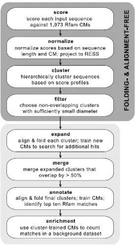
CHAPTER 2: AN EMPIRICAL STRUCTURE SPACE FOR FUNCTIONAL MOTIF ANALYSIS OF RNA... 43
2.1 Introduction ... 43
2.2 Results ... 47
2.2.1 Construction and normalization of the structural feature space ... 47
2.2.2 Suitability of the RESS for structure similarity analysis ... 50
2.2.3 Automated structural clustering for motif identification ... 51
2.2.4 Application of NoFold to novel motif discovery ... 57
2.3 Discussion ... 64
vii
2.5 References ... 90
CHAPTER 3: EXTENDING EMPIRICAL STRUCTURE SPACES TO PROTEIN FOLD RECOGNITION AND FUNCTION PREDICTION... 94
3.1 Introduction ... 94
3.2 Results ... 96
3.2.1 The protein empirical structure space (PESS) ... 96
3.2.2 Fold recognition performance ... 97
3.2.3 Proteome-scale fold prediction of human proteins ... 99
3.2.4 Finding missing hedgehog proteins in C. elegans ... 106
3.3 Discussion ... 107
3.4 Methods ... 109
3.5 References ... 126
CHAPTER 4: STRUCTURES AND PLASTICITY: ANALYSIS OF DENDRITICALLY TARGETED RNAS AND THE “LOCAL PROTEOME”... 129
4.1 Introduction ... 129
4.2 Results and Discussion ... 135
4.2.1 Gene-level localization ... 135
4.2.2 Differential localization of 3'UTR isoforms ... 138
4.2.3 Dendritic targeting motifs ... 143
4.2.4 Functional analysis of the “local proteome” using structure information ... 150
4.3 Conclusions ... 158
4.4 Methods ... 159
4.5 References ... 203
viii
LIST OF TABLES
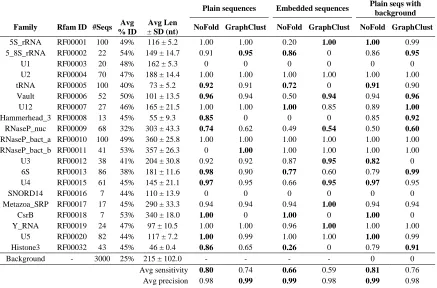
Table 2-1. Clustering sensitivity of NoFold and GraphClust for three test conditions on
the Rfam benchmark dataset. ... 87
Table 2-2. Clustering sensitivity and precision of NoFold and GraphClust for the synthetic structure benchmark. ... 88
Table 2-3. Summary of motifs identified in dendritic localization datasets. ... 89
Table 3-1. Overall % accuracy on three benchmarks using 10-fold cross validation. .... 124
Table 3-2. Putative structural matches to missing C. elegans Hh-related genes. ... 125
Table 4-1. Neurite-localized genes based on differential expression. ... 187
Table 4-2. Consistently observed genes in the neurites. ... 188
Table 4-3. Genes with differentially localized 3’UTR isoforms. ... 193
Table 4-4. Local proteome: predicted structures commonly found in synaptic proteins.194 Table 4-5. Local proteome: predicted transmembrane structures. ... 195
Table 4-6. Local proteome: predicted RNA-binding structures. ... 197
ix
LIST OF FIGURES
Figure 1-1. RNA structure. ... 29
Figure 1-2. Covariation in a multiple alignment of RNA sequences. ... 31
Figure 2-1. Normalization of the empirical feature space. ... 80
Figure 2-2. Structurally similar sequences are clustered together in the RESS. ... 82
Figure 2-3. Outline of the NoFold approach. ... 83
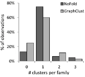
Figure 2-4. Distribution of the number of separate clusters assigned to each Rfam family for a given test. ... 84
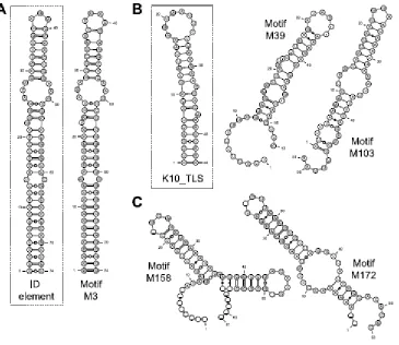
Figure 2-5. Consensus structures of motifs that are enriched in dendritically localized transcripts. ... 85
Figure 2-6. Potential translation initiation motifs. ... 86
Figure 3-1. Overview of PESS construction. ... 117
Figure 3-2. Classification and performance using the PESS. ... 118
Figure 3-3. Fold classification of the human proteome. ... 120
Figure 3-4. Analysis of unclassified human domains. ... 122
Figure 4-1. Sub-single cell profiling of soma and neurite RNA. ... 170
Figure 4-2. Overview of gene expression in individual soma and neurite samples. ... 171
Figure 4-3. Differentially expressed genes between soma and neurites. ... 173
Figure 4-4. Consistently observed genes in the neurites... 174
Figure 4-5. Alternative 3’UTR isoform usage in neurons. ... 175
Figure 4-6. Examples of genes with significantly differentially localized 3’ isoforms. . 177
Figure 4-7. The “selective neurite” regulatory pattern. ... 179
Figure 4-8. Potential GA-KT motifs formed by B1 and B2 SINE hairpins in localized genes. ... 180
x
Figure 4-10. Comparison of the most common structural folds represented in different proteome sets. ... 184
1
Chapter 1: Introduction
As an introduction to the computational structure analysis tools and biological
applications that will be presented in the main body of this thesis, I review here the basics
of ribonucleic acid (RNA) secondary structure, protein tertiary structure, and the
fundamental concepts of synaptic plasticity and long-term potentiation in neurons,
focusing in particular on areas where structure analysis can yield new insight into
biological function.
1.1.
RNA structure
RNAs are versatile macromolecules that play a wide variety of roles in the cell—
most notably as a mobile templates coding for proteins, but also sometimes as
independent regulatory or catalytic molecules [1,2]. RNAs self-base pair to form various
structures that help define their function and regulation. Below I review the basics of
2
1.1.1. Overview
RNA is a single-stranded polymer made up of a chain of individual nucleotides,
each composed of a ribose sugar with a phosphate group at the 5’ position, a nitrogenous
base at the 1’ position, and hydroxyl groups at the 2’ and 3’ positions. Nucleotides are
joined together by a phosphodiester bond between the phosphate group of one nucleotide
and the 3’ hydroxyl of another. Thus the final RNA polymer has directionality, where one
end has a free phosphate group (called the 5’ end) and the other end has a free hydroxyl
(called the 3’ end). The 5’ end is considered the “beginning” of the molecule, since
translation (the synthesis of protein from RNA) proceeds in a 5’ to 3’ direction.
There are four canonical types of bases used in RNA: adenine (A), guanine (G),
cytosine (C), and uracil (U). Certain bases can form hydrogen bonds with each other to
create base pairs. The standard “Watson-Crick” base pairs are G-C and A-U, but other
pairings, most notably G-U “wobble” pairs [3], are also possible under certain conditions.
Base pairing is energetically favorable, and therefore the single strand of a given RNA
will tend to form base-pairing interactions with itself when possible. This causes each
RNA to take on a shape determined by the base pairs that occur. The two-dimensional
conformation of an RNA that results from base pairing is generally referred to as its
“secondary structure”, whereas the linear sequence of nucleotides that make up the RNA
is called its “primary structure”.
RNA secondary structures can be broken down into a relatively small set of
building blocks. One of the most common building blocks is the stem-loop (or “hairpin”)
3
least three unpaired bases, where the single strand of RNA loops back around to pair with
itself at the stem (Fig. 1-1A). Stem-loops are often interrupted by interior loops, which
are regions of one or more unpaired bases within the stem; or by bulges, which are
interior loops where only one side of the stem is unpaired. Branches may also occur
where two or more stems split from a single stem, sometimes accompanied by internal
loop (Fig. 1-1A).
The definition of secondary structure is generally restricted to only base pairing
interactions that result in well-nested structures (i.e. interactions that do not cross over
each other) (Fig. 1-1B). However, RNA structure also has an important three-dimensional
component, referred to as its tertiary structure. For example, stem-type secondary
structures form a helix in three-dimensional space (Fig. 1-1C), and this helix can have
different properties and shapes depending on the combination of base pairs that form the
stem and the presence of bulges or interior loops [4]. Non-nested base pairing interactions
are also possible, including pseudoknots, which are regions of base pairing interactions
that cross over each other (Fig. 1-1D), and G-quadruplexes, which are formed by
interactions between repeated groups of guanines to form a four-stranded structure (Fig.
1-1E) [4].
1.1.2. RNA structure prediction
4
Until recently, experimental methods for probing RNA secondary structure were
relatively low-throughput. Classic methods include X-ray crystallography, nuclear
magnetic resonance (NMR) spectroscopy, single-strand RNA (ssRNA)- or double strand
RNA (dsRNA)-specific chemical modification followed by primer extension (e.g.
SHAPE [5]), and ssRNA/dsRNA nuclease cleavage followed by fragment size analysis
[6]. These methods, though accurate, are time consuming and difficult to apply to
multiple RNAs in parallel. New methods for structure probing combine various chemical-
and nuclease-based techniques with high-throughput RNA sequencing to greatly increase
the number of RNAs that can be probed at once [7]. Although these methods show great
promise, they do not always give complete information for all RNAs, and have not yet
been applied to all species. Because of this, computational structure prediction methods
continue to be developed to fill the holes in existing RNA structure data.
Computational methods
Given a set of parameter values defining the change in free energy associated with
different base pairs (i.e. their stability), and assuming that all secondary structures will be
well-nested, then the “optimal” secondary structure—that is, the structure with the
minimum free energy (MFE)—for any given RNA sequence can be found in using a
dynamic programming algorithm [8–12]. These thermodynamic modeling-based
approaches are still widely used today to predict secondary structure in the absence of
other sources of information. Although these methods are relatively fast, their main
5
external factors such as protein binding to the RNA or changes in environmental ion
concentration [1]. The differences between the MFE structure and true in vivo structure
are particularly apparent for longer (>700nt) sequences, for which only about 60% of
predicted base pairs are estimated to be correct on average [1].
One way to improve the accuracy of in silico secondary structure prediction is to
use comparative information across multiple homologous RNAs. If the structure of the
RNA is functionally important, it may show a pattern of conservation called
“covariation”. Covariation is when there are compensatory base changes that maintain
base-pairing potential of the sequence. In a multiple sequence alignment of homologous
RNAs, this manifests as columns of the alignment with pairing-compatible changes—for
example, when the base in one column changes from a G to an A, the base in the other
column changes from C to U (Fig. 1-2). Such a change maintains the ability of the RNA
to form a base pair between those particular bases. The observation of multiple
compensatory changes across evolution provides strong evidence for in vivo base-pairing
interactions, and can therefore be used to guide structure prediction [13–17]. Often, this is
used in combination with thermodynamic modeling to arrive at the final structure
prediction [18–21]. Although these covariation-based methods can be very accurate, they
are much more computationally intensive than thermodynamic modeling alone due to the
need to calculate a multiple alignment of the input sequences. This method is therefore
6
1.1.3. Structure-function relationships
One of the primary roles of RNA is to serve as a template for the creation of
proteins. Within protein-coding RNAs, also known as messenger RNAs (mRNAs), three
functionally distinct regions are defined: the coding region (CDS), which is the part of
the mRNA that is translated into protein; the 5’ untranslated region (UTR), which is
upstream of the CDS and is not translated; and the 3’UTR, which is downstream of the
CDS and also not translated. The 5’UTR is generally relatively short (a few hundred
nucleotides (nt)), but can occasionally contain sequence and structure motifs that help
recruit and position translational machinery, such as the ribosome, at the correct start site
of the CDS [22–24]. The 3’UTR, on the other hand, is often much longer (up to several
thousand nt) and contains a rich variety of sequence and structure motifs involved in
various aspects of mRNA regulation, including subcellular localization, translation, and
degradation [25].
There are several mechanisms by which secondary structures can play a functional
role in the mRNA. Most prominently, structures often serve as binding sites for
RNA-binding proteins (RBPs). Depending on the RBP, it may be the RNA structure itself that
is recognized (e.g. binding of the RBP Staufen to dsRNA [26]), or the structure may help
position a linear sequence of unpaired nucleotides (e.g. within a loop) into a more
favorable position for recognition [27]. Once bound, RBPs can initiate and regulate a
variety of different functions. For example, Staufen2 likely helps mediate dendritic
localization of the RNAs to which it binds [28,29]. Another example is the ADAR
7
perform RNA editing to change adenines to inosines [30]. Conversely, a secondary
structure can also function by occluding a binding site for an RBP or microRNA,
blocking those molecules from binding. In rare cases, secondary structures mediate
function by mimicking or replacing other molecules. For example, an mRNA from the
cricket paralysis virus contains an internal ribosome entry site (IRES) that mimics the
structure of tRNA-Met and forms a pseudoknot with the initiation codon. This allows the
virus to initiate translation in the absence of canonical initiation factors [31,32].
Another large class of RNA is non-coding RNA (ncRNA), which includes
functionally diverse subclasses such as microRNAs (miRNAs), transfer RNAs (tRNAs),
ribosomal RNAs (rRNAs), long non-coding RNAs (lncRNAs), among others [33]. For
these RNAs, structure is often a vital determinant of function [34]. For example, the
cloverleaf structure of tRNA is strongly conserved across species, despite substantial
variation on the sequence level (46% pairwise identity on average according to the Rfam
database [35]), which allows it to associate with the ribosome. In the case of ribozymes,
such as 23S rRNA, RNaseP, and self-splicing introns, the structure of the RNA actually
confers independent catalytic activity to the RNA [33]. For other ncRNAs, structure plays
the most important role during biogenesis. Examples of this are the hairpin structures of
pri- and pre-miRNA that are necessary for cleavage into mature miRNA by Drosha and
Dicer proteins [36]. There are many more examples of functional ncRNA structures in
the literature, and many families of such structures have been compiled into the Rfam
8
There are two particular ideas worth noting regarding the structure-function
relationship of RNA. The first is that if we know of a structure that plays a functional role
in one RNA, we can search the transcriptome for similar structures to identify other
RNAs that have a common function (in the case of ncRNAs) or are co-regulated by the
same RBP or pathway (in the case of mRNA regulatory motifs). This is the basis of the
Rfam database [35], which uses covariance models—a type of stochastic context-free
grammar that can model both sequence and secondary structure—to scan for new
instances of known functional structures. Secondly, and relatedly, if we know a set of
mRNAs are co-regulated, we can look for structural motifs shared between them to find
candidates for the regulatory element or RBP binding site. Computational methods for
performing this particular kind of analysis are currently lacking due to the difficulty of
obtaining accurate structure predictions for large datasets and the difficulty of measuring
the notion of similar secondary structures. This problem will be addressed in Chapter 2.
1.2.
Protein structure
Proteins are the main workhorses of the cell, participating in almost all aspects of
cellular function, including gene expression, energy production, signaling, catalysis,
transport, and cytoskeleton formation. Structure is an indispensable aspect of function for
almost all proteins, and even small disruptions of structure can lead to serious diseases
[37]. In this section, I review the basics of protein structure-function relationships and
9
1.2.1. Overview
A protein is composed of a linear chain of amino acid residues linked by peptide
bonds between the carboxyl group of one amino acid and the amino group of the next.
There are 20 canonical amino acids that vary in size, charge, hydrophobicity, polarity,
and modifiability. The unique combination and ordering of residues in a protein are the
basis for protein structure and function.
Protein structure is often described as having four levels: primary, secondary,
tertiary, and quaternary. The primary structure is simply the linear sequence of amino
acids making up the protein. The secondary structure is defined as the local patterns of
hydrogen bonding between a carboxyl oxygen and amino hydrogen of nearby residues.
The most common and stable secondary structures are the α-helix [38] and β-sheet [39],
but other conformations such as coils and turns are also observed. Tertiary structure is the
full three-dimensional conformation of the protein, which is stabilized by covalent
interactions, hydrogen bonds, hydrophobic interactions, van de Waals forces, electrostatic
interactions, and repulsive forces. It is the tertiary structure that is considered most
important for overall function of most proteins, although individual primary and
secondary features can also have functional roles. Finally, the quaternary structure refers
to the organization of multiple separate protein chains into a functional complex.
Many proteins have smaller subregions called domains. In the context of structural
biology, a domain is usually defined as a compact, stable, independent folding unit
[40]—that is, if the domain sequence were to be cleaved from the rest of the protein, it
10
evolutionary sequence analysis, a domain is defined as a conserved region of the protein
sequence, often with a conserved function (for example, the domains defined in the Pfam
database [41] are of this type). It is important to note that in practice these two definitions
often coincide, since structural domains are usually evolutionarily conserved and have a
specific function [40]. The definition of a structural domain is broader, however, because
it is possible for non-homologous sequences to have the same structure. In this thesis, I
will primarily use the word “domain” to refer to the union of these definitions, and
specify “structural domain” or “sequence domain” when distinction is necessary.
A remarkable feature of domains is their modularity. Most proteomes appear to be
composed of a finite library of domains that have been “mixed and matched” to produce
various functional combinations within multi-domain proteins [40]. Due to accumulated
sequence variation over time, the instances of a domain have varying levels of sequence
similarity across different proteins and species. Many domains have become so diverged
that it is impossible to recognize them based on sequence alone. In these cases, structural
information can be used to identify domains, because structure is usually more conserved
than sequence [42]. Given the complexity of relationships between domains, several
hierarchical classification schemes have been created to organize domain instances (that
is, individual observations of a domain in a protein) based on defined levels of similarity
and evolutionary relationship. The Structural Classification of Proteins (SCOP) database,
for example, manually curates groups of domains on four main levels: family,
superfamily, fold, and class [43]. “Families” group together homologous domains with
11
functional differences between members of a family, such as different binding
preferences for DNA-binding domains). “Superfamilies” group together families with
more divergent, but still recognizable, sequence similarities. Superfamilies also tend to
have a general conserved function. The next level is “fold”, which groups together
superfamilies with similar tertiary structures (that is, similar numbers and topological
arrangements of secondary structures). Folds are defined purely based on structure, and it
is not always clear if the constituent superfamilies are related evolutionarily or have
arrived at similar structures by convergent evolution. Nonetheless, members of a fold
typically still have similar coarse-grain functions, with the exception of some highly
diversified and prevalent “superfolds”, which have been adapted to a variety of distinct
purposes [44]. Interestingly, there appears to be a limited number of folds used by natural
proteins—only a little over 1,000 folds are currently defined, and the rate of new fold
discoveries has steadily declined over the past few years. Finally, the “class” level of
SCOP groups folds very roughly based on overall secondary structure composition and
other properties, such as all-α-helix, all-β-sheet, mixed-α-β, membrane proteins, and a
few others. Overall, this taxonomically-inspired classification scheme (and others, such
as CATH [45]) provides a convenient discretization of domain similarity that enables
analysis at defined levels of evolutionary and structural relationship.
1.2.2. Protein structure prediction
12
Protein tertiary structure can be experimentally determined (“solved”) using
several methods, most commonly X-ray crystallography and NMR spectroscopy. X-ray
crystallography requires purification and crystallization of the protein of interest, which is
then exposed to X-rays to obtain a diffraction pattern. This diffraction pattern is analyzed
to infer the location of atoms in the structure. Although crystallography can be very
accurate, it is limited by the difficulty of obtaining protein crystals. Proteins with flexible
domains are particularly difficult to crystalize, and must be split into non-flexible
fragments to obtain partial crystal structures. NMR spectroscopy, on the other hand, is
well-suited for flexible proteins, since it works on proteins in solution and does not
require crystallization. NMR spectroscopy measures atomic resonance while exposing the
protein to various radio frequencies in a strong magnetic field, which can be analyzed to
identify nearby atoms in the structure. This is then used to infer the three-dimensional
structure. The drawbacks of NMR spectroscopy are that it is generally limited to only
small proteins, cannot be used for insoluble proteins such as membrane proteins, and has
low spatial resolution. Recently, another method called Cryo-electron microscopy
(Cryo-EM) has improved in resolution to the point where it can be used for atomic-level
structure solving. Cryo-EM has promise to alleviate several of the difficulties facing
crystallography, since it freezes molecules rather than crystalizing them, but the method
is still under development [46]. Overall, all three methods are limited to various degrees
by expense and throughput capacity, and because of this only a fraction of known protein
sequences have been structurally characterized. This has motivated the development of a
13
Computational methods
Computational methods for predicting protein tertiary structure can generally be
divided into two categories: ab initio and template-based [47]. Ab initio (or de novo)
methods attempt to determine a protein’s structure directly from the sequence using
first-principles molecular dynamics simulations. However, due to the enormous search space
of possible three-dimensional conformations for an average-sized protein, ab initio
methods are generally only computationally feasible for the smallest proteins [48].
Therefore, template-based modeling has been the more popular method over the last two
decades.
Template-based modeling covers a wide variety of methods that make use of
currently known information about protein structures—e.g. experimentally solved protein
structures in the Protein Data Bank (PDB) [49]—as a starting point (or “template”) for
predicting the structures of new proteins. Template-based modeling can be subdivided
into two main types: homology modeling and threading. Homology modeling, also called
comparative modeling, uses sequence alignment methods to match a query sequence to
any homologous sequences within the database of structurally-solved proteins. These
methods work on the assumption that homologous proteins are likely to share a
conserved structure, and therefore the structure of the homolog can be used to predict the
structure of the query. Homology modeling methods such as HHPred [50]—which uses
hidden Markov model (HMM)-based profile-profile alignments to increase sensitivity—
14
challenge facing these methods is the difficulty of detecting more remote homologs—
those falling within the “twilight zone” of sequence similarity, usually <30% identity
[51]. This includes a large fraction of proteins at the current time, and has thus motivated
the second template-based method—threading. Threading or “fold recognition” methods
do not require homology or sequence similarity with a structurally solved protein in order
to work, but instead try to directly use structural information to find the best match for
the query. Briefly, threading comprises aligning a query sequence to a structural
“template”, defined in this context as the three-dimensional coordinates of atoms derived
from a known protein structure (usually with the side chains removed). The best
alignment between the query and structure is determined based on the compatibility of
residue contacts, secondary structures, solvent access, and other criteria. This process is
then repeated for every template in the database to identify which structure gives the most
thermodynamically favorable structure for that sequence. Although threading has the
advantage of working even in the absence of homology between the query and template,
it is limited by much greater computational costs than homology modeling. Nonetheless,
threading is much more tractable than ab initio methods, and thus has been used
extensively and to good success over the last several years [51].
More recently, a third category of methods has emerged that combines aspects of ab
initio and template-based methods [47]. These hybrid methods usually cut the protein
sequence into many smaller fragments, and then attempt to match each fragment to one
or more templates (which themselves are fragments of known structures). Once template
15
into a conformation that is energetically favorable for the protein as a whole. Using the
templates as a starting point greatly limits the search space, making the ab initio
simulations more tractable. I-TASSER [52] and Rosetta [53] are two examples of highly
successful hybrid methods. However, these methods are still too slow to be applied to
large scale projects, such as whole-proteome structure prediction.
1.2.3. Structure-function relationships
There is a strong association between structure and function among proteins.
Proteins with similar structure very often have similar function [54], and—to a lesser
extent—proteins with similar function may have similar structure. This has been shown
to hold true even for highly disparate amino acid sequences, and is the main motivation
behind the field of structural genomics, which makes extensive use of the experimental
and computation methods described above to make inferences about function based on
structural similarities between proteins on a genome scale [55].
There are limits to the amount of functional information that can be gained simply by
matching proteins to similar tertiary structures. For one thing, since structure prediction is
usually done on the level of individual domains, this information must be integrated to
understand the overall function of multi-domain proteins. Secondly, many of the nuances
of domain function are influenced by fine-grained differences in the arrangement of
secondary structures or by variation of specific residues in a binding pocket or enzymatic
active site. This is particularly evident in the case of “superfolds”; for example, the TIM
16
commission (EC) classes [44]. Finally, a large fraction of proteins include “intrinsically
disordered” regions that do not take on a well-defined native tertiary structure. These
regions often serve as flexible linkers between domains in multi-domain proteins, or may
only fold when bound by a cofactor [47,56]. The function of these regions is therefore not
amenable to typical structure-based analysis.
Despite these limitations, structure prediction has proved to be an extremely useful
first step towards a functional understanding of uncharacterized proteins [54]. Improving
the speed of methods for recognizing structural similarities, especially in the absence of
sequence similarity, will greatly increase our capability for genome-scale annotation of
protein function. A new approach to this problem will be discussed in Chapter 3.
1.3.
Neurons, plasticity, and structure
Neurons are highly polarized cells consisting of a cell body (soma), and long,
branched processes (usually a single axon and multiple dendrites). The flow of
information through the neuron typically proceeds from the dendrites, which receive
signals from other neurons at synapses; to the soma, which integrates signals; and finally
to the axon, which transmits signals to other neurons. Synapses show a remarkable ability
to remodel themselves in response to stimulation, becoming more or less responsive to
future inputs (synaptic plasticity). This is thought to be one of the mechanisms underlying
the larger scale phenomena of learning and memory in the brain. Here, I will survey
17
hippocampus, which have been studied extensively in this context, and highlight areas
where structure analysis can help further our understanding.
1.3.1. Components of pyramidal neurons
Pyramidal neurons exist in a wide variety of mammals and are generally found in
brain structures associated with complex cognitive function [57,58]. The morphology of
pyramidal neurons is characterized by a single axon with many branches that make
excitatory glutamatergic synapses with other neurons, as well as an extensive dendritic
arbor with mostly excitatory synaptic inputs [58]. Pyramidal neurons may also receive
some synaptic inputs on the axon and soma, which are typically inhibitory GABAergic
synapses [58].
An important set of substructures of pyramidal dendrites are the dendritic
spines—small, knob-like protrusions along the dendrites which are the site of most
glutamatergic synapses. Spines vary widely in size and shape [59] and show
morphological and functional plasticity over time [60–62]. A single pyramidal neuron
may have thousands of dendritic spines, occurring at a density of about 1-10 spines per
µm of dendritic length in mature neurons [59]. Although the precise purpose of spines is
unclear, one of their main functions is likely to compartmentalize synapses and help
prevent important molecules from diffusing away [63,64]. The spine neck may also serve
to modulate electrical conductance properties [65]. Abnormal spine morphology has been
observed in many neurological disorders, including Down Syndrome [66], Fragile X
18
Dendrites also contain a variety of organelles, including abundant mitochondria
[69], endoplasmic reticulum (ER) [70–73], Golgi “outposts” [70], and multivesicular
bodies [71,73]. An organelle called the “spine apparatus” has also been observed in
dendrites [74,75], which appears in 10-15% of mature hippocampal spines [71]; however,
the exact function of this organelle is not currently well understood. In addition to
organelles, many components of the translational machinery have been found in dendrites
at the base of spines, including tRNAs, polyribosomes, and initiation/elongation factors
[76–78].
1.3.2. Long-term potentiation
The idea that the plasticity of synapses could play a central role in learning and
memory was suggested over a century ago by Santiago Ramόn y Cajal [79]. In 1949,
Donald Hebb formalized a model of how synaptic plasticity relates to learning and
memory [80], but it was not until about 20 years later that substantial evidence for a
molecular basis of such a model was provided by the discovery of long-term potentiation
(LTP) [81,82]. These studies showed that stimulating excitatory hippocampal synapses
resulted in a long-lasting increase in synaptic strength of those synapses. Since then, LTP
has become an area of intense research in the field of neuroscience, and remains one of
the leading hypotheses of the molecular basis of learning and memory [83,84]. Although
there are now thought to be multiple forms of LTP, which depend on factors such as
brain region and stimulation frequency [84], I will focus here on N-methyl-D-aspartate
19
LTP is often described as having two stages: an early phase (E-LTP), usually
defined as the first 1-3 hours after stimulation; and a late phase (L-LTP), which requires
protein synthesis and gene transcription [85]. E-LTP is triggered by activation of
post-synaptic NMDA receptors (NMDARs), which open to allow calcium influx [84]. This
activates Ca2+/calmodulin-dependent protein kinase (CaMKII) [86], which causes a rapid
increase in α-Amino-3-hydroxy-5-methyl-4-isoxazoleprpionic acid receptors (AMPARs)
in the synapse membrane [87]. The exact mechanism by which CaMKII influences
AMPAR synaptic trafficking is currently unclear. Several early studies suggested that
CaMKII phosphorylates the carboxy-terminal tail (C-tail) of AMPAR subunit GluA1
and/or AMPAR-accessory proteins [84]. In contrast, a recent set of studies has suggested
that the C-tail of GluA1 is not needed for normal LTP, and furthermore, AMPARs can be
completely replaced with kainite receptors without a substantial impact on LTP [84,88].
There is also conflicting evidence about which other signaling cascades, besides that
mediated by CaMKII, might be important for LTP. Many molecules have been
discovered that seem to modulate LTP, but few besides CaMKII have been shown to be
vital [83]. These results show that despite substantial progress over the past 20 years,
there is still much that is not well understood about this process.
The second phase, L-LTP, is dependent on new protein translation. Furthermore,
this new translation often occurs in the dendrites themselves, in close proximity to the
activated synapse [89]. There is now substantial evidence that a subset of neuronal
mRNAs are actively localized to the dendrites, usually in a translationally repressed state,
20
of mRNA localization and local translation are discussed in more detail in the next two
sections. It is worth noting that there is also evidence for an important role of new
transcription for L-LTP [90], which will not be reviewed extensively here.
Beyond changing the molecular composition of the synapse, LTP also causes (and
possibly is perpetuated by) changes in the shape and size of the spine in which the
synapse is housed [69]. The mechanisms of how this occurs are still being investigated,
but filamentous actin (F-actin) polymerization dynamics likely play an important role
[69,84]. F-actin makes up one of the major structural components of spines, and
inhibition of actin polymerization prevents spine growth and LTP [91,92].
Activity-dependent cytoskeletal growth may be due to CaMKII activation of Rho GTPases, which
promote actin polymerization, although how this occurs is not known [84]. It is
hypothesized that these changes in structure may help promote AMPAR incorporation
into the synapse, and thus promotes LTP [84]. After increasing in size, the spine can be
further stabilized by cell adhesion molecules, such as N-cadherin, which has been shown
to increase after synaptic activity [69].
1.3.3. Importance of RNA localization and local translation
Direct evidence for the idea that new protein synthesis was required for memory
formation was first demonstrated in the 1960s, where it was shown that mice injected
with the protein synthesis inhibitor puromycin to the temporal lobe showed impaired
long-term memory formation if the injection was given within three days [93]. A large
21
synthesis in a variety of memory-related behaviors [94]. On the molecular level,
treatment with the protein synthesis inhibitor anisomycin was shown to inhibit spine
enlargement during LTP [95], lending further support that LTP might form the molecular
basis of learning and memory. However, these studies at first did not directly address the
question of where within the neuron this new protein synthesis was occurring, and it was
generally assumed that it would occur in the soma [85].
Following the discovery of polyribosomes [76] and multiple mRNAs [96–98] in the
dendrites, the idea that translation could occur locally in the dendrites began to gain
popularity. This model was attractive for several reasons. For one, it provided a simple
mechanism by which newly synthesized proteins could be sorted to the correct synapse:
synaptic activation could trigger translation of only those mRNAs in the vicinity of the
spine, thus causing a local increase in new proteins at the activated synapse. Other
theoretical benefits include reduced transport costs, faster response time, and prevention
of toxic ectopic protein expression [99,100]. Finally, in 1996, two studies provided direct
evidence that protein synthesis can in fact occur locally in isolated dendrites [101] and
hippocampal tissue slices [102].
Although local translation is now generally accepted as being important for lasting
synaptic potentiation [103], there is less known about exactly which mRNAs are
localized and what roles individual locally-translated proteins play in LTP. As techniques
for profiling and quantifying RNA have improved, estimates of the dendritic
transcriptome have expanded from a few RNAs [98] to a few hundred [104–107] to
22
“gold standard” localized RNAs, which have been observed by multiple labs and
methods to be robustly localized to the dendrites, such as CaMKIIα, β-actin, Arc, and
BC1. But overall, there has been surprisingly little concordance between different
analyses of the dendritic transcriptome, even when the same organism and brain region
are profiled. In terms of understanding the actual functional role of individual localized
mRNAs and their protein products, even more work remains to be done. To show
specifically that local translation of a particular protein is important for LTP, an ideal
experiment would disrupt only the local translation of that protein without altering its
somatic expression. So far, this has mostly been accomplished in a few isolated cases,
usually by abolishing the dendritic localization of the mRNA. For example, in mice
lacking the 3’UTR of CaMKIIα mRNA, which contains its dendritic targeting sequence,
it was shown that protein levels of CaMKII at the synapse were greatly reduced and
L-LTP was impaired [110]. Much more work remains to be done to understand the role of
the many potential locally-translated proteins in LTP.
1.3.4. Mechanisms of dendritic RNA localization: a role for structures
Proper localization of RNAs to the dendrites is a prerequisite for local translation,
and therefore for long-lasting synaptic potentiation. Dendritic localization is thought to be
mediated by specific RNA-binding proteins (RBPs) that recognize sequence or structure
motifs on their target RNAs [100,111,112]. These RBPs may recruit other proteins to the
RNA, forming a ribonucleoprotein complex (RNP). The RNP typically includes proteins
23
along microtubules in the soma and dendrites to bring the RNP to its destination. While
in the dendrites, RNA is mostly kept in a translationally repressed state by proteins in the
RNP [115–117]. This repression is then relieved when a nearby synapse is activated,
allowing for local production of proteins [117,118].
Interactions between RBPs and RNAs are vital for proper localization, and much
work has been done to try to identify the dendritic targeting elements (DTEs) on localized
RNAs that are recognized by RBPs. Identifying these DTEs would have benefits such as
(1) allowing us to predict additional localized RNAs based on the presence of similar
motifs, (2) enabling the identification of co-regulated groups of RNAs based on the
presence of shared DTEs, and (3) providing insights into how dysregulation of RBP
binding and RNA localization can lead to disease. Thus far, however, the identification of
DTEs has been challenging. Below I briefly outline what is known about the localization
and DTEs of a few of the most well-characterized dendritic RNAs and
localization-mediating RBPs.
BC1 RNA. Brain cytoplasmic RNA 1 (BC1) is a short (~150nt), structured
non-coding RNA that is dendritically localized [119] and plays a potential role in translational
regulation [120]. The stem loop structure at its 5’ end has been experimentally
determined [121] and is likely the DTE [122]. A particular part of the stem loop forms a
GA kink-turn motif and seems to be bound by hnRNP-A2, which mediates the
localization [123]. A type of short interspersed nuclear element (SINE) called the ID
element is derived from BC1 [124] and has also been shown to act as a DTE in several
24
Staufen. The Staufen family of proteins (Stau1 and Stau2) are RBPs that bind
dsRNA such as that found in stem-loop structures. Stau1 is ubiquitously expressed across
tissues and may play a role in L-LTP [127]. Stau2 protein is enriched in the brain [100],
shuttles to the dendrites in RNPs [128], and is likely involved in dendritic localization
[28]. Several secondary structures have been proposed to be bound by Stau2 [129], which
appear mostly sequence-independent.
ZBP1 and β-actin.A 54nt region in the 3’UTR of β-actin, known as the “zipcode”
sequence, is necessary and sufficient for its localization in several cell types [130].
Binding of zipcode-binding protein 1 (ZBP1, called IMP-1 in human) to the zipcode was
found to be important for both the localization and translational inhibition of β-actin
[131,132]. Later studies showed that most of the zipcode actually functions as a spacer
for two much shorter motifs that are bound by two KH domains of ZBP1, and that similar
bipartite motifs were conserved in other mouse/human mRNAs, making them potential
targets of ZBP1 as well [133].
FMRP. Fragile-X mental retardation protein (FMRP) is thought to play an
important role in translational repression of localized mRNAs and possibly also
modulates localization [116]. It appears to bind to a wide variety of localized RNAs,
including CaMKIIα, Map1b, PSD-95, and Fmr1 (its own mRNA) [100]. It has been
proposed to bind to G-quadruplexes through its RGG-box domain [134,135], although a
more recent study of FMRP binding using HITS-CLIP showed no enrichment for
25
hnRNP-A2. Heterogeneous ribonucleoprotein particle A2 (hnRNP-A2) binds to
known dendritically localized RNAs such as CaMKIIα and Arc [137] and is thought to be
directly involved in localization. Multiple motifs have been proposed to be recognized by
this RBP, including a pair of 11nt sequences (the hnRNP-A2 recognition element, A2RE)
first identified in the MBP mRNA in oligodendrocytes [138], G-quadruplex structures
and CGG repeats [139,140], and GA kink-turn structural motifs [123].
CaMKIIα.Although it is one of the most extensively studied dendritically localized
mRNAs, CaMKIIα still does not have a fully defined DTE. Most reports point to an
element in the 3’UTR, but there is conflicting evidence about the minimal element
needed for localization [110,123,141–143]. Implicated regions so far include both linear
sequences and secondary structures.
A common theme in many of these examples is the lack of consensus regarding the
location and nature (linear or structural) of DTEs on specific transcripts. Part of the
problem may be that some localized RNAs in fact have multiple DTEs, each regulating
distinct and/or redundant aspects of the localization process [99]. An interesting example
of this is BC1, which was shown to have two sub-motifs within its DTE: one that was
needed for nuclear export and another that was needed for transport to the distal dendrites
[123]. Adding to this difficulty, many DTEs are now known to have a secondary structure
component that is either central to or supports recognition by the RBP [144], which may
have contributed to conflicting reports in the past that mostly focused on linear sequence
DTEs. Given that there are hundreds or even thousands of localized RNAs in neurons, it
26
more likely explanation is that multiple localized RNAs share DTEs and are recognized
by the same RBP, but we are missing these signals due to a lack of tools that perform de
novo RNA structure motif discovery in large datasets.
1.3.5. Protein structures of the synapse
One of the gaps in our understanding of long-lasting synaptic potentiation is the
specific role of each locally-translated protein in this process. Although experimental
work will be needed to pick apart exact functions, we can make some initial guesses
using computational annotation methods. Structure-based functional annotation may be
of particular use in this case, given that there are a variety of important roles for protein
structures at the synapse. Examples include the PDZ domain in scaffold-associated
proteins [145]; cadherins, neurexins/neuroligins, ephBs/ephrin-B, and
immunoglobulin-containing cell adhesion folds at the synaptic junction [146]; transmembrane folds in
membrane-bound channels and receptors; kinase and phosphatase catalytic folds involved
in signaling and synaptic plasticity; and many others. Although many of the proteins
containing these structures are likely to be constitutively present at the spine or
post-synaptic density (surveyed in [147–149]), and thus may be primarily synthesized in the
soma, it would be interesting to see if a subpopulation of these proteins is locally
translated as well, and if new examples of these folds can be discovered. Furthermore,
recent genome-wide analyses of neurological diseases have revealed enrichment for
causative mutations in synaptic proteins in human and mouse [147,149], several of which
27
the structures of locally translated proteins will help guide future experimental work and
aid in predicting the functional impact of mutations.
1.4.
Overview of thesis
In this thesis, I present two new methods for structure-based analysis of
large-scale datasets based on the concept of empirical feature spaces—feature spaces defined
by examples of natural structures—and then apply these methods to address the questions
outlined above regarding the localization of RNA in the dendrites of neurons and the
possible roles of locally translated proteins.
In Chapter 2, I describe the RNA empirical structure space (RESS), which uses
Rfam covariance models to map uncharacterized RNAs to a structural feature space. I
will show that RNAs with similar structure cluster together within the RESS, even in the
absence of sequence similarity, and use this fact to develop a pipeline for de novo
secondary structure motif discovery that can be applied to finding functional motifs
enriched in co-regulated transcripts. Since this method scales linearly with increasing
input dataset size, it is feasible to run on thousands of sequences at once.
In Chapter 3, I describe the protein empirical structure space (PESS), which uses
threading against a small set of known structure templates to map uncharacterized protein
domain sequences to structural feature space. As with the RESS, the PESS clusters
protein sequences based on structure even in the absence of detectable sequence
similarity. I show that the PESS can be used for a variety of purposes including
28
distant homologs (or structural analogs) across species. This method saves substantial
amounts of time compared to traditional threading methods by using only a small library
of templates for threading, yet has accuracy on par with threading against a much larger
set.
In Chapter 4, I will combine experimental and computational methods, including
the two methods described above, to catalog the set of RNAs localized to the dendrites in
mouse hippocampal neurons, identify potential linear and structural localization signals,
and predict the functions of locally translated proteins based on domain-level structural
prediction. The results include findings that would be difficult to identify using
traditional sequence-based tools, demonstrating the utility of including structure-based
tools when performing functional analysis of RNA and protein.
Finally, in Chapter 5, I discuss some of the implications and future directions
suggested by this work, including several avenues where structure analysis may yield
29
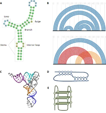
Figure 1-1. RNA structure.
(A) An example of RNA secondary structure, showing typical motifs. (B) A well nested
structure (top) and non-nested structure (bottom). The black horizontal lines indicate an
RNA sequence and the arches show base pairing. Red and orange arches highlight the
30
structure in (A). (C) An example of RNA tertiary structure. (Image from the public
domain.) (D) An example of a pseudoknot structure, which consists of non-nested base
pairing interactions. (E) An example of a G-quadruplex structure consisting of four
31
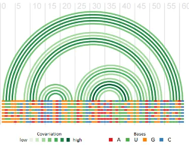
Figure 1-2. Covariation in a multiple alignment of RNA sequences.
Arches show base pairing interactions. Paired bases tend to show compensatory changes
that maintain pairing, whereas non-paired bases usually show uncorrelated variation.
Note that G-U pairs are generally considered compatible. Figure generated using R-chie
32
1.5.
References
1 Wan, Y. et al. (2011) Understanding the transcriptome through RNA structure.
Nat. Rev. Genet. 12, 641–655
2 Geisler, S. and Coller, J. (2013) RNA in unexpected places: long non-coding RNA functions in diverse cellular contexts. Nat. Rev. Mol. Cell Biol. 14, 699–712
3 Varani, G. and McClain, W.H. (2000) The G-U wobble base pair. EMBO Rep. 1, 18–23
4 Butcher, S.E. and Pyle, A.M. (2011) The Molecular Interactions That Stabilize RNA Tertiary Structure: RNA Motifs, Patterns, and Networks. Acc. Chem. Res.
44, 1302–1311
5 Wilkinson, K.A. et al. (2006) Selective 2′-hydroxyl acylation analyzed by primer extension (SHAPE): quantitative RNA structure analysis at single nucleotide resolution. Nat. Protoc. 1, 1610–1616
6 Kubota, M. et al. (2015) Progress and challenges for chemical probing of RNA structure inside living cells. Nat. Chem. Biol. 11, 933–941
7 Lu, Z. and Chang, H.Y. (2016) Decoding the RNA structurome. Curr. Opin. Struct. Biol. 36, 142–148
8 Zuker, M. and Stiegler, P. (1981) Optimal computer folding of large RNA
sequences using thermodynamics and auxiliary information. Nucleic Acids Res. 9, 133–148
9 Zuker, M. (1989) On finding all suboptimal foldings of an RNA molecule. Science
244, 48–52
10 Zuker, M. (2003) Mfold web server for nucleic acid folding and hybridization prediction. Nucleic Acids Res. 31, 3406–3415
11 Hofacker, I.L. (2003) Vienna RNA secondary structure server. Nucleic Acids Res.
31, 3429–3431
12 Hofacker, I.L. et al. (1994) Fast folding and comparison of RNA secondary structures. Monatshefte fur Chemie Chem. Mon. 125, 167–188
33
14 Hofacker, I.L. et al. (2002) Secondary Structure Prediction for Aligned RNA Sequences. J. Mol. Biol. 319, 1059–1066
15 Washietl, S. et al. (2005) Fast and reliable prediction of noncoding RNAs. Proc. Natl. Acad. Sci. 102, 2454–2459
16 Griffiths-Jones, S. (2003) Rfam: an RNA family database. Nucleic Acids Res. 31, 439–441
17 Yao, Z. et al. (2006) CMfinder--a covariance model based RNA motif finding algorithm. Bioinformatics 22, 445–52
18 Torarinsson, E. et al. (2007) Multiple structural alignment and clustering of RNA sequences. Bioinformatics 23, 926–32
19 Mathews, D.H. and Turner, D.H. (2002) Dynalign: an algorithm for finding the secondary structure common to two RNA sequences. J. Mol. Biol. 317, 191–203
20 Will, S. et al. (2007) Inferring noncoding RNA families and classes by means of genome-scale structure-based clustering. PLoS Comput. Biol. 3, e65
21 Sankoff, D. (1985) Simultaneous solution of the RNA folding, alignment and protosequence problems. SIAM J. Appl. Math. 45, 810–825
22 Shatsky, I.N. et al. (2010) Cap- and IRES-independent scanning mechanism of translation initiation as an alternative to the concept of cellular IRESs. Mol. Cells
30, 285–93
23 Wellensiek, B.P. et al. (2013) Genome-wide profiling of human cap-independent translation-enhancing elements. Nat. Methods 10, 747–50
24 Weingarten-Gabbay, S. et al. (2016) Systematic discovery of cap-independent translation sequences in human and viral genomes. Science (80-. ). 351, aad4939-aad4939
25 Tian, B. and Manley, J.L. (2016) Alternative polyadenylation of mRNA precursors. Nat. Rev. Mol. Cell Biol. DOI: 10.1038/nrm.2016.116
26 Heraud-Farlow, J.E. and Kiebler, M.A. (2014) The multifunctional Staufen proteins: conserved roles from neurogenesis to synaptic plasticity. Trends Neurosci. 37, 470–479
27 Li, X. et al. (2014) Finding the target sites of RNA-binding proteins. Wiley Interdiscip. Rev. RNA 5, 111–130