R E V I E W
Open Access
Insulin receptor signaling in the development of
neuronal structure and function
Shu-Ling Chiu
1, Hollis T Cline
2*Abstract
Sensory experience plays a crucial role in regulating neuronal shape and in developing synaptic contacts during brain formation. These features are required for a neuron to receive, integrate, and transmit signals within the neu-ronal network so that animals can adapt to the constant changing environment. Insulin receptor signaling, which has been extensively studied in peripheral organ systems such as liver, muscle and adipocyte, has recently been shown to play important roles in the central nervous system. Here we review the current understanding of the underlying mechanisms that regulate structural and functional aspects of circuit development, particularly with respect to the role of insulin receptor signaling in synaptic function and the development of dendritic arbor mor-phology. The potential link between insulin receptor signaling malfunction and neurological disorders will also be discussed.
Introduction
The human brain is made up of billions of neurons assembled into sophisticated circuits. Information received from sensory neurons is processed by neurons within distinct circuits to generate specific functional outputs, including cognitive decisions and behavior. A fascinating problem is how these huge numbers of neurons establish precise connections to assemble com-plex circuits during development. The neuron, the func-tional unit of the brain circuit, is a highly specialized cell composed of the cell body, the dendrite and the axon. The structure of the dendrite determines where and how an individual neuron can receive and integrate information from afferent neurons, whereas the morphology of the axon determines where processed information is sent to efferent neurons. Sites of contacts between the axon and dendrite, or synapses, mediate communication between neurons for proper information flow within the neuronal circuit. We will first review the current understanding of cellular and molecular mechanisms underlying synapse and dendrite develop-ment, then focus on recent evidence suggesting a func-tion for insulin receptor signaling in circuit funcfunc-tion and pathological brain diseases.
Synapse and dendrite development Synapse development
The number of synaptic contacts and the efficacy of synaptic transmission in the brain are dynamic through-out development and adulthood [1-3]. These dynamics are crucial for neurons to optimize connections in brain circuits during development. Synaptic plasticity is also important to optimize neuronal function in adults, for example, to adapt to our changing environment and to allow memories to form. Synapse development is a ser-ies of distinct processes, including synapse formation, synapse maturation and synapse maintenance. The mechanisms that regulate each of these processes are just starting to be unraveled.
Synapse formation
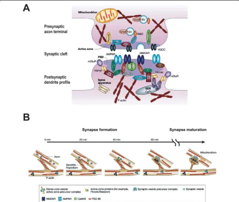
Synapses are specialized junctions between neurons where the presynaptic axon terminal is packed with synaptic vesicles and vesicle release machinery and the postsynaptic dendritic specialization contains transmem-brane neurotransmitter receptors, scaffold proteins and signaling machinery (Figure 1A; see [4] for detailed review). Time-lapse imaging in bothin vivoandin vitro preparations revealed that the temporal sequence of synapse formation is quite rapid. The first step involves the contact between dendrites and axons, which likely occurs by adhesive mechanisms. Second, the presynaptic specialization assembles quickly at sites of contact [5,6]. In fact, it is thought that components of the presynaptic
* Correspondence: [email protected]
2Departments of Cell Biology and Chemical Physiology, The Scripps Research
Institute, 10550 North Torrey Pines Road, La Jolla, CA 92037, USA
specialization are present in axons before synaptogenesis as packets of vesicle proteins and components of the active zone proteins [7,8], Finally, the postsynaptic spe-cialization, including the proteins postsynaptic density-95 (PSD-95), and neurotransmitter receptors, including N-methyl-D-aspartate (NMDA) receptors, are thought to arrive somewhat later during synapse formation (Figure 1B) [9]. Although the assembly of synapses is a
complex process, recent work has identified several mole-cules that are important in different steps of synapse for-mation [7,10]. For example, molecules that are present in gradients within target regions, such as ephrins, play an important role in directing axons and dendrites to the correct brain regions [11,12]. Adhesion molecules, such as cadherin, are thought to be important in establishing of the initial axodendritic contacts [13,14]. Some
Figure 1Schematic diagram of an excitatory synapse and the temporal sequence of synapse formation and maturation.(A)Synapses are specialized junctions between neurons composed of complex membrane and proteins. A synapse can be divided structurally into three parts: a presynaptic axon terminal packed with synaptic vesicles (SV) and release machinery, a synaptic cleft, and a postsynaptic dendritic counterpart filled with neurotransmitter receptors, scaffold proteins and signaling machinery.(B)Synapse formation is initiated by the contact between dendrites and axons, followed by the recruitment of presynaptic and postsynaptic specializations. Increases in synapse size and synaptic strength by accumulation of AMPA receptors at synapses are characteristics of synapse maturation. AMPAR,a -amino-3-hydroxy-5-methyl-4-isoxazole propionic acid receptor; CaMKII, Calcium calmodulin dependent kinase type II; CASK, calcium calmodulin-dependent serine kinase; GKAP, guanylate kinase-associated protein; GRIP, glutamate receptor-interacting protein; InsP3R, inositol triphosphate receptor; mGluR,
transsynaptic molecules, such as neuroligin and neurexin, are crucial in bidirectional signaling and the recruitment of both pre- and postsynaptic proteins to new synapses [15,16]. In addition to molecular players, neuronal activ-ity appears to be another key regulator in the formation of nascent synapses [17-20].
Synapse maturation
Synapse maturation is characterized by an increase in the morphological size and transmission strength of the synapse, which includes changes in both the presynaptic axon terminal and the postsynaptic dendrite. From the presynaptic point of view, a prominent ultrastructural feature of synaptic maturation is the increase in the number of synaptic vesicles per terminal [21-23], which likely contributes to the increase in probability of trans-mitter release in mature synapses [17]. Transmission at immature glutamatergic synapses is mainly mediated by NMDA receptors, which shift their kinetics by replacing NMDA receptor subunit 2B-containing receptors with NMDA receptor subunit 2A-containing receptors [24,25]. These immature synapses can be silent or have low synaptic strength at resting membrane potentials because of voltage-dependent magnesium blockade of the NMDA receptor. As the synapses mature,a -amino-3-hydroxy-5-methyl-4-isoxazole propionic acid (AMPA) receptors are recruited to the postsynaptic membrane, and in addition to NMDA receptors, provide fully func-tional glutamatergic synaptic transmission [26-29]. Neu-ronal activity reportedly induces synapse maturation by promoting the incorporation of NMDA receptor subunit 2A-containing NMDA receptors into synaptic sites. Furthermore, activity recruits AMPA receptors to the postsynaptic site to activate silent synapses and increase the strength of synaptic transmission (Figure 1B) [4,26,27].
Synapse maintenance or synapse elimination
The precise connectivity required for circuit function relies not only on the formation of new contacts but also the maintenance of the correct synapses. In fact, the density of synapses formed at early stages of devel-opment is far greater than the density retained at later stages, indicating that only selective synapses are stabi-lized and maintained during development [30]. The importance of synapse maintenance is well documented at the neuromuscular junction, where each muscle fiber is temporarily innervated by multiple motor axons but only one input becomes stabilized while others are eliminated [31,32]. A reduction in synapse density has also been demonstrated in various regions of the central nervous system (CNS) [33-37], suggesting synapse elimi-nation could be a common process for refining the brain circuits during development. For instance, climb-ing fiber to Purkinje cell synapses in cerebellum undergo synapse elimination at early postnatal ages in mammals.
Although the detailed mechanisms regulating synapse elimination and maintenance remain largely unknown, neuronal activity appears to contribute to the mainte-nance of correct synapses while weaker synapses are usually eliminated [34,38,39]. Additionally, molecular players such as insulin-like growth factor (IGF)-2, com-plement protein 1q, major histocompatibility complex protein, protein kinase Cg, metabotropic glutamate receptor subtype 1, and glutamate receptor (GluR) delta 2 subunit reportedly regulate synapse maintenance or elimination [40-45].
Dendrite development
The architecture of the dendritic arbor contributes to the precise patterning of synaptic connections required for normal circuit function. Dendritic structure not only determines which axons are potential presynaptic part-ners, but also determines how the inputs are integrated. The marriage of single cell labeling and in vivo time-lapse imaging has made it possible to explore the cellu-lar mechanisms underlying dendritic development [19,20,46,47]. Advances in microscopy, cell biology and molecular genetic methods have paved the way for sig-nificant advances in our understanding of the mechan-isms behind the molecular and activity-dependent regulation of dendrite development.
Cellular mechanisms
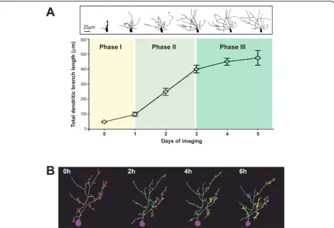
Imaging optic tectal neuronsin vivoinXenopustadpoles showed that dendritic arbor elaboration goes through distinct phases (Figure 2A) [19,48]. Many newly differ-entiated neurons first undergo axonogenesis with only little elaboration of the dendritic arbor. About one day after evidence of morphological differentiation, neurons go into a rapid dendritic arbor growth phase for a few days until they enter the third phase, characterized by a slower dendritic arbor growth rate and more stable den-dritic arbors. During the rapid denden-dritic arbor growth period, one might think that the growth of the dendritic arbor can be easily achieved by continuously lengthen-ing pre-existlengthen-ing dendrites and sproutlengthen-ing new dendritic branches; however, time-lapse imaging at intervals of minutes to hours reveals that dendritic growth is highly dynamic, consisting of not only branch addition and extension, but also retraction and loss of dendritic branches (Figure 2B) [18,49-52]. It is worth noting that these dynamics in dendritic morphogenesis persist in mature neurons when their overall structure is stable, although at a slower rate [20,53-55]. Therefore, it is very likely that mechanisms that regulate dendritic dynamics early during development may also play a role in dendri-tic plasdendri-ticity later in life.
Molecular mechanisms
cytoskeleton provides the fundamental support of the dendritic structure. Filopodia are thin, highly motile actin-based protrusions and some of them are trans-formed into more stable microtubule-based dendritic branches. The Rho family of small GTPases, including Rac, RhoA, and Cdc42, regulate the rearrangement of cytoskeleton and participate in distinct aspects of den-drite morphogenesis [56,57]. For example, Rac and Cdc42 activity promote dendritic arbor dynamics by increasing the rate of actin polymerization, whereas increased RhoA activity inhibits dendritic arbor growth in Xenopus tectal neurons [57]. Consistently, several guanine exchange factors that activate Rac, such as Tiam1 [58] and STEF [14,59,60], have been shown to regulate neurite formation whereas Rho-specific guanine exchange factors, such as KIAA0380 [14], and Rho-spe-cific GTPase activating proteins, such as p190 RhoGAP [61], which activate or inactivate Rho, respectively, have been shown to regulate neurite retraction [14,59,60]in
vitro. Interestingly, there is considerable crosstalk among these Rho GTPases. RhoA activity was increased by Rac activation and Cdc42 inhibition, whereas Rac was inhibited by activation of Rho in Xenopus tectal neurons in vivo[62]. This tight cross-regulation of Rho GTPases seems to work together to determine the struc-ture of the dendritic tree. What controls the activity of Rho GTPases is a key question to understand the under-lying mechanisms in dendritic morphogenesis.
In theXenopus visual system visual activity promotes dendritic arbor growth through mechanisms that require both glutamate receptor activity and Rho GTPase activ-ity in Xenopus tectal neurons [63]. Accordingly, the working hypothesis is that glutamate receptor activity promotes dendritic growth by elevating Rac and Cdc42 activities, leading to increased branch dynamics, and concurrently decreasing RhoA activity to relieve its inhi-bition on branch extension [63]. In addition to Rho GTPases, several other molecular mechanisms, including
signaling through neurotrophins [64,65], CPG15 [66] and calcium calmodulin dependent kinase type II [67], or local protein synthesis, mediated by cytoplasmic poly-adenylation [47], have been shown to regulate dendritic arbor development in an activity-dependent manner. These data suggest that dendritic arbor growth is orga-nized by signals from their surrounding environment. Synaptic function and dendritic development
During circuit development, the increase in synapse number and synaptic strength occur concurrently with the elaboration of dendritic arbors, suggesting a coordi-nated regulation of synaptic function and dendritic devel-opment. Almost two decades ago, Vaughn first proposed the‘synaptotrophic hypothesis’, which states that the sta-bilization of synapses might stabilize the dendritic branches and thereby explain the coordinated develop-ment of synapses and dendritic arbors [68]. Recent research has provided new supporting evidence for this hypothesis. Adhesion molecules, which play important roles in the initial assembly and stabilization of the synapses, regulate dendritic arbor development in mam-mals and flies [69,70]. Moreover, live imaging of synapse formation and dendrite growth in zebrafish showed that the presence of the synapses associated with the stabiliza-tion of terminal dendrites [71]. On the other hand, block-ade of synaptic transmission or synapse maturation reduces dendritic arbor elaboration and blocks activity-dependent dendritic growth inXenopus[18,50,52,63,72]. It is interesting to note that decreasing GABAergic trans-mission also changes the pattern of dendritic arbor growth and blocks visual experience-dependent struc-tural plasticity [73]. These data suggest that synaptic con-tacts and synaptic transmission regulate the growth and elaboration of complex dendritic arbors in sculpting circuit function during development.
The insulin receptor
The insulin receptor is a receptor tyrosine kinase well studied with regard to its function in the regulation of peripheral glucose metabolism. Although expression of the insulin receptor in the brain was discovered decades ago [74,75], insulin receptor function in this classic
‘insulin-insensitive’ organ remains largely unknown. Recent studies in neuronal cell culture suggest that insu-lin receptor signainsu-ling regulates several neuronal func-tions, including spine density and neurite growth [76,77]; however, the role of insulin receptor signaling in controlling structure and function of CNS circuit development has not yet been widely exploredin vivo.
Structure and signaling of the insulin receptor in peripheral tissues
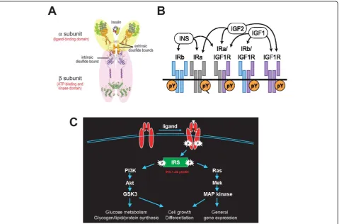
The insulin receptor was first found as a homodimer, with extrinsic disulfide bonds to generate the functional
receptor. Each monomer of the insulin receptor is com-posed of one aand onebsubunit bridged by an intrin-sic disulfide bond [78,79]. The 135-kDaasubunit is the extracellular ligand binding portion, whereas the 95-kDa b subunit consists of an extracellular, a single trans-membrane, and an intracellular kinase domain. Ligand binding to thea subunits activates the intrinsic kinase activity located in thebsubunits and subsequently initi-ates a cascade of phosphorylation events that leads to different biological functions (Figure 3A) [80].
Crystal structures of the unphosphorylated and phos-phorylated kinase domains of the insulin receptor have provided detailed information on how insulin receptor kinase activity is initiated. The kinase domain is com-posed of two lobes, the amino- and carboxy-terminal lobes, with an activation loop in between. In the unpho-sphorylated state, the activation loop traverses the cleft between two lobes such that both ATP binding and pro-tein substrate-binding sites are blocked. More specifi-cally, while residues in the beginning of the activation loop restrict the access of ATP to its binding sites on the insulin receptor, tyrosine 1162, one of the three phosphorylation sites in the activation loop, binds to the active site and competes with the kinase substrates [81]. Autophosphorylation of tyrosine 1158, 1162 and 1163 in the activation loop of the kinase domain causes rearrangement of the activation loop and reorientation of the amino- and carboxy-terminal lobes of the kinase, which is necessary for productive ATP binding. Tyrosine 1163 is the key phosphotyrosine in stabilizing the con-formation of this phosphorylated activation loop, whereas tyrosine 1158 is accessible for interaction with downstream signaling proteins [81]. The knowledge of insulin receptor structure not only provides valuable understanding on how insulin receptor signaling is transduced but also allows functional analysis following the generation of various mutants of the putative ATP binding site or phosphorylation sites [82-85].
Unlike other receptor tyrosine kinases, most functions of the insulin receptor require accessory molecules known as insulin receptor substrates (IRSs) - for example IRS1-4- to engage multiple downstream signaling [86,87]. Two major cellular signaling pathways, phosphoinositide-3 kinase (PIphosphoinositide-3K)/Akt and the Ras/mitogen-activated pro-tein kinase (MAPK) pathways, can be activated by the kinase activity of insulin receptor. These cascades regu-late diverse cellular processes, such as gene expression, protein synthesis, and vesicle trafficking, which result in the regulation of glucose, lipid and protein metabolism, cell growth and differentiation (Figure 3C) [88,89].
Diversity of the insulin receptor
diversity in its protein structure and function. First, alternative splicing produces two isoforms of insulin receptor, IRa, an exon-11 lacking form, and IRb, an exon-11 containing form in a tissue-specific manner. Moreover, post-translational glycosylation contributes to different modifications of these receptors in different cell types or tissues [90]. Furthermore, assembly of hybrids between different isoforms and heterodimers with homologous IGF-1 receptor results in further diversity [91,92]. Although different ligand binding affi-nity and selectivity have been reported for insulin recep-tors, the physiological significance of the splice variance, post-translational modification and homo- or hetero-dimerization between different insulin receptors and IGF-1 receptor remain largely unknown (Figure 3B) [86,93]. Neurons have mainly the IRa isoform with less glycosylation compared to glial cells or peripheral tissues [94]. The different properties of neuronal insulin
receptors might suggest different roles of insulin recep-tors in the CNS. Interestingly, IRa binds insulin or IGF-2 with comparable affinity [95] and hybrids of IRa with the IGF-1 receptor binds IGF-1, IGF-2 and insulin with similar affinity [86,93]. Taken together, these data suggest that, in addition to insulin, IGF-1 and IGF-2 are potential ligands for the insulin receptor in the brain. The capability of neuronal insulin receptors to interact with various ligands suggests that insulin receptors may play versatile functions in the CNS.
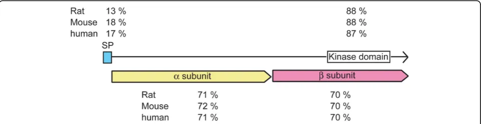
In contrast to other species, Xenopus laevishas two insulin receptor genes, which we isolated from brain cDNA libraries. At the nucleotide level, these two Xeno-pus brain insulin receptors are highly similar to each other (93.6% identity) and are splice variants homolo-gous to a human brain isoform of insulin receptor lacking exon-11 [96]. At the amino acid level, the corre-sponding peptides of these two Xenopus insulin
receptors, termed IR1 and IR2, share overall 95% iden-tity and 97% similarity. Since only one insulin receptor gene has been reported in human or other vertebrates, the two copies of insulin receptor genes potentially result from the tetraploid nature of theX. laevis genome [97]. Alignment of the amino acid sequence ofIR1, the more abundant Xenopus insulin receptor, with other species showed that theXenopusinsulin receptor shares overall identities of 70%, 69% and 69% with those of human, rat and mouse, respectively (Figure 4). Detailed alignments of different domains of insulin receptor further revealed that the kinase domain of the Xenopus insulin receptor shares the highest identity of 87 to 88% with that of human, rat and mouse compared to other regions (Figure 4). In addition, the potential ATP bind-ing site and phosphorylation sites on the activation loop of the kinase domain [81] are remarkably identical to those of human, rat and mouse, suggesting that these amino acids may play a functional role in insulin recep-tor action and thus are well conserved in evolution. This also suggests that it is suitable to be mutated for studying insulin receptor functionin vivo.
Expression pattern of insulin receptor in the brain The insulin receptor is distributed in a widespread, but selective, pattern in the brain, including olfactory bulb, cerebral cortex, hypothalamus, hippocampus and cere-bellum as reported in rodents [74,75,90]. The expression level of the insulin receptor is developmentally regu-lated, being higher at early stages and lower in the adult. At the cellular level, the insulin receptor is enriched in neurons compared to glia [74]. Subcellularly, the insulin receptor is a component of synapses, where it concentrates at the postsynaptic density (PSD) in cul-tured hippocampal neurons [98]. These data together suggest that the insulin receptor is in the right place at the right time to regulate the initial neuronal develop-ment by regulating synaptic function in the CNS.
Although the IGF-1 receptor, which can dimerize with the insulin receptor and affect its ligand affinity and spe-cificity, as mentioned previously, shows a similar distri-bution in the brain as the insulin receptor, it also exhibits a distinct expression pattern compared to the insulin receptor when examined in detail locally [99]. For example, both receptors are highly expressed in hip-pocampus; however, insulin receptor mRNA is more abundant in the CA1 region whereas IGF-1 receptor mRNA is greater in the CA3 region, implying that insu-lin/IGF-1 receptor signaling may play distinct roles in subregions of the hippocampus.
Function of insulin receptor in circuit development and plasticity
Brain insulin receptor signaling reportedly plays diverse roles in the CNS, including regulation of synaptic plasti-city [100-106], dendritic outgrowth [77,107], and involve-ment in neuronal survival [108,109], life span [110-114], learning and memory [115-117], and neurological disor-ders [118-121]. A role for insulin receptor signaling in synaptic function and dendritic morphogenesis, there-fore, makes it a potential regulator of circuit development and circuit function.
Synaptic function
Recent work suggests insulin receptor signaling is involved in postsynaptic neurotransmitter receptor traf-ficking. For excitatory receptors, insulin accelerates cla-thrin-dependent endocytosis of GluR2 subunit-containing AMPA receptors and contributes to long-term depression [100,102,122-124]. In contrast, insulin reportedly acceler-ates GluR1 subunit-containing AMPA receptor insertion into the membrane in a GluR1 subunit-dependent man-ner in cultured hippocampal neurons [104]. Therefore, the physiological significance of insulin receptor signaling in AMPA receptor-mediated transmission is somewhat controversial and needs to be further studied in vivo. Moreover, insulin promotes the delivery of NMDA
receptors to the cell surface by exocytosis inXenopus oocytes expressing recombinant NMDA receptor [105]. For inhibitory receptors, insulin rapidly recruits type A g-aminobutyric acid (GABAA) receptors to the postsynap-tic membrane in cultured hippocampal neurons [106]. These data suggest that insulin receptor signaling is cap-able of regulating both excitatory and inhibitory synaptic transmission in the CNS. In addition, brief incubation of insulin results in increased protein synthesis of PSD-95, a dendritic scaffolding protein that associates neurotrans-mitter receptors and cytoskeletal elements at synapses in hippocampal slices and synaptosomes [125], also suggest-ing that insulin receptor signalsuggest-ing can potentially regulate structural aspects of synaptic function, synaptogenesis and synapse maturation.
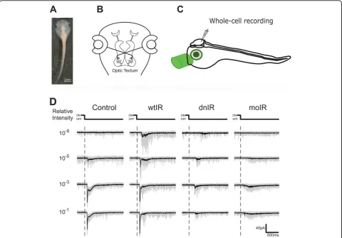
Recently, our laboratory provided direct in vivo evidence for the function of insulin receptor signaling in both the structure and function of brain circuit devel-opment in the visual system of liveXenopus tadpoles [84]. The retinotectal circuit ofXenopus (see schematic in Figure 5), in which tectal neurons receive direct visual input from the retinal ganglion cells in the eye [126], is a powerful experimental system to study both structural [47,52,63,73] and functional plasticity [17,36,47,73,127-130]in vivo. By taking advantage of the Xenopusvisual circuit as anin vivoexperimental system amenable to molecular manipulation, electrophysiology and a variety of imaging methods, we showed that the insulin receptor is required for optic tectal neurons to receive normal levels of visual input within the retino-tectal circuit [84]. Reduced insulin receptor phosphory-lation by ectopic expression of dominant negative insulin receptor (dnIR), which contains a point mutation to abolish insulin receptor binding to ATP, or decrease insulin receptor protein by morpholino-mediated knock-down in tectal neurons, severely decreases their gluta-matergic synaptic input and reduces their responses to natural light stimuli (Figure 5). Few studies have made a direct comparison between the effects of protein knock-down and dominant negative interference with signaling. It is interesting to note that decreasing insulin receptor signaling either by expression of a dominant negative receptor or by morpholino-mediated knockdown leads to a comparable magnitude of functional impairment in visual system processing, suggesting that the presence of the protein itself does not play a role in visual system development independent of its kinase-dependent signaling.
Dendritic morphogenesis
Several molecules downstream of the insulin receptor, including both the Ras/MAPK and PI3K/Akt/mamma-lian target of rapamycin (mTOR) pathways, have been implicated in excitatory synaptic connectivity as well as dendritic structure [131,132]. IRSp53, a novel insulin
receptor substrate enriched in the brain, where it loca-lizes to synapses as a component of the PSD [98], is par-ticularly interesting. Structural analysis predicted that IRSp53 contains several protein-protein interaction domains, including an amino-terminal F-actin bundling domain [133,134], a central Cdc42/Rac interactive bind-ing (CRIB) motif [135], a Src homology region 3 (SH3) domain [76,136,137], a proline rich SH3-binding domain [136], a proline-rich WW-binding motif [136], and a carboxy-terminal postsynaptic density-95/discs large/ zona occudens-1 (PDZ) domain [76,138]. Biochemical studies showed that it directly interacts with PSD scaf-fold proteins, Shank and PSD-95 [76,137-139], small GTPases such as Rac and Cdc42 [77,139-141], and actin regulators such as WAVE2 and Mena [140,141]. These data together suggest a link between insulin receptor signaling and the structural stabilization of excitatory synaptic contacts through the association of synaptic scaffolding proteins and the cytoskeleton. In fact, these ideas were further supported by the findings that over-expression of IRSp53 can increase spine density in cul-tured hippocampal neurons [76] and induce filopodium formation and neurite outgrowth in N1E-115 neuroblas-toma cells [77,142], whereas RNA interference knock-down of IRSp53 protein decreases spine density and alters spine morphogenesis [76]. Another line of evi-dence supporting the idea that insulin receptor plays a role in dendritic arbor development comes from trans-genic mice lacking IGF-1, a potential ligand for insulin receptor and IGF-1 receptor heterodimer receptors in the brain. Pyramidal neurons from the IGF-1 null mice showed significant reduction in dendritic arbor length and complexity as well as spine density [107].
Experience-dependent dendritic plasticity
Activity shapes synaptic connectivity and dendritic mor-phogenesis in the CNS, particularly in sensory regions. Interestingly, insulin is released from neurons upon depolarization [143,144] and IRSp53 translocates to synapses in response to activity [145], suggesting that insulin receptor signaling may increase in an activity-dependent manner. Consistent with this idea, we have shown recently that insulin receptor signaling plays an important role in visual experience-dependent structural plasticity [84]. More specifically, enhanced visual stimu-lation normally induces tectal neurons to increase their rate of dendritic growth by increasing branch length extension and branch tip stabilization. In the absence of insulin receptor signaling, however, more branches shorten and more branches are lost during the period of visual stimulation.
Insulin receptor signaling and synaptic structure
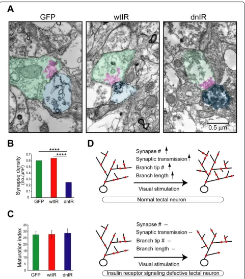
neurons to receive proper glutamatergic synaptic input and undergo activity-dependent dendritic arbor growth. To probe the role of insulin receptor signaling in devel-opmental plasticity of the glutamatergic synapse, we examined the spontaneous AMPA receptor-mediated miniature excitatory postsynaptic currents (mEPSC) in dnIR-expressing neurons. We found that they have much lower mEPSC frequency but equivalent amplitude compared to controls, indicating that either presynaptic vesicle release probability or synapse number is reduced in dnIR-expressing neurons. Because the paired-pulsed ratio with retinal ganglion cell axon stimulation in dnIR-expressing neurons did not change, it is unlikely that the lower mEPSC frequency in dnIR-expressing neurons is due to low probability of release. To test whether synaptic contacts onto dnIR-expressing tectal neurons were changed in dnIR-expressing neurons, we
used electron microscopy to estimate synapse density on tectal neurons. This methodology gives both definite identification of synaptic contacts onto transfected neu-rons and ultrastructural information about both pre-and post-synaptic profiles [23]. We estimated synapse density by measuring the number of green fluorescent protein (GFP)-labeled synapses normalized to the total area of GFP-labeled dendritic profiles and found that dnIR-expressing dendrites had less than half of the synapse density of GFP-labeled neuron controls, although no changes in other ultrastructural features or synapse maturation were observed (Figures 6A-C) [84]. These observations, according to both electrophysiologi-cal and ultrastructural data, together with decreased dendritic plasticity in dnIR-expressing neurons, suggest that insulin receptor signaling maintains both synaptic contacts and the branches they sit on, which in turn
promotes dendritic branch elaboration with visual experience.
Our observations are consistent with the synapto-trophic hypothesis, which states that the formation and maintenance of synapses promote the stabilization of dendritic branches and that dendritic arbor growth cor-relates positively with the number and strength of synapses [18]. In the optic tectum of Xenopus, visual experience increases dendritic arbor growth rate, retino-tectal synaptogenesis and retinoretino-tectal synaptic strength [17,52,63,128,146]. Similarly, in zebrafish, synapses appear to stabilize growing dendrites and promote further dendrite branch growth in tectal neurons [71]. Conversely, blocking synapse maturation by interfering with AMPA receptor trafficking into synapses reduces dendritic arbor elaboration and completely blocks the visual stimulation-induced increase in dendritic arbor growth [52]. Therefore, the visual stimulation-induced increase in synapse number and strength [17] appears to stabilize newly extended dendritic branches. The failure of neurons with decreased insulin receptor signaling to increase their growth rate in response to visual stimula-tion could be a result of their low synapse density. One potential mechanism by which a lower synapse density could affect experience-dependent structural plasticity is that these neurons do not form and maintain synapses on newly added branches, and they are consequently retracted. The alternative, but not mutually exclusive, mechanism is based on the fact that, in these experi-ments, we transfected single tectal neurons within an otherwise normal optic tectum. Therefore, while sur-rounding tectal cells, which have twice the synapse den-sity of the dnIR-expressing neurons, respond to visual stimulation normally and can increase their synapses and promote dendritic growth, the single dnIR-expres-sing neuron, which responds to visual inputs very weakly, may not be able to compete with normal neigh-boring tectal neurons for retinal inputs. Consequently, this might lead to branch length retraction and branch loss in the insulin receptor signaling deficient neurons.
Unexpectedly, we found that dnIR-expressing neurons can still elaborate their dendrites over a period of sev-eral days even when synapse density is low during early development. A similar observation was reported with manipulation of levels of the neurotrophin brain-derived neurotrophic factor, which significantly changed synapse number but not dendritic arbor morphology [147]. In the case of insulin receptor signaling where experience-dependent structural plasticity is decreased when assayed over a period of 4 hours, these daily imaging data suggest that under conditions of decreased synaptic input, alternative mechanisms participate in dendritic arbor growth control.
Insulin receptor signaling and neurological diseases Emergent evidence suggests an association of insulin receptor signaling with several neurological disorders. Although the role that the insulin receptor may play in these disorders is still a puzzle, enhanced brain insulin receptor signaling has been used to treat schizophrenia patients early in the mid-20th century [148,149] and insulin sensitizing drugs are now in clinical trials for the treatment of Alzheimer’s disease [150-152], highlighting its importance in both neuronal developmental and degenerative diseases.
Neurodegenerative diseases
Reduced mRNA and protein levels have been reported in postmortem material from patients with neurodegen-erative disorders, for example Alzheimer’s sisease [118,153] and Parkinson’s disease [119], implying a role for insulin receptor signaling in neurodegenerative dis-eases. Among these, Alzheimer’s disease is the best-stu-died neurodegenerative disease with respect to insulin receptor signaling.
Alzheimer’s disease, the most common brain degen-eration characterized clinically by progressive decline of memory and pathologically by loss of synapses, forma-tion of neurofibrillary tangles and neuritic plaques, has been extensively studied with respect to insulin receptor signaling. Insulin receptor signaling inhibits a key event in the formation of neurofibrillary tangles by reducing tau protein phosphorylation [154,155]. Additionally, insulin receptor signaling prevents plaque formation by modulating amyloidb (Ab) release [156] and degrada-tion [157-160].
Although tangle formation and amyloid deposits are useful diagnostic markers, synapse loss is more robustly correlated with cognitive deficits than any other patho-logical lesion observable in Alzheimer’s patients [161-164]. Progressive accumulation and toxicity of Ab oligomers is the leading hypothesis for etiology of Alz-heimer’s disease [163]. Interestingly, the Ab oligomer induces glutamatergic synapse loss [165,166], which in addition to cholinergic synapses seems to be most severely affected in Alzheimer’s disease patients [167,168].
interacting with the insulin receptor and interfering with insulin receptor signaling. Our data further support the idea that synapse loss resulting from reduced insulin receptor signaling will decrease experience-dependent structural plasticity and ultimately lead to deficits in cir-cuit function, including information processing and inte-gration. By contrast, reduced IGF-1 receptor function also reportedly decreases Ab toxicity and ameliorates neuronal/synaptic loss in animal models of Alzheimer’s disease [174,175]. The seemingly opposite outcomes from decreased insulin receptor and IGF-1 receptor sig-naling implies that either they initiate different pathways or they share the same signaling pathway but bi-direc-tionally regulate Abtoxicity and synaptic loss in Alzhei-mer’s disease.
Neuronal developmental disorders
Several neuronal developmental disorders are thought to be associated with insulin receptor signaling malfunc-tion. For instance, schizophrenia is a chronic neurodeve-lopmental disorder that affects approximately 1.1% of the US population, and decreased insulin receptor pro-tein and activity and altered downstream signaling have been reported in post-mortem schizophrenia patients [121]. Although the underlying mechanism is poorly understood, insulin treatment of schizophrenic patients was initiated during the 1930s and reportedly gives effective clinical results, [148,149]. Surprisingly, schizo-phrenia and Alzheimer’s disease share some early patho-logical hallmarks, such as impaired synaptic connectivity [176-178] and abnormal dendritic structure [179,180], that eventually result in impaired circuit function and aberrant cognitive behavior.
Another example is tuberous sclerosis (TSC), a genetic disorder resulting from mutation in one of the two tumor suppressor genes,TSC1or TSC2, that often give rise to multiple neurological deficits such as epilepsy, mental retardation and autism. Interestingly, loss of TSC function decreases synaptic transmission and alters spine morphology through the mTOR pathway, which overlaps with insulin receptor signaling [181]. One potential etiology for TSC-related neuronal deficits could be their ability to negatively regulate insulin receptor signaling in the brain as reported in the fly and in mammalian cell lines [182,183].
It is now recognized that molecules that regulate aging can also affect early neuronal development. For example, cyclin-dependent kinase 5, which plays roles in neuronal migration in the developing CNS, is also involved in the pathology of Alzheimer’s disease [184]. Insulin receptor signaling, therefore, might participate at both ends of the story: early development as well as later neurodegen-erative diseases.
Perspectives
Accumulating data support the idea that insulin recep-tor signaling plays a prominent role in both structural and functional aspects of circuit development. The detailed cellular and molecular mechanisms by which insulin receptor signaling control synaptic function and dendritic structure are still to be determined. Besides the role of insulin receptor signaling in circuit forma-tion, insulin receptor signaling has been linked to sev-eral neurological disorders. Whether failures in synaptic function and dendritic structure caused by decreased insulin receptor signaling contribute to brain diseases later in life is an important issue to address.
Synapse formation or maintenance?
cellular mechanism of insulin receptor signaling in regu-lating synapse connectivity.
Endogenous ligand and receptor composition
Insulin is thought to be the primary ligand for the insu-lin receptor; however, IGF-2 also reportedly binds the homodimer of the insulin receptor splice variant in the brain [95]. Moreover, the discovery that the insulin receptor and IGF-1 receptor heterodimerize expands the potential ligands for insulin receptor heterodimers in the brain to include insulin, IGF-2, IGF-1 and possibly others [86]. Several potential ligands - for example, mammalian insulin and nematode insulin and IGFs -have been reported to affect synaptic transmission and plasticity, dendrite structure, whole animal lifespan and behaviors in various model systems [101,105-107,186-189]. The identity of the primary ligand(s) that activate insulin receptor signaling and reg-ulate synapse number, where the ligands are found in the brain and how are they regulated are all important questions requiring further exploration.
At the receptor level, it is important to investigate the composition of the receptor dimer since it determines the specificity and affinity of the ligand(s) and might initiate distinct downstream signaling pathways. Our strategy of using dnIR expression can potentially disrupt three types of receptor signaling according to the recep-tor composition: the insulin receprecep-tor homodimer; the insulin receptor-IGF-1 receptor heterodimer; and the IGF-1 receptor homodimer. It is interesting to note that when antisense morpholino oligonucleotides were used to specifically knockdown insulin receptor but not IGF-1 receptor, morpholino-transfected neurons show a similar deficit in visual responses recordedin vivo com-pared to dnIR-expressing neurons. This result indicates that the insulin receptor, instead of the IGF-1 receptor, plays major roles in visual circuit function. Nevertheless, whether the insulin receptor executes its function through the insulin receptor homodimer or the insulin receptor/IGF-1 receptor heterodimer is still an open question. Traditional co-immunoprecipitation of the insulin receptor dimers from brain lysate might help in deciphering the receptor composition if one can develop specific antibodies to differentiate these two structurally similar receptors. Alternatively, molecular tools for example, morpholino or RNA interference -to specifically knockdown the insulin recep-tor, the IGF-1 receptor alone, or both together may provide further insight.
Molecular mechanisms
The decrease in insulin receptor signaling by dnIR expression affects visual responses in tectal neurons to the same extent as morpholino-mediated knockdown of
insulin receptor protein, indicating that kinase activity of the insulin receptor plays a major role in insulin receptor function. What are the downstream cascades activated by insulin receptor kinase activity in the CNS? Studies in peripheral tissues have demonstrated that MAPK or Akt are major pathways downstream of the insulin receptor [88]. Whether MAPK or Akt pathways underlie insulin receptor-mediated circuit development needs to be further explored. In addition to these gen-eral pathways, some molecules appear to be more speci-fic to insulin receptor signaling, for example, IRSs [190]. As mentioned before, IRSp53 is a great candidate to execute insulin receptor function at excitatory synapses by regulating the actin cytoskeleton through a pathway that requires its coupling with activated Rho GTPase [77,140,141]. Whether this effect on actin cytoskeleton originates from insulin receptor signaling would be interesting to know. Recently, the phosphorylation sites of IRSp53 that specifically respond to insulin receptor signaling have been discovered [191]. Mutations of these sites would allow us to understand the interplay between the insulin receptor, IRSp53 and RhoGTPases in the structural aspects of circuit development.
Neurological disorders
schizophrenia [195]. Further research on this type of transgenic system will provide insight into the physiolo-gical function of the insulin receptor in the development of the normal brain as well as the etiology of neurologi-cal diseases.
Abbreviations
Ab: amyloidb; AMPA:a-amino-3-hydroxy-5-methyl-4-isoxazole propionic acid; CNS: central nervous system; dnIR: dominant negative insulin receptor; GABA:g-aminobutyric acid; GFP: green fluorescent protein; GluR: glutamate receptor; IGF: insulin-like growth factor; IR: insulin receptor; IRS: insulin receptor substrate; MAPK: mitogen-activated protein kinase; mEPSC: miniature excitatory postsynaptic currents; mTOR: mammalian target of rapamycin; NMDA: N-methyl-D-aspartate; PI3K: phosphoinositide-3 kinase; PSD: postsynaptic density; PSD-95: postsynaptic density protein-95; TSC: tuberous sclerosis.
Acknowledgements
The authors thank current and past members of the Cline Lab for insightful discussions, particularly Emiliano Rial Verde for encouraging the work. Supported by grants from the NIH (EY11261 and DP1OD000458 to HTC) and an Elizabeth Sloan Livingston Foundation Fellowship (S-LC).
Author details
1Watson School of Biological Sciences and Cold Spring Harbor Laboratory,
Cold Spring Harbor, NY 11724, USA.2Departments of Cell Biology and
Chemical Physiology, The Scripps Research Institute, 10550 North Torrey Pines Road, La Jolla, CA 92037, USA.
Authors’contributions
S-LC and HTC discussed the content and wrote the manuscript.
Competing interests
The authors declare that they have no competing interests.
Received: 14 December 2009 Accepted: 15 March 2010 Published: 15 March 2010
References
1. Trachtenberg JT, Chen BE, Knott GW, Feng G, Sanes JR, Welker E, Svoboda K:Long-termin vivoimaging of experience-dependent synaptic plasticity in adult cortex.Nature2002,420:788-794.
2. Holtmaat A, Wilbrecht L, Knott GW, Welker E, Svoboda K: Experience-dependent and cell-type-specific spine growth in the neocortex.Nature 2006,441:979-983.
3. Chen C, Regehr WG:Developmental remodeling of the retinogeniculate synapse.Neuron2000,28:955-966.
4. Li Z, Sheng M:Some assembly required: the development of neuronal synapses.Nat Rev Mol Cell Biol2003,4:833-841.
5. Ahmari SE, Buchanan J, Smith SJ:Assembly of presynaptic active zones from cytoplasmic transport packets.Nat Neurosci2000,3:445-451. 6. Bresler T, Shapira M, Boeckers T, Dresbach T, Futter M, Garner CC,
Rosenblum K, Gundelfinger ED, Ziv NE:Postsynaptic density assembly is fundamentally different from presynaptic active zone assembly.
J Neurosci2004,24:1507-1520.
7. Jin Y, Garner CC:Molecular mechanisms of presynaptic differentiation.
Annu Rev Cell Dev Biol2008,24:237-262.
8. Ziv NE, Garner CC:Cellular and molecular mechanisms of presynaptic assembly.Nat Rev Neurosci2004,5:385-399.
9. Gerrow K, Romorini S, Nabi SM, Colicos MA, Sala C, El-Husseini A:A preformed complex of postsynaptic proteins is involved in excitatory synapse development.Neuron2006,49:547-562.
10. McAllister AK:Dynamic aspects of CNS synapse formation.Annu Rev Neurosci2007,30:425-450.
11. Dufour A, Seibt J, Passante L, Depaepe V, Ciossek T, Frisen J, Kullander K, Flanagan JG, Polleux F, Vanderhaeghen P:Area specificity and topography of thalamocortical projections are controlled by ephrin/Eph genes.
Neuron2003,39:453-465.
12. O’Leary DD, Wilkinson DG:Eph receptors and ephrins in neural development.Curr Opin Neurobiol1999,9:65-73.
13. Benson DL, Colman DR, Huntley GW:Molecules, maps and synapse specificity.Nat Rev Neurosci2001,2:899-909.
14. Togashi H, Abe K, Mizoguchi A, Takaoka K, Chisaka O, Takeichi M:Cadherin regulates dendritic spine morphogenesis.Neuron2002,35:77-89. 15. Scheiffele P:Cell-cell signaling during synapse formation in the CNS.
Annu Rev Neurosci2003,26:485-508.
16. Levinson JN, El-Husseini A:Building excitatory and inhibitory synapses: balancing neuroligin partnerships.Neuron2005,48:171-174. 17. Aizenman CD, Cline HT:Enhanced visual activityin vivoforms nascent
synapses in the developing retinotectal projection.J Neurophysiol2007,
97:2949-2957.
18. Cline H, Haas K:The regulation of dendritic arbor development and plasticity by glutamatergic synaptic input: a review of the synaptotrophic hypothesis.J Physiol2008,586:1509-1517.
19. Cline HT:Dendritic arbor development and synaptogenesis.Curr Opin Neurobiol2001,11:118-126.
20. Wong RO, Ghosh A:Activity-dependent regulation of dendritic growth and patterning.Nat Rev Neurosci2002,3:803-812.
21. Blue ME, Parnavelas JG:The formation and maturation of synapses in the visual cortex of the rat. II. Quantitative analysis.J Neurocytol1983,
12:697-712.
22. Mohrmann R, Lessmann V, Gottmann K:Developmental maturation of synaptic vesicle cycling as a distinctive feature of central glutamatergic synapses.Neuroscience2003,117:7-18.
23. Li J, Cline HT:The accumulation of dense core vesicles in developing optic tectal synapses inXenopus laevisincreases following visual deprivation.J Comp Neurol2010.
24. Hestrin S:Developmental regulation of NMDA receptor-mediated synaptic currents at a central synapse.Nature1992,357:686-689. 25. Shi J, Aamodt S, Constantine-Paton M:Temporal correlations between
functional and molecular changes in NMDA receptors and GABA neurotransmission in the superior colliculus.J Neurosci1997,
17:6264-6276.
26. Isaac JT, Nicoll RA, Malenka RC:Evidence for silent synapses: implications for the expression of LTP.Neuron1995,15:427-434.
27. Liao D, Hessler NA, Malinow R:Activation of postsynaptically silent synapses during pairing-induced LTP in CA1 region of hippocampal slice.Nature1995,375:400-404.
28. Wu G, Malinow R, Cline HT:Maturation of a central glutamatergic synapse.Science1996,274:972-976.
29. Washbourne P, Bennett JE, McAllister AK:Rapid recruitment of NMDA receptor transport packets to nascent synapses.Nat Neurosci2002,
5:751-759.
30. Luo L, O’Leary DD:Axon retraction and degeneration in development and disease.Annu Rev Neurosci2005,28:127-156.
31. Walsh MK, Lichtman JW:In vivotime-lapse imaging of synaptic takeover associated with naturally occurring synapse elimination.Neuron2003,
37:67-73.
32. Nguyen QT, Lichtman JW:Mechanism of synapse disassembly at the developing neuromuscular junction.Curr Opin Neurobiol1996,6:104-112. 33. Rakic P, Bourgeois JP, Eckenhoff MF, Zecevic N, Goldman-Rakic PS:
Concurrent overproduction of synapses in diverse regions of the primate cerebral cortex.Science1986,232:232-235.
34. Hashimoto K, Kano M:Functional differentiation of multiple climbing fiber inputs during synapse elimination in the developing cerebellum.
Neuron2003,38:785-796.
35. Nakamura H, O’Leary DD:Inaccuracies in initial growth and arborization of chick retinotectal axons followed by course corrections and axon remodeling to develop topographic order.J Neurosci1989,9:3776-3795. 36. Tao HW, Poo MM:Activity-dependent matching of excitatory and
inhibitory inputs during refinement of visual receptive fields.Neuron 2005,45:829-836.
37. Chen C, Regehr WG:Presynaptic modulation of the retinogeniculate synapse.J Neurosci2003,23:3130-3135.
38. Colman H, Nabekura J, Lichtman JW:Alterations in synaptic strength preceding axon withdrawal.Science1997,275:356-361.
40. Kano M, Hashimoto K, Chen C, Abeliovich A, Aiba A, Kurihara H, Watanabe M, Inoue Y, Tonegawa S:Impaired synapse elimination during cerebellar development in PKC gamma mutant mice.Cell1995,83:1223-1231. 41. Kano M, Hashimoto K, Kurihara H, Watanabe M, Inoue Y, Aiba A,
Tonegawa S:Persistent multiple climbing fiber innervation of cerebellar Purkinje cells in mice lacking mGluR1.Neuron1997,18:71-79.
42. Kashiwabuchi N, Ikeda K, Araki K, Hirano T, Shibuki K, Takayama C, Inoue Y, Kutsuwada T, Yagi T, Kang Y,et al:Impairment of motor coordination, Purkinje cell synapse formation, and cerebellar long-term depression in GluR delta 2 mutant mice.Cell1995,81:245-252.
43. Huh GS, Boulanger LM, Du H, Riquelme PA, Brotz TM, Shatz CJ:Functional requirement for class I MHC in CNS development and plasticity.Science 2000,290:2155-2159.
44. Ishii DN:Relationship of insulin-like growth factor II gene expression in muscle to synaptogenesis.Proc Natl Acad Sci USA1989,86:2898-2902. 45. Stevens B, Allen NJ, Vazquez LE, Howell GR, Christopherson KS, Nouri N,
Micheva KD, Mehalow AK, Huberman AD, Stafford B, Sher A, Litke AM, Lambris JD, Smith SJ, John SW, Barres BA:The classical complement cascade mediates CNS synapse elimination.Cell2007,131:1164-1178. 46. Hua JY, Smith SJ:Neural activity and the dynamics of central nervous
system development.Nat Neurosci2004,7:327-332.
47. Bestman JE, Cline HT:The RNA binding protein CPEB regulates dendrite morphogenesis and neuronal circuit assemblyin vivo.Proc Natl Acad Sci USA2008,105:20494-20499.
48. Wu GY, Zou DJ, Rajan I, Cline H:Dendritic dynamicsin vivochange during neuronal maturation.J Neurosci1999,19:4472-4483.
49. Dailey ME, Smith SJ:The dynamics of dendritic structure in developing hippocampal slices.J Neurosci1996,16:2983-2994.
50. Rajan I, Cline HT:Glutamate receptor activity is required for normal development of tectal cell dendritesin vivo.J Neurosci1998,18:7836-7846. 51. Ewald RC, Van Keuren-Jensen KR, Aizenman CD, Cline HT:Roles of NR2A
and NR2B in the development of dendritic arbor morphologyin vivo.J Neurosci2008,28:850-861.
52. Haas K, Li J, Cline HT:AMPA receptors regulate experience-dependent dendritic arbor growthin vivo.Proc Natl Acad Sci USA2006,
103:12127-12131.
53. Akaaboune M, Culican SM, Turney SG, Lichtman JW:Rapid and reversible effects of activity on acetylcholine receptor density at the
neuromuscular junctionin vivo.Science1999,286:503-507.
54. Lee WC, Chen JL, Huang H, Leslie JH, Amitai Y, So PT, Nedivi E:A dynamic zone defines interneuron remodeling in the adult neocortex.Proc Natl Acad Sci USA2008,105:19968-19973.
55. Lee WC, Huang H, Feng G, Sanes JR, Brown EN, So PT, Nedivi E:Dynamic remodeling of dendritic arbors in GABAergic interneurons of adult visual cortex.PLoS Biol2006,4:e29.
56. Van Aelst L, Cline HT:Rho GTPases and activity-dependent dendrite development.Curr Opin Neurobiol2004,14:297-304.
57. Li Z, Van Aelst L, Cline HT:Rho GTPases regulate distinct aspects of dendritic arbor growth inXenopuscentral neuronsin vivo.Nature Neurosci2000,3:217-225.
58. Tolias KF, Bikoff JB, Burette A, Paradis S, Harrar D, Tavazoie S, Weinberg RJ, Greenberg ME:The Rac1-GEF Tiam1 couples the NMDA receptor to the activity-dependent development of dendritic arbors and spines.Neuron 2005,45:525-538.
59. Matsuo N, Hoshino M, Yoshizawa M, Nabeshima Y:Characterization of STEF, a guanine nucleotide exchange factor for Rac1, required for neurite growth.J Biol Chem2002,277:2860-2868.
60. Brouns MR, Matheson SF, Settleman J:p190 RhoGAP is the principal Src substrate in brain and regulates axon outgrowth, guidance and fasciculation.Nat Cell Biol2001,3:361-367.
61. Brouns MR, Matheson SF, Settleman J:p190 RhoGAP is the principal Src substrate in brain and regulates axon outgrowth, guidance and fasciculation.Nat Cell Biol2001,3:361-367.
62. Li Z, Aizenman CD, Cline HT:Regulation of Rho GTPases by crosstalk and neuronal activityin vivo.Neuron2002,33:741-750.
63. Sin WC, Haas K, Ruthazer ES, Cline HT:Dendrite growth increased by visual activity requires NMDA receptor and Rho GTPases.Nature2002,
419:475-480.
64. Balkowiec A, Katz DM:Activity-dependent release of endogenous brain-derived neurotrophic factor from primary sensory neurons detected by ELISAin situ.J Neurosci2000,20:7417-7423.
65. Marshak S, Nikolakopoulou AM, Dirks R, Martens GJ, Cohen-Cory S: Cell-autonomous TrkB signaling in presynaptic retinal ganglion cells mediates axon arbor growth and synapse maturation during the establishment of retinotectal synaptic connectivity.J Neurosci2007,
27:2444-2456.
66. Nedivi E, GY W, Cline HT:Promotion of dendritic growth by CPG15, an activity-induced signaling molecule.Science1998,281:1863-1866. 67. Wu G-Y, Cline HT:Stabilization of dendritic arbor structurein vivoby
CaMKII.Science1998,279:222-226.
68. Vaughn JE:Fine structure of synaptogenesis in the vertebrate central nervous system.Synapse1989,3:255-285.
69. Shima Y, Kengaku M, Hirano T, Takeichi M, Uemura T:Regulation of dendritic maintenance and growth by a mammalian 7-pass transmembrane cadherin.Dev Cell2004,7:205-216.
70. Ye B, Jan YN:The cadherin superfamily and dendrite development.
Trends Cell Biol2005,15:64-67.
71. Niell CM, Meyer MP, Smith SJ:In vivoimaging of synapse formation on a growing dendritic arbor.Nat Neurosci2004,7:254-260.
72. Rajan I, Witte S, Cline HT:NMDA receptor activity stabilizes presynaptic retinotectal axons and postsynaptic optic tectal cell dendritesin vivo.J Neurobiol1999,38:357-368.
73. Shen W, Da Silva JS, He H, Cline HT:Type A GABA-receptor-dependent synaptic transmission sculpts dendritic arbor structure inXenopus
tadpolesin vivo.J Neurosci2009,29:5032-5043.
74. Unger J, McNeill TH, Moxley RT, White M, Moss A, Livingston JN:
Distribution of insulin receptor-like immunoreactivity in the rat forebrain.Neuroscience1989,31:143-157.
75. Havrankova J, Roth J, Brownstein M:Insulin receptors are widely distributed in the central nervous system of the rat.Nature1978,
272:827-829.
76. Choi J, Ko J, Racz B, Burette A, Lee JR, Kim S, Na M, Lee HW, Kim K, Weinberg RJ, Kim E:Regulation of dendritic spine morphogenesis by insulin receptor substrate 53, a downstream effector of Rac1 and Cdc42 small GTPases.J Neurosci2005,25:869-879.
77. Govind S, Kozma R, Monfries C, Lim L, Ahmed S:Cdc42Hs facilitates cytoskeletal reorganization and neurite outgrowth by localizing the 58-kD insulin receptor substrate to filamentous actin.J Cell Biol2001,
152:579-594.
78. Cheatham B, Kahn CR:Cysteine 647 in the insulin receptor is required for normal covalent interaction between alpha- and beta-subunits and signal transduction.J Biol Chem1992,267:7108-7115.
79. Seino S, Bell GI:Alternative splicing of human insulin receptor messenger RNA.Biochem Biophys Res Commun1989,159:312-316.
80. De Meyts P, Whittaker J:Structural biology of insulin and IGF1 receptors: implications for drug design.Nat Rev Drug Discov2002,1:769-783. 81. Hubbard SR, Wei L, Ellis L, Hendrickson WA:Crystal structure of the
tyrosine kinase domain of the human insulin receptor.Nature1994,
372:746-754.
82. Ebina Y, Araki E, Taira M, Shimada F, Mori M, Craik CS, Siddle K, Pierce SB, Roth RA, Rutter WJ:Replacement of lysine residue 1030 in the putative ATP-binding region of the insulin receptor abolishes insulin- and antibody-stimulated glucose uptake and receptor kinase activity.Proc Natl Acad Sci USA1987,84:704-708.
83. Kanezaki Y, Obata T, Matsushima R, Minami A, Yuasa T, Kishi K, Bando Y, Uehara H, Izumi K, Mitani T, Matsumoto M, Takeshita Y, Nakaya Y, Matsumoto T, Ebina Y:K(ATP) channel knockout mice crossbred with transgenic mice expressing a dominant-negative form of human insulin receptor have glucose intolerance but not diabetes.Endocr J2004,
51:133-144.
84. Chiu SL, Chen CM, Cline HT:Insulin receptor signaling regulates synapse number, dendritic plasticity, and circuit functionin vivo.Neuron2008,
58:708-719.
85. Jacob KK, Whittaker J, Stanley FM:Insulin receptor tyrosine kinase activity and phosphorylation of tyrosines 1162 and 1163 are required for insulin-increased prolactin gene expression.Mol Cell Endocrinol2002,
186:7-16.
86. White MF:Insulin signaling in health and disease.Science2003,
302:1710-1711.
88. Saltiel AR, Kahn CR:Insulin signalling and the regulation of glucose and lipid metabolism.Nature2001,414:799-806.
89. Schulingkamp RJ, Pagano TC, Hung D, Raffa RB:Insulin receptors and insulin action in the brain: review and clinical implications.Neurosci Biobehav Rev2000,24:855-872.
90. Wozniak M, Rydzewski B, Baker SP, Raizada MK:The cellular and
physiological actions of insulin in the central nervous system.Neurochem Int1993,22:1-10.
91. Benyoucef S, Surinya KH, Hadaschik D, Siddle K:Characterization of insulin/ IGF hybrid receptors: contributions of the insulin receptor L2 and Fn1 domains and the alternatively spliced exon 11 sequence to ligand binding and receptor activation.Biochem J2007,403:603-613. 92. Slaaby R, Schaffer L, Lautrup-Larsen I, Andersen AS, Shaw AC, Mathiasen IS,
Brandt J:Hybrid receptors formed by insulin receptor (IR) and insulin-like growth factor I receptor (IGF-IR) have low insulin and high IGF-1 affinity irrespective of the IR splice variant.J Biol Chem2006,281:25869-25874. 93. Pandini G, Frasca F, Mineo R, Sciacca L, Vigneri R, Belfiore A:
Insulin/insulin-like growth factor I hybrid receptors have different biological
characteristics depending on the insulin receptor isoform involved.J Biol Chem2002,277:39684-39695.
94. Heidenreich KA, Zahniser NR, Berhanu P, Brandenburg D, Olefsky JM:
Structural differences between insulin receptors in the brain and peripheral target tissues.J Biol Chem1983,258:8527-8530.
95. Frasca F, Pandini G, Scalia P, Sciacca L, Mineo R, Costantino A, Goldfine ID, Belfiore A, Vigneri R:Insulin receptor isoform A, a newly recognized, high-affinity insulin-like growth factor II receptor in fetal and cancer cells.Mol Cell Biol1999,19:3278-3288.
96. Kenner KA, Kusari J, Heidenreich KA:cDNA sequence analysis of the human brain insulin receptor.Biochem Biophys Res Commun1995,217:304-312. 97. Hughes MK, Hughes AL:Evolution of duplicate genes in a tetraploid
animal,Xenopus laevis.Mol Biol Evol1993,10:1360-1369.
98. Abbott MA, Wells DG, Fallon JR:The insulin receptor tyrosine kinase substrate p58/53 and the insulin receptor are components of CNS synapses.J Neurosci1999,19:7300-7308.
99. Bondy CA, Bach MA, Lee W-H:Mapping of brain insulin and insulin-like growth factor receptor gene expression byin situhybridization.
Neuroprotocols1992,1:240-249.
100. Beattie EC, Carroll RC, Yu X, Morishita W, Yasuda H, von Zastrow M, Malenka RC:Regulation of AMPA receptor endocytosis by a signaling mechanism shared with LTD.Nat Neurosci2000,3:1291-1300. 101. Huang CC, You JL, Lee CC, Hsu KS:Insulin induces a novel form of
postsynaptic mossy fiber long-term depression in the hippocampus.Mol Cell Neurosci2003,24:831-841.
102. Lin JW, Ju W, Foster K, Lee SH, Ahmadian G, Wyszynski M, Wang YT, Sheng M:
Distinct molecular mechanisms and divergent endocytotic pathways of AMPA receptor internalization.Nat Neurosci2000,3:1282-1290.
103. Ma XH, Zhong P, Gu Z, Feng J, Yan Z:Muscarinic potentiation of GABA(A) receptor currents is gated by insulin signaling in the prefrontal cortex.J Neurosci2003,23:1159-1168.
104. Passafaro M, Piech V, Sheng M:Subunit-specific temporal and spatial patterns of AMPA receptor exocytosis in hippocampal neurons.Nat Neurosci2001,4:917-926.
105. Skeberdis VA, Lan J, Zheng X, Zukin RS, Bennett MV:Insulin promotes rapid delivery of N-methyl-D-aspartate receptors to the cell surface by exocytosis.Proc Natl Acad Sci USA2001,98:3561-3566.
106. Wan Q, Xiong ZG, Man HY, Ackerley CA, Braunton J, Lu WY, Becker LE, MacDonald JF, Wang YT:Recruitment of functional GABA(A) receptors to postsynaptic domains by insulin.Nature1997,388:686-690.
107. Cheng CM, Mervis RF, Niu SL, Salem N Jr, Witters LA, Tseng V, Reinhardt R, Bondy CA:Insulin-like growth factor 1 is essential for normal dendritic growth.J Neurosci Res2003,73:1-9.
108. Mielke JG, Taghibiglou C, Wang YT:Endogenous insulin signaling protects cultured neurons from oxygen-glucose deprivation-induced cell death.
Neuroscience2006,143:165-173.
109. Valenciano AI, Corrochano S, de Pablo F, de la Villa P, de la Rosa EJ:
Proinsulin/insulin is synthesized locally and prevents caspase- and cathepsin-mediated cell death in the embryonic mouse retina.J Neurochem2006,99:524-536.
110. Kenyon C, Chang J, Gensch E, Rudner A, Tabtiang R:AC. elegansmutant that lives twice as long as wild type.Nature1993,366:461-464.
111. Kappeler L, De Magalhaes Filho C, Dupont J, Leneuve P, Cervera P, Perin L, Loudes C, Blaise A, Klein R, Epelbaum J, Le Bouc Y, Holzenberger M:Brain IGF-1 receptors control mammalian growth and lifespan through a neuroendocrine mechanism.PLoS Biol2008,6:e254.
112. Kimura KD, Tissenbaum HA, Liu Y, Ruvkun G:daf-2, an insulin receptor-like gene that regulates longevity and diapause inCaenorhabditis elegans.
Science1997,277:942-946.
113. Tatar M, Kopelman A, Epstein D, Tu MP, Yin CM, Garofalo RS:A mutant
Drosophilainsulin receptor homolog that extends life-span and impairs neuroendocrine function.Science2001,292:107-110.
114. Wolkow CA, Kimura KD, Lee MS, Ruvkun G:Regulation ofC. elegans life-span by insulinlike signaling in the nervous system.Science2000,
290:147-150.
115. Dou JT, Chen M, Dufour F, Alkon DL, Zhao WQ:Insulin receptor signaling in long-term memory consolidation following spatial learning.Learn Mem2005,12:646-655.
116. Wickelgren I:Tracking insulin to the mind.Science1998,280:517-519. 117. Zhao W, Chen H, Xu H, Moore E, Meiri N, Quon MJ, Alkon DL:Brain insulin
receptors and spatial memory. Correlated changes in gene expression, tyrosine phosphorylation, and signaling molecules in the hippocampus of water maze trained rats.J Biol Chem1999,274:34893-34902. 118. Steen E, Terry BM, Rivera EJ, Cannon JL, Neely TR, Tavares R, Xu XJ,
Wands JR, de la Monte SM:Impaired insulin and insulin-like growth factor expression and signaling mechanisms in Alzheimer’s disease–is this type 3 diabetes?J Alzheimers Dis2005,7:63-80.
119. Moroo I, Yamada T, Makino H, Tooyama I, McGeer PL, McGeer EG, Hirayama K:Loss of insulin receptor immunoreactivity from the substantia nigra pars compacta neurons in Parkinson’s disease.Acta Neuropathol1994,87:343-348.
120. Takahashi M, Yamada T, Tooyama I, Moroo I, Kimura H, Yamamoto T, Okada H:Insulin receptor mRNA in the substantia nigra in Parkinson’s disease.Neurosci Lett1996,204:201-204.
121. Zhao Z, Ksiezak-Reding H, Riggio S, Haroutunian V, Pasinetti GM:Insulin receptor deficits in schizophrenia and in cellular and animal models of insulin receptor dysfunction.Schizophr Res2006,84:1-14.
122. Man HY, Lin JW, Ju WH, Ahmadian G, Liu L, Becker LE, Sheng M, Wang YT:
Regulation of AMPA receptor-mediated synaptic transmission by clathrin-dependent receptor internalization.Neuron2000,25:649-662. 123. Zhou Q, Xiao M, Nicoll RA:Contribution of cytoskeleton to the
internalization of AMPA receptors.Proc Natl Acad Sci USA2001,
98:1261-1266.
124. Wang Z, Meyer RL:Fine retinotopic organization of optic terminal arbors in the tectum of normal goldfish.Vis Neurosci2000,17:723-735. 125. Lee CC, Huang CC, Wu MY, Hsu KS:Insulin stimulates postsynaptic
density-95 protein translation via the phosphoinositide 3-kinase-Akt-mammalian target of rapamycin signaling pathway.J Biol Chem2005,
280:18543-18550.
126. Holt CE:A single-cell analysis of early retinal ganglion cell differentiation inXenopus: from soma to axon tip.J Neurosci1989,9:3123-3145. 127. Engert F, Tao HW, Zhang LI, Poo MM:Moving visual stimuli rapidly induce
direction sensitivity of developing tectal neurons.Nature2002,
419:470-475.
128. Zhang LI, Tao HW, Poo M:Visual input induces long-term potentiation of developing retinotectal synapses.Nat Neurosci2000,3:708-715. 129. Pratt KG, Aizenman CD:Homeostatic regulation of intrinsic excitability
and synaptic transmission in a developing visual circuit.J Neurosci2007,
27:8268-8277.
130. Pratt KG, Dong W, Aizenman CD:Development and spike timing-dependent plasticity of recurrent excitation in theXenopusoptic tectum.
Nat Neurosci2008,11:467-475.
131. Wu GY, Deisseroth K, Tsien RW:Spaced stimuli stabilize MAPK pathway activation and its effects on dendritic morphology.Nat Neurosci2001,
4:151-158.
132. Dunah AW, Hueske E, Wyszynski M, Hoogenraad CC, Jaworski J, Pak DT, Simonetta A, Liu G, Sheng M:LAR receptor protein tyrosine phosphatases in the development and maintenance of excitatory synapses.Nat Neurosci2005,8:458-467.