This is the peer-reviewed, manuscript version of an article published in Veterinary Journal. The version of record is available from the journal site: http://doi.org/10.1016/j.tvjl.2017.08.007. © 2017. This manuscript version is made available under the CC-BY-NC-ND 4.0 license http://creativecommons.org/licenses/by-nc-nd/4.0/.
The full details of the published version of the article are as follows:
TITLE: Dogs with macroadenomas have lower body temperature and heart rate than dogs with microadenomas
AUTHORS: Ghita Benchekroun, Loic Desquilbet, Michael E. Herrtage, Nick D. Jeffery, Dan Rosenberg, Nicolas Granger
JOURNAL: Veterinary Journal PUBLISHER: Elsevier
Short communication
Dogs with macroadenomas have lower body temperature and heart rate than dogs with microadenomas
Ghita Benchekrouna, Loic Desquilbetb, Michael E. Herrtagec, Nick D. Jefferyd, Dan Rosenberge and Nicolas Grangerf,*
a: Unité de Médecine Interne, Université Paris-Est, Ecole Nationale Vétérinaire d’Alfort,
Maisons-Alfort, France;
b: UMR-CNRS-MNHN 7179, 1 avenue du Petit Château, 91800 Brunoy, France; Ecole Nationale
Vétérinaire d'Alfort, 7 avenue du Général de Gaulle, F-94704 Maisons-Alfort, France;
c: Department of Veterinary Medicine, The Queen’s Veterinary School Hospital, University of
Cambridge, United Kingdom;
d: Small Animal Clinical Sciences, Texas A&M University, College Station, TX 77843, USA; e: Micen Vet Centre, Créteil, France;
f: The Royal Veterinary College, Hawkshead Lane, Hatfield, Hertfordshire, AL9 7TA, United
Kingdom.
* Corresponding author.
Highlights
Cut-off values of 38.3°C and 84 bpm allowed discrimination between
pituitary macroadenomas and microadenomas.
Fifty nine per cent of dogs with pituitary macroadenomas had a body
temperature ≤ 38.3°C.
Forty one per cent of dogs with pituitary macroadenomas had a heart
rate ≤ 84 bpm.
Seventy five per cent of Cushing dogs with vague neurological signs
had a heart rate ≤ 84 bpm or a body temperature ≤ 38.3°C.
Abstract
Pituitary macroadenomas compress the hypothalamus, which partly regulates heart rate and body
temperature. The aim of this study was to investigate whether heart rate and / or body temperature
could aid in clinically differentiating dogs with macroadenomas from dogs with microadenomas
(i.e. small non-compressive pituitary mass). Two groups of dogs diagnosed with
pituitary-dependent hyperadrenocorticism (i.e. Cushing’s disease) were included. Heart rate and body
temperature were collected on initial presentation before any procedure. Dogs with
macroadenoma had a significantly lower heart rate and body temperature (p < 0.01) compared to
dogs with microadenoma. We suggest that the combined cut-off values of 84 beats per minutes
and 38.3°C in dogs with Cushing’s disease, especially with vague neurological signs (9 of 12 dogs
= 75%), might help to suspect the presence of a macroadenoma.
Pituitary-dependent hyperadrenocorticism (i.e. Cushing’s disease) in dogs can be caused by
large (macroadenoma) or small (microadenoma) pituitary tumours. In addition to endocrine
manifestations, macroadenomas can cause neurological signs because they expand into the
pituitary stalk and compress and invade the hypothalamus, third ventricle and thalamus (Behrend
2015). It is often difficult to suspect a macroadenoma because early neurological signs (such as
lethargy, reduced appetite, delayed response to stimulation, loss of interest in household activities
and episodes of disorientation) are non-specific and perceived as ‘ageing’ by owners. More
definitive neurological signs eventually occur (ataxia, tetraparesis, head pressing, pacing, circling,
etc.), probably when the brain compensatory mechanisms become overwhelmed (Nelson et al
1989, Wood et al 2007).
Previous studies reported dysfunction of the autonomic nervous system in dogs with
macroadenomas, including impaired heart rate and body temperature regulation, which might
result from the compression of the hypothalamus (Verstraete and Thoonen 1939, De Lahunta and
Glass 2015).
We hypothesized that dogs with macroadenomas would have lower heart rates and body
temperature and that such findings could help differentiate macroadenomas from microadenomas
in dogs with pituitary hyperadrenocorticism. Therefore, we compared heart rates and body
temperatures between dogs with pituitary macroadenomas and microadenomas.
Dogs presented to the Internal Medicine Unit, National Veterinary School of Alfort (France)
and at the Queen’s Veterinary School Hospital, University of Cambridge (United Kingdom) were
All dogs were diagnosed with hyperadrenocorticism using adrenocorticotropic hormone
(ACTH) stimulation test, low-dose dexamethasone suppression test, ACTH plasma concentration
measurement, serum biochemistry, hematology and adrenal glands imaging with CT or
ultrasound. All dogs underwent brain imaging by CT or magnetic resonance imaging (MRI). We
took advantage of the routine practice, in our institutions, to combine pituitary and adrenal CT
scans in dogs investigated for hyperadrenocorticism. We diagnosed macroadenomas in dogs that
had a pituitary height / brain area ratio (P/B) ≥ 0.40 x 10⁻² mm-1, as previously described(Granger
et al 2005). Conversely dogs diagnosed with microadenomas had a P/B ratio < 0.40 x 10⁻² mm-1.
For all dogs, attending clinicians recorded heart rate, body temperature and body weight
within 2 weeks prior to brain imaging. In dogs presenting with obvious neurological signs, a
neurology specialist performed the neurological examination and recorded any neurological
deficit identified through history or physical examination. In dogs without obvious neurological
signs, an internal medicine specialist performed the clinical examination. We did not consider
autonomic nervous system signs (low body temperature or low heart rate) as neurological signs
at the time of the study. The data were analysed with SAS using a p value of 0.05 to denote
significance. Medians and interquartile ranges are reported for quantitative data. We investigated
the association between macroadenoma and body temperature and between macroadenoma and
heart rate by using a multivariate linear regression, including potential confounders (association
with body temperature (or heart rate) with a p value < 0.20). The candidate variables included as
Mann-Whitney test and proportions with Fisher’s Exact Test. The association between 2
quantitative variables was tested with Spearman’s correlation test. Finally, we examined the
optimal threshold values for heart rate and body temperature using Receiver Operating
Characteristic analysis.
We recruited 59 dogs into the study: 29 in the macroadenoma group and 30 in the
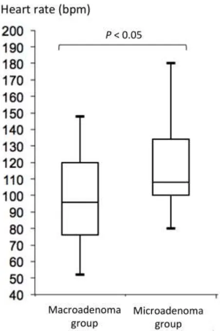
microadenoma group (Table 1). Dogs with macroadenomas had lower heart rates and lower body
temperatures than dogs with microadenomas (P<0.05 for both comparisons; Figure 1 and 2).
Plotting the heart rate against the body temperature resulted in more widespread dispersion of the
plot of the macroadenoma group compared to the microadenoma group (Figure 3).
The crude difference estimated by the univariate linear regression between the two groups
(macroadenoma versus microadenoma)was estimated to be – 0.52°C (p < 0.01) and –19 bpm (p
< 0.01) respectively for body temperature and heart rate. In both models, only the body weight
was identified as a potential confounder. After adjustment for body weight, the temperature
difference ∆ = – 0.65 °C and heart rate difference ∆ = – 18 bpm remained significant (p < 0.01
and p = 0.04).
The most discriminating thresholds for both parameters were determined from receiver
operating characteristic (ROC) curves (Table 2). Using these thresholds identified with ROC (84
bpm and 38.3°C), a greater proportion of dogs with macroadenomas had either a heart rate ≤ 84
with microadenomas had both a heart rate ≤84 bpm and a body temperature ≤ 38.3°C, whereas 11
dogs with macroadenomas exhibited both of these autonomic signs (Table 3).
No dogs with microadenomas exhibited neurological deficits. Similarly, 9/29 dogs with
macroadenomas exhibited no neurological deficits. In the remaining 20 dogs, 12 exhibited vague
neurological signs (i.e., only altered mentation and behavioral changes); 9 (75%) of these dogs
had a heart rate ≤ 84 bpm or a body temperature ≤ 38.3°C, suggesting autonomic nervous system
dysfunction. The remaining 8 dogs with macroadenomas presented with circling, cortical
blindness and partial seizures.
Our data show that the heart rate and body temperature are lower in dogs with
macroadenomas compared to dogs with microadenomas, and that dogs with hyperadrenocorticism
presenting with both a heart rate ≤ 84 bpm and a body temperature ≤ 38.3°C are very likely to
have macroadenomas responsible for their endocrine disease. However, the heart rate and the
body temperature had a low sensitivity, i.e., a third of dogs (34%) with a macroadenomas had a
heart rate > 84 bpm or a body temperature > 38.3°C.
Diencephalic lesions can cause hypothermia or hyperthermia in humans (Carmel 1985) and
this might occur in animals. Healthy, smaller-sized or older animals are more likely to develop
hypothermia (Lee 2017), therefore we tested and excluded age and bodyweight as potential
confounding factors. One possible cause for the difference in heart rate between the two groups
(Fitch et al, 1977). Another possible explanation would be a destruction of the hypothalamus by
the macroadenoma. Concurrent measurement of the blood pressure in our study dogs would have
helped to explore the possible causes of bradycardia, such as the Cushing’s reflex. One of the
potential weaknesses of the study is that there is an association between HR and BW in dogs
(Hezzel et al, 2013). Moreover, in our study, dogs with macroadenomas were larger. We included
body weight into our modelling as a potential confounding factor, but the difference in heart rate
remained significant after adjustment for body weight. Other limitations to this study include the
fluctuating nature of the heart rate and body temperature and the non-standardized method of
measurement of heart rate and body temperature. However, we sought to reproduce the routine
clinical situation where vital parameters are measured only once during a consultation. Therefore,
we purposely collected data only at initial presentation. Standardized methods of measurement of
both parameters as well as repetitive measures would require a prospective study.
Because the diagnostic value of these parameters will always remain limited in dogs with
clear forebrain neurological dysfunction, it would be of interest to evaluate the temperature and
heart rate of dogs with macroadenomas and hyperadrenocorticism, but without obvious
neurological signs. We had too few such dogs for meaningful comparison.
In summary, our data revealed that dogs with macroadenomas had a lower heart rates and
body temperatures than dogs with microadenomas. In the context of a case diagnosed with
hyperadrenocorticism, the finding of a heart rate ≤ 84 bpm or a body temperature ≤ 38.3°C should
Conflict of interest statement
The authors disclose no conflict of interest.
Acknowledgments
The authors would like to thank the referring veterinary surgeons of the dogs described in this
paper, the staff of the Queen’s Veterinary School Hospital and of the National Veterinary School
of Alfort and Dr Mathilde Duchaussoy-Granger DVM Dip.APhys MRCVS, for their help.
References
Behrend EN. Canine hyperadrenocorticism, 2015. In: Feldman EC, Nelson RW, Reusch CE,
Scott-Moncrieff JCR, editors. Canine and Feline Endocrinology. Saunders Elsevier, St.
Louis, MO, USA: 377-451.
Carmel PW, 1985. Vegetative dysfunctions of the hypothalamus. Acta Neurochir (Wien );
75(1-4):113-121.
de Lahunta A, Glass E, Kent M, 2015. Diencephalon. In: de Lahunta A, Glass E and Kent M,
editors. Veterinary neuroanatomy and clinical neurology. St Louis: Saunders: 499-508.
Fitch W, McDowall DG, Keaney NP, Pickerodt VW, 1977. Systemic vascular responses to
increased intracranial pressure. 2. The 'Cushing' response in the presence of intracranial
space-occupying lesions: systemic and cerebral haemodynamic studies in the dog and the
Granger N, de Fornel P, Devauchelle P, et al, 2005. Plasma opiomelanocortin,
pro-adrenocorticotropin hormone, and pituitary adenoma size in dogs with Cushing's disease. J
Vet Intern Med; 19(1):23-28.
Hezzell MJ1, Dennis SG, Humm K, et al, 2013. Relationships between heart rate and age,
bodyweight and breed in 10,849 dogs. J Small Anim Pract; 54(6):318-24.
Lee JA. Hypothermia, 2017. In: Ettinger SJ, Feldman EC, Côté E, editors. Textbook of Veterinary
Internal Medicine. Saunders Elsevier, St. Louis, MO, USA: 203.
Nelson RW, Ihle SL, Feldman EC, 1989. Pituitary macroadenomas and macroadenocarcinomas
in dogs treated with mitotane for pituitary-dependent hyperadrenocorticism: 13 cases
(1981-1986). J Am Vet Med Assoc; 194(11):1612-1617.
Verstraete A, Thoonen J, 1939. Twee nieuwe gevallen van hypophysaire stoornissen bij den hond.
Vlaams diergeneesk Tijdschr; 8:304-314.
Wood FD, Pollard RE, Uerling MR, Feldman EC, 2007. Diagnostic imaging findings and
endocrine test results in dogs with pituitary-dependent hyperadrenocorticism that did or did
not have neurologic abnormalities: 157 cases (1989-2005). J Am Vet Med Assoc;
Table 1
Signalment (body weight, age and sex) of dogs with macroadenomas and microadenomas.
Body weight (kg)
Median 25th Percentile
75th
Percentile Min Max
Macroadenoma 22.5 12.8 35 5 44
Microadenoma 7 5 12 3 27
Age (years)
Median 25th Percentile
75th
Percentile Min Max
Macroadenoma 9 8 12 5 15
Microadenoma 11 9 12 7 14
Male Female
Macroadenoma 23 6
Table 2
Results of the receiver operating characteristic curve analysis for body temperature (°C: degree
Celsius) and heart rate (bpm: beats per minute).
Area under the curve (95% CI) Youden index Associated criterion Sensitivity (95% CI) Specificity (95% CI)
Body Temperature (°C) 0.79
(0.66-0.88) 0.49 ≤ 38.3
0.62
(0.42 – 0.79)
0.86
(0.69 – 0.96)
Heart rate (bpm) 0.70
(0.57-0.81) 0.38 ≤ 84
0.41
(0.23 – 0.61)
0.97
Table 3: Pituitary height/brain area (P/B) ratio, neurological signs, body temperature (°C: degree
P/B ratio Body temperature Heart rate Heart rate and body temperature
Median Range Median Range
Dogs with T ≤ 38.3°C Number (%)
Median Range
Dogs with HR ≤ 84 bpm Number (%)
Dogs with T ≤ 38.3°C or HR ≤
84 bpm Number (%)
Dogs with T ≤ 38.3°C and HR ≤
84 bpm Number (%)
Macroadenoma group
N= 29 dogs 1 0.41-1.71 38.3 37-39.2 18 (59%) 96 52-148 12 (41%) 19 (66%) 11 (38%)
No neurological sign
N= 9 dogs 0.6 0.41-1.35 38.5 37.6-39 4 (44%) 92 72-140 4 (44%) 4 (44%) 4 (44%)
Vague neurological signs (altered mentation and behavioural changes) N= 12 dogs
0.8 0.45-1.28 38.2 37.7-39.2 8 (67%) 93 60-120 5 (42%) 9 (75%) 4 (33%)
Evident neurological signs (circling, cortical blindness, partial seizures) N = 8 dogs
1 0.58-1.71 38.3 37-39.1 6 (75%) 98 52-148 3 (38%) 6 (75%) 3 (38%)
Microadenoma group
Figure legends
Figure 1: Box plot of body temperature in 29 dogs with macroadenomas and 30 dogs with
microadenomas. The box represents interquartile range (25th to 75th percentile). The bars represent
Figure 2: Box plot of heart rate in 29 dogs with macroadenomas and 30 dogs with
Figure 3: Plot of the heart rate (x-axis) against the body temperature (y-axis) for dogs with
macroadenoma (solid circle) and dogs with microadenomas (open circle). Dogs in the
microadenoma group are mainly located in the upper right quadrant. Note the dispersion of the
data in the macroadenoma group towards the left with 12 dogs with a HR ≤ 84 bpm and 18 dogs
with a BT ≤ 38.3°C but only one dog in the microadenoma group with a HR ≤ 84 bpm and 4 with
a BT ≤ 38.3°C. Dashed lines on both plots represent the cut off value found using ROC curves