_____________________________________________________________________________________________________
*Corresponding author: E-mail: [email protected];
Foreign Body Impacted in the Submassetric
Region-A Case Report
Chaitra Patil
1*, Narasimhamurthy Srinath
1,
Doddarayapete Narayanaswamy Umashankar
1and Mahesh Kumar
11Department of Maxillofacial Surgery, Krishnadevaraya College of Dental Sciences and Hospital,
Bangalore, India.
Authors’ contributions
This work was carried out in collaboration between all authors. All authors read and approved the final manuscript.
Article Information
DOI: 10.9734/AJCRS/2018/41857
Editor(s):
(1)Dr. Ivan Felismino Charas dos Santos, University Estadual Paulista "Júlio de Mesquita Filho", Brazil.
Reviewers:
(1) Lauritano Dorina, University of Milan-Bicocca, Italy. (2)V. Swathi, Rajiv Gandhi University of Health Sciences, India. (3)F. Armando Montesinos, National Autonomous University of Mexico, Mexico. Complete Peer review History:http://www.sciencedomain.org/review-history/25292
Received 2nd May 2018 Accepted 10th June 2018 Published 28th June 2018
ABSTRACT
A foreign body is an object lying partially or completely within the body that originated from the external environment. Foreign bodies are generally encountered in the orofacial region following trauma or iatrogenic procedures. If untreated can lead to serious complications like pain, swelling and infection. Here is a case report of a retained foreign body in the orofacial region of 32-year-old male patient. This paper highlights the problems associated with diagnosis, localising and managing unlikely foreign bodies at unusual facial sites.
Keywords: Foreign bodies; cellulitis; swelling; crepitation.
1. INTRODUCTION
Foreign bodies are often found in facial wounds but rarely reported in the literature [1]. Some authors believe that the head and neck region is most frequently affected by trauma and facial involvement is very common due to the exposure of face [2]. The foreign bodies encountered in the orofacial region are commonly associated with morbidity and mortality. The foreign bodies usually are the result of trauma or iatrogenic procedures. Most commonly found foreign bodies in the orofacial region are metallic
objects, restorative materials, obturation
materials, wooden pieces, glass pieces, broken instruments, needles, etc [3]. These foreign bodies may be challenging to surgeon due to their size, accessibility, proximity to the vital structures. Diagnoses of foreign bodies are often made accidentally on radiographic examination or may be due to the symptoms associated with it. Their identification and removal from the tissue are often necessary. Prompt diagnosis and surgical removal of such foreign bodies will greatly minimize the associated complications which may include; allergic reactions, cellulitis, abscess, necrotizing fasciitis and osteomyelitis.
2. CASE REPORT
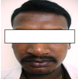
A 32-year-old male reported to the department of oral and maxillofacial surgery Krishnadevaraya College of dental science and hospital Bangalore with a chief complaint of pain and swelling in the lower left back region of the face since 8 days. The patient gave a history of trauma 14 years back in the left lower posterior region of the face following which he fell on a glass bottle in the same region. He was taken to a nearby hospital where he got the primary treatment for the same. On inspection, there was a diffuse swelling in the left posterior mandibular ramus region. There was a linear scar measuring approximately 2-3 cm in the same region since 10 years. On palpation, the swelling was tender and firm in consistency, with crepitation.
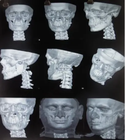
A plain radiograph (PA mandible Fig. 5) was requested and it revealed a small radio-opaque mass on the lower left ramus region measuring about 2-3 mm. For further detailed picture patient was advised to get a CT-scan with 3D reconstruction (Fig. 6) which revealed two well defined foreign objects in the same region. The patient was not aware of the foreign body in the maxillofacial region.
Fig. 1. Frontal view showing mild swelling on the lower faceregion
Fig. 2. Profile view showing a scar on the lower left face region
Fig. 3. Glass pieces were located in the submassetric region
Fig. 5. PA mandible view showing a radiopaque Mass on the left side of ramus
region
Fig. 6. 3D CT scan showing two foreign bodies in the left submassetric region
The patient was admitted to the ward for surgical removal of the foreign bodies under general anaesthesia. Standard skin preparation was done, a left mandibular vestibular incision was
given in the 3rd molar region extending up to the
anterior border of the ramus. Full-thickness mucoperiosteal flap was reflected and the foreign bodies were located in the submassetric region, deep in the masseter muscle. Masseter muscle was reflected from the lateral surface of the ramus. The two glass pieces were successfully
retrieved through intra-oral approach. Thorough debridement was done and hemostasis was achieved. Patient had an Uneventful recovery
and was discharged after 24 hours
postoperatively.
3. DISCUSSION
Incorporation of the Foreign materials in the body can be deliberate or accidental. The diagnosis and early detection of foreign bodies are usually
based on the patient’s history, clinical
examination and the various radiological imaging methods such as the plain radiographs, computed tomography, magnetic resonance imaging and ultrasound [4]. Foreign bodies possess a great potential for late complications
like pain, swelling, cellulitis, abscess,
osteomyelitis.
Initial evaluation of patients with skin puncture wounds should be completed with a high suspicion for a foreign body. Patients also present for evaluation several months or even years after the initial injury, and consequently, the clinical evaluation may fail to elicit a history of antecedent skin puncture.
Surgical removal of FB is important because it may serve as unrecognized foci of infection. Superficial foreign bodies are usually easy to remove if seen. However, penetrating foreign bodies are more difficult to remove. The accurate localization is essential, in particular when the foreign body is in a critical location, it may be located in an air-filled cavity such as the maxillary sinus, in soft tissue such as the tongue or between bone and muscle.
Various imaging modalities like conventional plain radiographs, CT, MRI & ultrasonography are used to detect foreign bodies. Conventional plain radiography is usually the preferred imaging
method for detecting foreign bodies.
Conventional plain radiographs can determine a foreign body’s position and help radiologists to determine whether the object is in a critical location or not. Although it is used frequently, additional imaging modalities may be needed for exact location [5].
However, metallic artefacts are an important source of error when detecting foreign bodies with CT imaging. If a foreign body’s composition is initially unknown, MRI cannot be used as the first diagnostic tool, because artefacts related to the foreign body’s composition hinder the clear demonstration of iron, glass, graphite and even plastic [7].
Ultrasonography might be useful for locating superficial foreign bodies;however, it might be unsuitable for those located deep and inside the air-filled cavities [8].
CT can be used to detect deeply seated foreign bodies because it reproduces accurate location,
position, size, and shape of them [5].Therefore,
some authors have suggested that CT is the standard imaging technique for observing foreign
bodies [6].Thus of all the imaging modalities in
disposal to a craniofacial surgeon CT remains the less expensive and more readily available and faster to localize a foreign bodies.
Superficial located foreign body in the
craniofacial region can be removed under local anaesthesia. However deeper FB is preferentially removed under GA. Surgical access to the FB can be achieved through the existing skin laceration or in deeply placed FB can be accessed by intra-oral or extraoral incisions.
Selection of the antibiotics as prophylaxis for the surgical retrieval will depend on its location and communications with oral cavity, nasal cavity and proximity to the meninges. Foreign bodies in orbit generally have higher morbidity than other sites, requiring more aggressive medical management.
4. CONCLUSION
In conclusion, the following factors should be considered in the management of FB.
Foreign bodies can be detected with plain
radiography, CT scans, MRI and
ultrasonography. Among all the imaging
techniques CT is the gold standard for visualization of foreign bodies. Access to the foreign bodies depends on its location and surgical access can be gained through intra-oral or extra-oral approach. If there is an existing scar access can also be gained through it. Thorough debridement of the wound with proper irrigation should be carried out followed by closure.
Routine postoperative screening and
radiography should be done.
CONSENT
As per international standard or university standard, patient’s written consent has been collected and preserved by the authors.
ETHICAL APPROVAL
As per international standard or university standard written ethical approval has been collected and preserved by the authors.
COMPETING INTERESTS
Authors have declared that no competing interests exist.
REFERENCES
1. Cavalcante WC, Coelho HA, Neto AIT,
Santos LCS, Carvalho MC. Corpo
estranho na intimidade dos ossos da face: Relato de caso. Revista de Cirurgia e Traumatologia BucoMaxilo-Facial. 2010; 10:97–102.
2. Cohen MA, Boyes-Varley G. Penetrating
injuries to the maxillofacial region. Journal of Oral and Maxillofacial Surgery. 1986; 44(3):197–202.
3. Ghom A, Gupta M, Khatri P, Khandewal A,
Debta FM. Foriegn bodies in the oral and maxillofacial region: Report of two cases. Journal of Indian Academy of Oral Medicine and Radiology. 2011;23(4):630-632.
4. Aregbesola SB, Ugboko V. Unusual
foreign bodies in the orofacial soft tissue spaces. A report of three cases. Nigerian
Journal of Clinical Practice. 2013;
16(3):381-385.
5. Eggers G, Welzel T, Mukhamadiev D,
Wortche R, Hassfeld S, Muhling J. X-ray-based volumetric imaging of foreign
bodies: A comparison of computed
tomography and digital volume
tomography. J Oral Maxillofac Surg. 2007; 65:1880–1885.
6. Eggers G, Mukhamadiev D, Hassfeld S.
Detection of foreign bodies of the head with digital volume tomography. Dentomaxillofac Radiol. 2005;34:74–79.
7. Lagalla R, Manfre L, Caronia A,
an in vitro model of the orbit and in pig eyes. Eur Radiol. 2000;10:1338–1341.
8. Stockmann P, Vairaktaris E, Fenner M,
Tudor C, Neukam FW, Nkenke E.
Conventional radiographs: Are they still the standard inlocalization of projectiles? Oral Surg Oral Med Oral Pathol Oral Radiol Endod. 2007;104:71–75.
_________________________________________________________________________________ © 2018 Patil et al.; This is an Open Access article distributed under the terms of the Creative Commons Attribution License (http://creativecommons.org/licenses/by/4.0), which permits unrestricted use, distribution, and reproduction in any medium, provided the original work is properly cited.
Peer-review history: