Case Report
Pulmonary Silicosis Presents with Pleural Effusion
Mohsin Salih,
1Tarake Aljarod,
2Mohamed Ayan,
3Melnick Jeffrey,
1and Bobby H. Shah
11Internal Medicine Department, St. Luke’s Hospital, Chesterfield, MO 63017, USA 2Internal Medicine Department, Morehouse School of Medicine, Atlanta, GA 30310, USA 3Internal Medicine, Creighton University Medical Center, Omaha, NE 68102, USA
Correspondence should be addressed to Mohamed Ayan; [email protected]
Received 18 June 2015; Revised 20 August 2015; Accepted 20 August 2015
Academic Editor: Reda E. Girgis
Copyright © 2015 Mohsin Salih et al. This is an open access article distributed under the Creative Commons Attribution License, which permits unrestricted use, distribution, and reproduction in any medium, provided the original work is properly cited.
Silica and silicate mineral dust inhalation can cause a variety of histopathological changes in the lungs and pleura. These include pulmonary silicotic nodules, interstitial infiltrate, fibrosis, and pleural thickening. Pleural effusion is an extremely rare presentation of silicosis. To our best knowledge, there have been only 2 cases of silicosis with pleural effusion reported in medical literature. Herein, we describe a case of a 77-year-old male with almost 50 years’ history of occupational silica exposure. He presented with a 4-week history of exertional shortness of breath. He is a lifetime nonsmoker, with no known other significant pulmonary disease. He had chest X-ray which showed a right lung infiltrate and bilateral pleural thickening and effusion. Chest CT showed moderate-sized bilateral pleural effusion and thickening with multiple bilateral intrapulmonary nodules seen. He had undergone extensive workup and was diagnosed with silicosis.
1. Introduction
Various pleural involvements such as pleural thickening and progressive multifocal fibrosis (PMF) associated pleural invaginations are well-recognized complications associated with silicosis, particularly advanced pulmonary silicosis. However, pleural effusion is not a well-recognized finding in patients with silicosis. To the best of our knowledge, there have been only 2 cases reported in the medical literature that described pulmonary silicosis presented with pleural effu-sion. Herein, we describe a case of a 77-year-old gentleman who presented with shortness of breath and bilateral pleural effusion. The patient had undergone extensive workup and was diagnosed with pulmonary silicosis.
2. Case Presentation
A 77-year-old gentleman with history of coronary artery disease, diabetes mellitus, and hypertension presented to the emergency department with worsening shortness of breath. He reported that he has been having some chronic baseline dyspnea on exertion which has been stable until 4-5 days prior to his presentation when it started to get worse with
simple tasks such as getting dressed and showering. He denied any cough, sputum production, hemoptysis, paroxys-mal nocturnal dyspnea, orthopnea, leg swelling, fever, chills, weight loss, or night sweating. He is a lifetime nonsmoker, with no other significant pulmonary history other than his occupational exposure. He had worked for at least 50 to 60 years carving and grinding stones, including granite and numerous other stones. Although he has been wearing a respirator for a large part of his more adult life, there was quite some time, including numerous years, where he wore no respiratory protection whatsoever. He has no tubercular risk factors other than silica exposure. There was no history of TB exposure. His past medical history is notable for coronary artery disease, diabetes, chronic kidney disease, and hypertension. There was no family history of lung cancer. On examination, he was afebrile and tachypneic with respiratory rate of 30. His oxygen saturation was 95% on room air at rest. On chest exam, he had signs of bilateral pleural effusion. Pertinent lab studies showed WBC count of 9 k/𝜇L, creatinine of 2.8 mg/dL, albumin of 3.6 g/dL, total protein of 7.4 g/dL, and BNP of 216, and the arterial blood gas was normal. He had chest X-ray which showed a right lung infiltrate and moderate right pleural effusion, small left pleural effusion, bilateral
2 Case Reports in Medicine
Figure 1: Chest X-ray showed a right lung infiltrate and moderate right pleural effusion, small left pleural effusion, bilateral small irregular opacities, and pleural thickening.
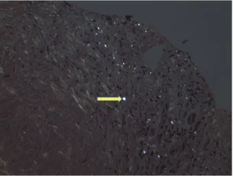
small irregular opacities, and pleural thickening (Figure 1). Chest CT showed moderate-sized bilateral pleural effusion and thickening with pulmonary nodules. There is mild interstitial fibrosis. Some of the nodules are pleural-based (Figure 2). A few nodules do have a small amount of asso-ciated calcification. The largest nodule measures 6 mm. No progressive massive fibrosis (PMF) is defined as the presence of pneumoconiotic nodules more than 2 cm in diameter on CT image. Transthoracic echocardiography showed normal ejection fraction of 60–65% and no significant valvular lesion. He subsequently had a thoracentesis. The pleural fluid was clearly exudative with LDH effusion/LDH serum ratio of 2.15 and pleural fluid LDH of 1205 IU/L. The pleural fluid analysis showed also pH of 7.17, RBC count of 60,000/microL, and WBC count of 871/microL which were mainly lymphocytes. Flow cytometry did not show any evidence of lymphoma. The pleural fluid Gram staining was negative for any bacte-ria. Histoplasma antigen and histoplasma immunodiffusion were negative. The PDD test was negative. Cultures of the pleural fluid and BAL were unremarkable for any bacteria, TB, or fungi. He had video-assisted lung biopsy, chest tube placement, and mechanical pleurodesis. Lung biopsy (thoracoscopic wedge resection) showed a silicotic nodule within lung parenchyma composed mainly of bundles of interlacing collagen. There is minimal inflammatory reaction (Figure 3). Lung biopsy under polarized microscopy showed bright white silica crystals of varying sizes (Figure 4). Pleural biopsy showed fibrinous exudate with organizing fibrosis and mild acute and chronic inflammation and hyperplasia of mesothelial cells. Pleural biopsy culture for tuberculosis was negative.
3. Discussion
Silicosis is an occupational lung disease caused by inhalation of free crystalline silicon dioxide or silica. The pulmonary silicosis is a well-known occupational lung disease caused by silica inhalation; however, new cases of pulmonary silicosis are still seen in clinical practice. Phagocytosis of crys-talline silica in the lung causes lysosomal damage, activating
Figure 2: Chest CT showed moderate-sized bilateral pleural effu-sion and thickening with pulmonary nodules. There is mild inter-stitial fibrosis. There are multiple bilateral intrapulmonary nodules seen. Some of the nodules are pleural-based. A few nodules do have a small amount of associated calcification.
Figure 3: Lung biopsy showed a silicotic nodule within lung paren-chyma composed mainly of bundles of interlacing collagen. There is minimal inflammatory reaction.
Figure 4: Lung biopsy underpolarized microscopy showing bright white silica crystals of varying sizes.
the NALP3 inflammasome and triggering the inflammatory cascade with subsequent fibrosis [1].
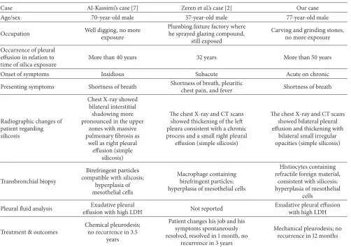
Table 1: Summary of the reported cases of silicosis presenting with pleural effusion.
Case Al-Kassimi’s case [7] Zeren et al.’s case [2] Our case
Age/sex 70-year-old male 57-year-old male 77-year-old male
Occupation Well digging, no more
exposure
Plumbing fixture factory where he sprayed glazing compound,
still exposed
Carving and grinding stones, no more exposure
Occurrence of pleural effusion in relation to time of silica exposure
More than 40 years 32 years More than 50 years
Onset of symptoms Insidious Subacute Acute on chronic
Presenting symptoms Shortness of breath Shortness of breath, pleuritic
chest pain, and fever Shortness of breath
Radiographic changes of patient regarding silicosis
Chest X-ray showed bilateral interstitial shadowing more pronounced in the upper
zones with massive pulmonary fibrosis as
well as right pleural effusion (simple
silicosis)
The chest X-ray and CT scans showed thickening of the left pleura consistent with a chronic process and a small right pleural
effusion (simple silicosis)
The chest X-ray and CT scans showed bilateral pleural effusion and thickening with
bilateral small irregular opacities (simple silicosis)
Transbronchial biopsy
Birefringent particles compatible with silicosis;
hyperplasia of mesothelial cells
Macrophage containing birefringent particles; hyperplasia of mesothelial cells
Histiocytes containing refractile foreign material,
consistent with silicosis; hyperplasia of mesothelial
cells
Pleural fluid analysis Exudative pleural
effusion with high LDH Not reported
Exudative pleural effusion with high LDH
Treatment & outcomes
Chemical pleurodesis; no recurrence in 3.5
years
Patient changes his job and his symptoms spontaneously resolved, resolved in 1 month, no
recurrence in 3 years
Mechanical pleurodesis; no recurrence in 12 months
[2, 3]. Extrapulmonary involvement of multiple organs was reported including liver, kidney, spleen, hilar and extratho-racic lymph nodes, and tonsillar and peritoneal involvement. Tuberculosis is a well-established complication of silicosis. Silicosis was also found to be associated with some chronic kidney diseases [4], rheumatoid arthritis, systemic sclerosis, and a human carcinogen [5, 6].
Diagnosis of silicosis generally relies on 3 key elements: a history of substantial exposure to silica dusts with appropriate latency from the time of first exposure, compatible radio-logical features, and exclusion of other competing diagnoses, such as miliary tuberculosis, fungal infections, sarcoidosis, idiopathic pulmonary fibrosis, other interstitial lung diseases, and primary or secondary lung cancers [1]. Several pul-monary clinical presentations have been described including acute, accelerated, and chronic silicosis. The onset of the pre-senting symptoms in relation to the first exposure to silica and the radiological features play an important role differentiating the above-mentioned pulmonary involvements.
As mentioned above, pleural involvement in silicosis is well recognized especially in advanced pulmonary silicosis. However, pleural silicosis is still less common and less empha-sized in medical literature than asbestosis as an occupational lung disease that is associated with pleural plaque, pleural
effusion, and diffuse pleural thickening [7]. The pleural disease in silicosis has not, to our knowledge, been extensively described in the literature as a possible underlying cause for pleural effusion in patients with significant occupational exposure to silica or silicates. After doing a thorough medical literature review, with the exception of 2 case reports and 1 retrospective study that evaluated the extent of pleural dis-eases in autopsy-proven silicosis, we did not find any review articles or online resources that mentioned pleural effusion as one of the clinical manifestations with which silicosis patients can present (Table 1) [2, 7]. Al-Kassimi reported a case of advanced pulmonary silicosis who was found to have pleural effusion upon presentation. The pleural biopsy was significant for mesothelial hyperplasia [7]. Zeren et al. also reported unusual presentation of silica-associated pleural effusion and pleuritis indicating preferential involvement of the pleura. Like our case, the pleural biopsy reported by Zeren et al. showed pleural thickening and fibrosis. However, it was different from our case and the case reported by Al-Kassimi because there was no lung involvement at both radiological and pathological levels.
4 Case Reports in Medicine
the patients studied. Sixty-eight percent (26 of 38) of the patients with pleural effusion had possible/probable cause for their effusion that was not silicosis (pneumonia, cardiac failure, hypoalbuminemia, pneumothorax, and malignancy) and only 11% of the silicosis patients had pleural effusion that is thought to be related to silicosis (no other causes have been found) [8]. In any case of silicosis, based on this case series, the majority of pleural effusions associated with silicosis will be due to a cause other than silicosis. In addition to pleural effusion, Arakawa et al. described some other pleural diseases in silicosis such as pleural thickening and PMF associated pleural invagination. In advanced silicosis, the pleural surface is thickened and is fibrotic with typical silicotic nodules and plaques. However, it was a retrospective review of medical records and so there was no uniform approach to work up the causes of the effusion.
This patient met diagnostic criteria for pulmonary sili-cosis mentioned above in terms of his job-related prolonged exposure to silica, bilateral pulmonary nodules on both CXR and CT scan, and absence of other alternative conditions such as primary or secondary malignancies, TB, or fungal infections. This patient likely had chronic silicosis given his remote history of silica exposure and his chronic history of dyspnea. Probably, the moderate-sized pleural effusion is what makes his shortness of breath worsen and present in acute pattern as we ruled out all possible infectious causes.
4. Conclusion
Different pleural disease can occur in silicosis particularly in advanced stages. Although pleural effusion is unusual in silicosis it is a possible cause if infectious, malignant, cardiovascular, renal, and connective tissue disease causes have been ruled out.
Statement of Human Rights
This paper does not contain any studies with human partici-pants performed by any of the authors.
Conflict of Interests
The authors declare that they have no relevant conflict of interests for this case.
Acknowledgment
The authors would like to thank Dr. Melnick Jeffrey from the radiology department for helping them acquire appropriate images.
References
[1] C. C. Leung, I. T. S. Yu, and W. Chen, “Silicosis,”The Lancet, vol. 379, no. 9830, pp. 2008–2018, 2012.
[2] E. H. Zeren, T. V. Colby, and V. L. Roggli, “Silica-induced pleural disease: an unusual case mimicking malignant mesothelioma,”
Chest, vol. 112, no. 5, pp. 1436–1438, 1997.
[3] J. E. Craighead, J. Kleinerman, J. Z. Abraham et al., “Diseases associated with exposure to silica and nonfibrous silicate min-erals,”Archives of Pathology and Laboratory Medicine, vol. 112, no. 7, pp. 673–720, 1988.
[4] National Institute for Occupational Safety and Health,Health Effects of Occupational Exposure to Respirable Crystalline Silica, Department of Health and Human Services, Cincinnati, Ohio, USA, 2002.
[5] A. Makol, M. J. Reilly, and K. D. Rosenman, “Prevalence of connective tissue disease in silicosis (1985–2006)—a report from the state of michigan surveillance system for silicosis,”The American Journal of Industrial Medicine, vol. 54, no. 4, pp. 255– 262, 2011.
[6] National Toxicity Program, Ed.,Report on Carcinogens, Depart-ment of Health and Human Services, Public Health Ser-vice, Research Triangle Park, NC, USA, 11th edition, 2005, http://ntp.niehs.nih.gov/pubhealth/roc/roc13/index.html. [7] F. A. Al-Kassimi, “Pleural effusion in silicosis of the lung,”
British Journal of Industrial Medicine, vol. 49, no. 6, pp. 448– 450, 1992.
Submit your manuscripts at
http://www.hindawi.com
Stem Cells
International
Hindawi Publishing Corporationhttp://www.hindawi.com Volume 2014
Hindawi Publishing Corporation
http://www.hindawi.com Volume 2014
INFLAMMATION
Hindawi Publishing Corporation
http://www.hindawi.com Volume 2014
Behavioural
Neurology
Endocrinology
International Journal ofHindawi Publishing Corporation
http://www.hindawi.com Volume 2014 Hindawi Publishing Corporation
http://www.hindawi.com Volume 2014
Disease Markers
Hindawi Publishing Corporation
http://www.hindawi.com Volume 2014
BioMed
Research International
Oncology
Journal ofHindawi Publishing Corporation
http://www.hindawi.com Volume 2014
Hindawi Publishing Corporation
http://www.hindawi.com Volume 2014
Oxidative Medicine and Cellular Longevity
Hindawi Publishing Corporation
http://www.hindawi.com Volume 2014
PPAR Research
The Scientific
World Journal
Hindawi Publishing Corporation
http://www.hindawi.com Volume 2014
Immunology Research
Hindawi Publishing Corporation
http://www.hindawi.com Volume 2014
Journal of
Obesity
Journal ofHindawi Publishing Corporation
http://www.hindawi.com Volume 2014
Hindawi Publishing Corporation
http://www.hindawi.com Volume 2014
Computational and Mathematical Methods in Medicine
Ophthalmology
Journal ofHindawi Publishing Corporation
http://www.hindawi.com Volume 2014
Diabetes Research
Journal ofHindawi Publishing Corporation
http://www.hindawi.com Volume 2014
Hindawi Publishing Corporation
http://www.hindawi.com Volume 2014
Research and Treatment
AIDS
Hindawi Publishing Corporation
http://www.hindawi.com Volume 2014
Gastroenterology Research and Practice
Hindawi Publishing Corporation
http://www.hindawi.com Volume 2014
Parkinson’s
Disease
Evidence-Based Complementary and Alternative Medicine
Volume 2014 Hindawi Publishing Corporation