Nirjhar Chatterjee et al JMSCR Volume 08 Issue 08 August 2020 Page 157
Research Paper
Neonatal Sepsis: Role of Inerleukin-6 and Tumour Necrosis Factor-α in
Rapid Diagnosis and Its Comparison with Automated Blood Culture
Authors
Nirjhar Chatterjee
1*, Anuradha De
2, Sushma Malik
3, Jayanthi Shastri
41
Assistant Professor, Department of Microbiology, TNMC & Nair Hospital Mumbai
2
Professor, Department of Microbiology, TNMC & Nair Hospital Mumbai
3
Professor and Head, Department of Pediatrics, TNMC & Nair Hospital Mumbai
4
Professor and Head, Department of Microbiology TNMC & Nair Hospital Mumbai *Corresponding Author
Nirjhar Chatterjee Abstract
Context: Neonatal sepsis is a clinical syndrome of bacteraemia characterized by systemic signs and symptoms of infection in the first month of life. Blood culture is the mainstay of diagnosis but it has a long turnaround time and a delay in instituting therapy affects outcome. Taking this into perspective, the present study was undertaken to find out the importance of two inflammatory markers in the rapid diagnosis of neonatal sepsis.
Aim: To compare the utility of Interleukin-6 and Tumour necrosis factor-α with automated blood culture in rapid diagnosis of neonatal sepsis.
Settings and Design: A prospective cross-sectional study of one year (from September 2017 to August 2018) was carried out in Neonatal Intensive Care Unit of this institute.
Methods and Material: Sixty neonates with clinically suspected sepsis were included. Blood cultures were processed in BACTEC 9120 system. Enzyme Linked Immunosorbent Assay was performed for IL-6 and TNF-α. Values were compared to blood culture positivity.
Statistical Analysis:Sensitivity, Specificity, Mean, Student unpaired T test, Chi square test and p- values
Results: In this study, sensitivity and negative predictive value of both IL-6 and TNF-α was 100%. Statistically IL-6 was shown to be a better marker than TNF-α in the rapid diagnosis of neonatal sepsis, both in early- and late-onset. IL-6 and TNF-α jointly showed a high sensitivity and were comparable in specificity to blood culture, as compared to any of these two markers alone.
Conclusions: This study highlights the importance of IL-6 and TNF-α estimation for rapid diagnosis of neonatal sepsis, enabling effective management and better prognosis.
Keywords:Neonatal sepsis; Interleukin-6; Tumour necrosis factor-α.
Introduction
Neonatal sepsis is a clinical syndrome of bacteraemia characterized by systemic signs and symptoms of infection in the first month of life.[1] The underdeveloped immune system predisposes
preterm newborns to infection which rapidly evolve into generalized sepsis. Neonatal sepsis can be divided into two main classes depending on the onset of symptoms related to sepsis - Early onset sepsis (EOS) within the first 72 hours of life and
http://jmscr.igmpublication.org/home/ ISSN (e)-2347-176x ISSN (p) 2455-0450
Nirjhar Chatterjee et al JMSCR Volume 08 Issue 08 August 2020 Page 158
Late onset sepsis (LOS) after 72 hours.[2] As per National Neonatal Perinatal Database (2002-03),[3] the comparative incidence of EOS versus LOS in India was 67% : 31.6%.
Blood culture is the gold standard for its diagnosis. Drawbacks of blood culture are that culture reports are time-consuming (available only after 48-72 hours) and has unacceptably low sensitivity.[2] Other laboratory tests (CRP, micro-ESR, etc.) lack sensitivity or specificity. So the neonates with clinical suspicion of infection are empirically treated with antibiotics. To avoid unnecessary treatment of non-infected neonates, an early, rapid, sensitive and specific laboratory test would be helpful to guide clinicians in neonatal units to decide whether or not to start antibiotics.
In sepsis, cytokines are involved in activating vascular and endothelial defence mechanisms, which changes perfusion and vascular resistance in microcirculation.[4]
Though certain studies are done abroad on the role of interleukins, tumour necrosis factor-α and other biomarkers in the diagnosis of neonatal sepsis([5],[6]) there are few documented studies from India.[7] Lack of information on Indian population validated the need of this study.
Aim and Objectives
1. To find out an appropriate and accurate rapid diagnostic test for neonatal sepsis. 2. To compare the utility of Interleukin-6
and/or Tumour necrosis factor-α with automated blood culture in rapid diagnosis of neonatal sepsis.
Subjects and Methods
This prospective cross-sectional study was carried out in the Neonatal Intensive Care Unit (NICU) of this institute for one year (from September 2017 to August 2018).
Total sixty neonates, with the assent of their parents, admitted in NICU of this hospital with clinical suspicion of sepsis, with any two of four blood parameters i.e. increased blood count > 11000/dl or decreased blood count < 5000/dl, neutropenia in
absolute neutrophil count < 1000/dl, C-reactive protein > 5mg/dl, Micro Erythrocyte sedimentation rate of > postnatal day + 3mm, were included in the study.([2],[6],[8]) The neonates who were already on antibiotics and immunocompromised (i.e. neonate whose parents are HIV positive, neonate with leucopoenia / aplastic anaemia / bone marrow aplasia or dysplasia, neonate whose mother was diagnosed of TORCH infection during pregnancy or at the time of delivery), were excluded from the study.
At or around the time of admission, detailed history of these neonates and provisional diagnosis were recorded in a structured questionnaire. Two ml blood was collected in the BACTEC Peds Plus bottle from all these neonates and processed in BACTEC 9120 automated blood culture system for blood culture in the Microbiology Laboratory of this institute. Flash positive bottles were subcultured on Blood agar and MacConkey agar plates and Sabouraud’s dextrose agar slants without antibiotics to confirm growth. Additional 2 ml blood that was collected in plain vacutainers was rotated in 3500 rpm for 4 minutes to separate the serum and it was kept at -20°C.
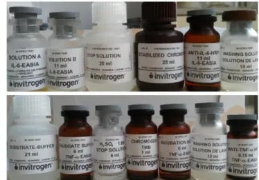
Enzyme Linked Immunosorbent Assay (ELISA) was performed with the serum sample for IL-6 by KAC1261 kit and TNF-α by KAC1751 kit (both marketed by Thermo Fisher Scientific, Waltham, MA, USA and manufactured by BioSource Europe S.A. Rue de l'Industrie, Nivelles, Belgium) (Figure 1).
Nirjhar Chatterjee et al JMSCR Volume 08 Issue 08 August 2020 Page 159
A standard curve was constructed using all standard points for which absorbances were below the limit of linearity of reader used. The OD was plotted on the ordinate against the standard concentrations on the abscissa using linear graph paper and the curve was drawn by connecting the plotted points with straight lines. IL-6 and TNF-α concentrations of samples or controls were determined for which absorbance is no greater than those of the last standard plotted at 450 nm. The minimum normal level of IL-6 and TNF-α as per kit literature was 3-8.5 pg/ml and 6pg/ml respectively. As kit literature cut off is not standardized in Indian population, so in this study, cut off value of IL-6 and TNF-α negative was taken as 331.77 pg/ml and 65.83 pg/ml respectively (mean value for culture negative neonates). Both IL-6 and TNF-α were compared with the automated blood culture to identify their utility in the diagnosis of neonatal sepsis.
As for statistical analysis, continuous variables were summarized by using summary statistics (number of observations, mean and standard deviation). Categorical values were summarized by using frequencies and percentages. Comparison was made using Student Unpaired t test, Chi square test and/or Fisher Exact Test according to the applicability. Sensitivity, specificity, positive predictive value (PPV) and negative predictive value (NPV) were calculated by using appropriate formulas. All p-values were reported based on two-sided significance test and all the statistical tests were interpreted at 5% level of significance.
Results
A total of 60 clinically suspected cases of neonatal septicaemia fulfilling the inclusion criteria were included. Out of 60 patients, males predominated (65%), with Male:Female 1.86:1. Neonates were distributed according to onset of sepsis. Maximum 61.67% (37/60) was EOS, rest (38.33%) LOS. Blood culture positivity was compared and it was noted that of all the blood cultures, 41.67% (25/60) showed growth. Out of which maximum (64%, 16/25) were yeast, 24% (6/25) were Gram negative
bacilli (GNB) and 12% (3/25) were gram positive cocci (GPC).
Out of 60 neonates, in 46.67% cases, value of IL-6 was ≥ 1001 pg/ml, in 5% cases between 501-1000 pg/ml and in 48.33% ≤500 pg/ml. Mean value of IL-6 was 1491.00 ± 330.57 pg/ml (range 552 - 1756 pg/ml) among culture positive cases, which was highly significant (p value = 0.001), as compared to culture negative neonates (331.78 ± 79.48 pg/ml, range 14 - 1438 pg/ml). Mean value of IL-6 was 961.00 ± 160.77 pg/ml among LOS, which was highly significant (p value = 0.001) as compared to 739.01 ± 113.42 pg/ml among EOS cases.
Out of all neonates, in 16.67% cases, value of TNF-α was ≥ 1001 pg/ml, in 15% cases between 501-1000 pg/ml and in 68.33%, ≤500 pg/ml. Mean value of TNF-α was 845.20 ± 61.49 pg/ml; range 314 - 1148 pg/ml among culture positives, which was highly significant (p value=0.001) as compared to 65.83 ± 6.75 pg/ml (range 18 - 186 pg/ml) among culture negatives. Mean value of TNF-α was 582.00 ± 97.68 pg/ml among LOS, which was highly significant (p value=0.001) as compared to 271.57 ± 61.19 pg/ml among EOS.
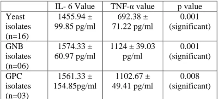
Comparison of mean IL-6 value with mean TNF- α value in different types of organisms isolated is depicted in Table 1.
Table 1: Comparison of Mean IL-6 Value with Mean TNF- α Value in Different Types of Isolates
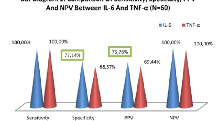
Sensitivity, specificity, PPV and NPV of IL-6 and TNF-α are compared in Bar diagram 1.
IL- 6 Value TNF-α value p value Yeast
isolates (n=16)
1455.94 ± 99.85 pg/ml
692.38 ± 71.22 pg/ml
0.001 (significant)
GNB isolates (n=06)
1574.33 ± 60.97 pg/ml
1124 ± 39.03 pg/ml
0.001 (significant)
GPC isolates (n=03)
1561.33 ± 154.85pg/ml
1102.67 ± 49.41 pg/ml
Nirjhar Chatterjee et al JMSCR Volume 08 Issue 08 August 2020 Page 160
Comparison of sensitivity, specificity, PPV and NPV between IL-6 or TNF-α alone and with both markers combined is shown in Bar diagram 2.
Comparison of statistical values of IL-6 in comparison to blood cultures in EOS and LOS is depicted in Bar Diagram 3.
Comparison of Sensitivity, Specificity, PPV and NPV of TNF-α in comparison to blood culture in EOS and in LOS is explained in Bar Diagram 4.
Comparison of sensitivity specificity PPV and NPV between IL-6 and TNF- α in EOS and LOS is depicted in Table-2.
Table 2: Comparison of Specificity and PPV of IL-6 & TNF-α in EOS and LOS cases
*Not Significant
Discussion
Sepsis continues to be a major cause of morbidity and mortality in neonates. Signs and symptoms are often misguiding due to their nonspecific nature and with their late occurrence, it becomes a major challenge when it comes to the diagnosis of neonatal sepsis. Though blood culture is still considered the gold standard but it having very low sensitivity and false negativity with a turnaround time of 48-72 hours, is losing its place in rapid diagnosis of neonatal sepsis. Several studies with cytokines like interleukin-6 (IL-6), tumour necrosis factor α (TNF-α), interleukin-8 (IL-8), interleukin-2 soluble receptor and interleukin-1β (IL-Iβ) have shown promising results in rapid diagnosis with early rise, before the newborn develops signs or Sensitivity Specificity PPV NPV
100,00%
77,14% 75,76%
100,00% 100,00%
68,57% 69,44%
100,00%
Bar Diagram 1: Comparison Of Sensitivity, Specificity, PPV And NPV Between IL-6 And TNF-α (N=60)
IL-6 TNF-α
Specificity and PPV of IL-6 were more as compared to TNF- α but difference was not statistically significant (p values 0.420 and 0.509 respectively).
Sensitivity Specificity PPV NPV
68,57% 69,44% 100,00%
77,14% 75,76%
100,00% 82,61%
86,21%
Bar Diagram 2: Comparison Of Sensitivity, Specificity, PPV And NPV Between IL-6 / Tnf-α Alone With IL-6 And
Tnf-α Combined
TNF-α IL-6 IL-6 & TNF-α Combined
Specificity and PPV of these markers combined were more compared to either IL-6 or TNF- α independently but none of it was statistically significant. (p value= 0.047 &0.018 for IL-6 and 0.070 & 0.096 for TNF- α respectively)
Sensitivity Specificity PPV NPV 100%
73,08% 61,11%
100% 100%
88,89% 93,33% 100%
Bar Diagram 3: Comparison of Sensitivity, Specificity, PPV & NPV of IL-6 in comparison to blood culture in
EOS and LOS
EOS LOS
Specificity of IL-6 in LOS was more compared to EOS though it was not statistically significant (p= 0.880). PPV in LOS was highly significant (p = 0.045) compared to EOS.
Sensitivity Specificity PPV NPV 100%
57,69%
50,00%
100% 100%
45,45%
70,00%
100%
Bar Diagram 4:Comparison of Sensitivity, Specificity, PPV &NPV of TNF-α in comparison to blood culture in
EOS and LOS
EOS LOS
Specificity of TNF-α in EOS was more compared to LOS. PPV in LOS was more as compared to EOS. None statistically significant (p value = 0.12, 0.32 respectively)
Specificity % p
value
PPV % p
value
IL-6 TNF-α IL-6 TNF-α
EOS (n=37)
73.08 57.69 0.18* 61.11 50.00 0.41*
LOS (n=23)
Nirjhar Chatterjee et al JMSCR Volume 08 Issue 08 August 2020 Page 161
symptoms and even before general laboratory tests become positive.
This study was done over a period of one year (September 2017 to August 2018). A total of 60 clinically suspected cases of neonatal sepsis admitted in NICU of this institute were included in this study.
The sex distribution showed that males predominated in this study group (65%), which is comparable to the study done by Verma et al[7] where M:F ratio was 1.87:1 and Sarangi et al[9] where M:F ratio was 1.6: 1. In comparison to female counterpart, the male neonates have less immunological protection, because the regulating factors for the gamma globulin synthesis are probably situated on X chromosomes.[10]
In the present study, early onset sepsis (61.67%) was found to be more common than late onset sepsis (38.33%), which was very similar to the study by Verma et al[7] and Jeyaganguly et al,[11] where the incidence of EOS & LOS were 69.03% & 30.96% and 59% & 41% respectively, signifying more chances of intrauterine infection of neonates in these studies. This is in contrast to the study by Sarangi et al, where they found majority (68%) was LOS.[9]
In the present study, 41.67% patients showed growth in blood culture. Similar findings are observed by a study in Tamilnadu,[11] who found a 43.33% culture positivity. In other studies done by Sarangi et al,[9] Caldas et al,[13] Vedavati et al[12] and Mahale et al,[14] the positivity rate was 32.8%, 51.23%, 64% and 24% respectively. Blood culture is a time-consuming investigation with variable positivity and low specificity but it is still considered as a gold standard in the diagnosis of neonatal sepsis.
In the current study mean value of IL-6 in culture positive neonates was highly statistically significant than in culture negatives. These values are much higher than that was found by other studies (Table 3).
Table 3: Mean value of IL-6 in different studies
High mean value in this study in contrast to the low mean values of other studies([4],[5], [15]-[19]) is most likely due to different days of sample collection, as in the present study, samples were collected on the day of diagnosis of clinical sepsis, whereas the other studies mentioned above have given a mean value of IL-6 from blood sample collected on different days after onset of sepsis. It has been shown by these studies that the mean value decreases as the day of sample collection is delayed, thereby decreasing the overall mean.[4]
All blood samples in this study were collected from peripheral veins, whereas in other studies collections from the same neonate were from multiple sites like cord blood, peripheral vein and arterial blood. There are reports showing that the value of cytokines changes depending on the site of collecting samples, e.g. cord blood sample has a much lesser value of cytokines in comparison to peripheral venous blood sample.[5]
It is to be noted that culture proven sepsis had higher values of IL-6 and TNF-α, than culture negative sepsis in all the above studies, including the present study. This can be explained by the fact that these cytokines are secreted as an early immune response to any infection or inflammation, leading to increased level of these biomarkers in culture proven sepsis cases, as compared to the levels in
Study/Year/Locati on
Mean value in Culture positives
Mean value in Culture negatives
Kocabas et al/ 2007/Turkey[15]
22.9 ± 20.0 pg/ml 2.37 ± 2.32 pg/ml
Kurt et al/ 2007/Turkey[4]
193.95 ± 74.11 pg/ml
155.42 ± 70.06 pg/ml
Prasant et al/ 2013/Karnataka[5]
185.54 ± 157.31 pg/ml
56.87 ± 97.28 pg/ml
Sugitharini et al/ 2013/ Tamilnadu[16]
320.9 ± 43.38 pg/mL
-
Ganesan et al/ 2016/Tamilnadu[1
7]
179.642± 163.09 pg/ml (range 52.92 - 655
pg/ ml)
24.257 ± 13.653 pg/ml (range 18 - 366 pg
/ml) Chatterjee et al/
2017/WB[18]
68.94 ± 36.32 pg/ml 8.26 ± 3.82 pg/ml
Rashwan et al/ 2018/Egypt [19]
83.40 ± 25.68 pg/ml 72.54 ± 27.68 pg/ml
Present study /2018/Maharashtr
a
1491.00 ± 330.57 pg/ml (range 552 - 1756
pg/ml)
331.78 ± 79.48 pg/ml (range 14 - 1438
Nirjhar Chatterjee et al JMSCR Volume 08 Issue 08 August 2020 Page 162
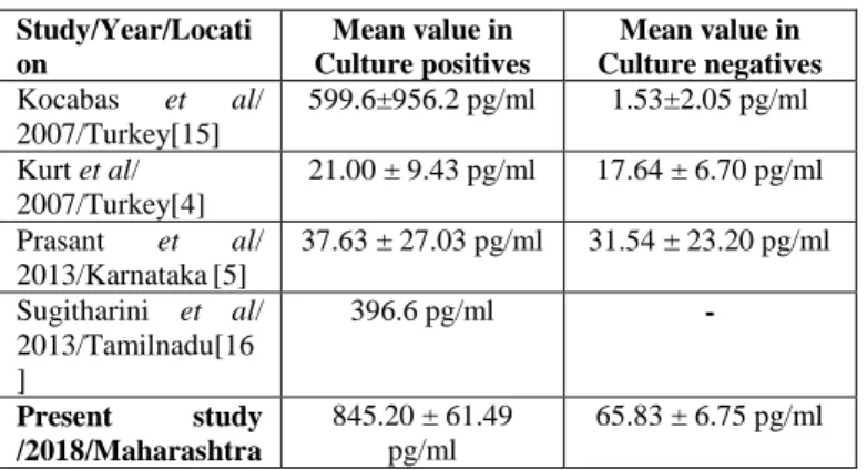
culture negative sepsis, as was also pointed out by Kurt et al[4] and Prasant et al[5] in their studies. In this study, value of IL-6 in LOS was highly statistically significant in comparison to EOS. Similar findings are seen in other studies. Rashwan et al[19] showed value of 83.58 ± 67.25 pg/ml in LOS versus 77.76 ± 60.25 pg/ml in EOS. Kocabas et al[15] showed value of 29.4±22.5 pg/ml in LOS and 16.5±15.5 pg/ml in EOS. This is probably due to the fact that in these studies, culture positivity was more in LOS than EOS, leading to increased mean value of IL-6 in LOS. IL-6 values were more in all culture positive cases, as explained earlier. Mean value of TNF-α in this study was highly statistically significant in culture positive neonates in comparison to culture negative neonates. Table 4 shows the mean values of TNF- α in different studies, including the present one.
Table 4: Mean value of TNF- α in different studies
Higher value of TNF-α is seen in the present study, as specific values differ due to collection time and site variability, as explained earlier.([4],[5]) However, as was seen with IL-6, culture proven sepsis had higher values than culture negative sepsis in TNF-α also in all the above studies,[(4)-[6],[19]) including the present study.
The higher mean value of TNF-α in LOS compared to EOS shown in the present study was highly statistically significant. Similar finding was reported by Kocabas et al,[15] where value of TNF-α in LOS was reported as 747.6±1020.2 pg/ml and 451.6±903.7 pg/ml in EOS. The reason is already explained previously with respect to IL-6.
In the present study, mean IL-6 value in samples with specific organisms isolated was highly
statistically significant as compared to TNF-α value (Table 1). This can be explained that the individual values of IL-6 are much higher than the individual values of TNF-α and therefore, the values in culture positives with specific organisms were also higher in IL-6 than in TNF-α.
Sensitivity, specificity, PPV and NPV of IL-6 in different studies are depicted in Table 5.
Table 5: Sensitivity specificity, PPV and NPV of IL-6 in different studies
Though in other studies including the present study, the four variables were within a wide range but most studies have emphasized IL-6 as a promising marker for rapid diagnosis of neonatal sepsis.
Sensitivity, specificity, PPV and NPV of TNF-α in different studies are shown in Table 6.
Table 6: Sensitivity, specificity, PPV and NPV of TNF-α in different studies
The explanation for the similarity of the above values in the present study to another Indian study[5] and a stark dissimilarity to studies done abroad might be the limitation of this study, i.e. having lesser sample size and absence of a comparative control group. The day of collection of blood and
Study/Year/Locati on
Mean value in Culture positives
Mean value in Culture negatives
Kocabas et al/ 2007/Turkey[15]
599.6±956.2 pg/ml 1.53±2.05 pg/ml
Kurt et al/ 2007/Turkey[4]
21.00 ± 9.43 pg/ml 17.64 ± 6.70 pg/ml
Prasant et al/ 2013/Karnataka[5]
37.63 ± 27.03 pg/ml 31.54 ± 23.20 pg/ml
Sugitharini et al/ 2013/Tamilnadu[16 ]
396.6 pg/ml -
Present study
/2018/Maharashtra
845.20 ± 61.49 pg/ml
65.83 ± 6.75 pg/ml
Study/Year/Place Sensitiv ity%
Specificity %
PPV% NPV%
Kocabas et al/ 2007/Turkey[15]
96.20 89.70 86.20 96.10
Caldas et al/ 2008/Brazil[13] 64.30-77.50 75.00 - 87.00 81.80 - 91.20 54.50 - 69.00 Prasant et al/
2013/Karnataka[5] 54.00 – 78.00 78.00 – 96.00 78.00 – 93.30 67.60 – 78.00 Sonawane et al
/2015/Maharashtra[2 0]
95.83 87.50 92.00 93.33
Ganesan et al/ 2016/Tamilnadu [17]
100.00 62.86 27.78 100.00
Das et al/
2016/WB[21]
76.00 70.59 - -
Rashwan et al/ 2018/Egypt[19]
82.35 100 - -
Present study/
2018/Maharashtra
100.00 77.14 75.76 100.00
Study/Year/Place Sensitivity
%
Specificity %
PPV% NPV%
Kocabas et al/ 2007/Turkey[15]
100.00 96.60 96.20 96.50
Caldas et al/ 2008/Brazil[13] 62.20 - 74.30 81.80 - 95.20 86.70 - 95.80 58.8-66.7
Prasant et al
/2013/Karnataka[5] 68.90 - 88.00 40.00 – 62.00 56.70 - 62.70 63.60 - 76.90
Present study
/2018/Maharashtr a
Nirjhar Chatterjee et al JMSCR Volume 08 Issue 08 August 2020 Page 163
sample site variability, as explained previously for IL-6, also may be the contributing factors. Therefore, these findings are required to be confirmed by future studies with larger sample size and also comparing these values with healthy controls.
In this study, IL-6 had a better specificity and PPV than TNF-α. (Bar Diagram 1). Similar to this, an Indian study from Karnataka[5] showed IL-6 had higher specificity and NPV than TNF-α. However, sensitivity and PPV of IL-6 was almost similar to TNF-α in their study (Tables 5 & 6). In contrast to this, a study from Turkey[15] reported increased values for these four variables in TNF-α than in IL-6. Another study from Brazil[4] showed increased specificity and NPV for TNF-α values than for IL-6, though sensitivity and PPV of both these biomarkers were almost similar (Tables 5 & 6). This discrepancy is most likely due to different geographical locations and different cut-off values used in different studies according to the different kits used. According to the values obtained in the present study and the study from Karnataka,5 IL-6 can be the preferable cytokine for predicting neonatal sepsis in resource limited settings in India. In the present study, specificity and PPV of IL-6 and TNF-α in combination were more in comparison to any of the markers alone (Bar Diagram 2). Caldas et al[13] also reported that value of all these four parameters increase (87%, 94.1%, 95%, 90%) when these biomarkers are used in combination, than any of them used alone (Tables 5 & 6). Thus it can be said that, if a set of biomarkers are required to be chosen, both these cytokines in combination will be a much better predictor than either of them used alone.
In the present study, specificity and PPV of IL-6 in LOS was more than in EOS (Bar Diagram 3). In contrast, another Indian study from Uttar Pradesh,[22] reported a decrease in sensitivity in LOS (73.6% ) than in EOS (86.3%). A study by Tam et al,[23] did not show any significant difference of sensitivity and specificity in EOS versus LOS (Sensitivity 54–84% versus 44–100%, specificity of 70–100% versus 74–93%).
In the present study, specificity of TNF-α was more in EOS and PPV was better in LOS (Bar Diagram 4). A study from Minneapolis,[23] reported comparable values in EOS and LOS. Sensitivity was 75% versus 60–100% and specificity was 88% versus 79–86% in EOS versus LOS.
In this study, specificity and PPV of IL-6 was more than TNF-α in both EOS and LOS (Table 2). So, it can be concluded that be it early onset or late onset sepsis, IL-6 can be chosen as a dependable marker. A study from Minneapolis,[23] refute this finding by reporting nearly equal statistical variables for IL-6 and TNF-α in EOS and LOS. Therefore, to answer which cytokine gives a better predictability with respect to type of sepsis (EOS/LOS), a bigger multicentre study with control group is needed. The two biomarkers IL- 6 and TNF-α can be predictors of neonatal sepsis, as evident from this study. IL-6 was shown to be a better marker than TNF-α both in early- and late-onset neonatal sepsis in this study. Therefore, in resource limited settings, only IL-6 can be used as a diagnostic marker.
Conclusion
This study also proved that both IL-6 and TNF-α jointly have high sensitivity and comparable specificity to that of blood culture, as compared to any of these two markers alone.
Identification of different biomarkers with sensitivity and specificity comparable to blood culture – the gold standard, would assist in early detection of neonatal sepsis. Faster diagnosis will help to avoid unnecessary antibiotic therapy, which leads to development of drug-resistant organisms and increased load on health care system.
Acknowledgement
Staff Research Society, TNMC & Nair Hospital for their grant.
References
1. Kliegman RM, Stanton BF, GemeIII JWS, Schor NF. Infections of the Neonatal Infant. In Nelson’s Textbook of Pediatrics: 19th
Nirjhar Chatterjee et al JMSCR Volume 08 Issue 08 August 2020 Page 164
(First South Asian Edition). Vol 1. (Elsevier, New Delhi) 2016:909-25
2. Paul VK, Bagga A. Newborn Infants. In Ghai Essential Paediatrics: 8th Ed. (CBS Publishers & Distributors Pvt Ltd, New Delhi) 2013:162-4
3. National Neonatal Prenatal Database, NNPD NETWORK, Indian Council of Medical research, New Delhi 2002-03:13-19, 23-70 4. Kurt ANC, Aygun AD, Godekmerdan A,
Kurt A, Dogan Y. Serum IL-1β, IL-6, and TNF-α Levels in early diagnosis and management of neonatal sepsis. In Mediators of inflammation. Hindawi Publishing Corporation, London, UK 2007;31397:1-5
5. Prashant A, Vishwanath P, Kulkarni P, Narayana PS, Gowdara V, Nataraj SM, et al. Comparative assessment of cytokines and other inflammatory markers for the early diagnosis of neonatal sepsis - A case control study. PLoS One 2013;8:1-9
6. Carton J, Tchernia G, Celton JL, Damay M, Cheron G, Farrokhi P, et al. Alloimmmune neonatal neutropenia. American Journal of Hematology and Oncology 1991;13:21-5 7. Verma P, Berwal PK, Nagaraj N, Swami S,
Jivaji P, Narayan S. Neonatal sepsis: epidemiology, clinical spectrum, recent antimicrobial agents and their antibiotic susceptibility pattern. International Journal of Contemporary Pediatrics 2015;2:176-80 8. MacDonald MG, Seshia MMK. Avery’s
Neonatology - Pathophysiology and
Management of the Newborn: 7th Ed (Wolter Kluwer, Philadelphia, USA) 2017:1253-63
9. Sarangi KK, Pattnaik D, Mishra SN, Nayak MK, Jena J. Bacteriological profile and antibiogram of blood culture isolates done by automated culture and sensitivity method in a neonatal intensive care unit in a tertiary care hospital in Odisha, India. International
Journal of Advances in Medicine
2015;2:387-92
10.Rozanska AJ, Wojkowska MP, Adamski M,
Borszewska KE, Gulczyńska M,
Nowiczewski, et al. Infections and risk-adjusted length of stay and hospital mortality in Polish Neonatology Intensive Care Units. International Journal of Infectious Diseases 2015;35:87-92
11.Jeyaganguli D, Velvizhi G, Sucilathangam G, Revathy C. Diagnostic Value of C - Reactive Protein and Haematological Markers in Neonatal Sepsis. International Journal of Current Microbiology and Applied Sciences 2018;11:722-7
12.Vedavati BI, Kumari BA. Role of Sepsis Screen Tests in Predicting Neonatal Septicemia. International Journal of Current
Microbiology and Applied Sciences
2018;7:2522-7
13.Caldas JPS, Marba STM, Blolla MHSL, Calil R, Morais SS, et al. Accuracy of white blood cell count, C-reactive protein, interleukine-6 and tumor necrosis factor alpha for diagnosing late neonatal sepsis. The Journal of Pediatrics 2008;84:536-42 14.Mahale RP, Rao MR, Shivapp SG.
Evaluation of Procalcitonin, CRP and Blood Culture in the Diagnosis of Neonatal Sepsis.
International Journal of Current
Microbiology and Applied Sciences
2018;7:2747-55
15.Kocabas E, Sarkcoglu A, Aksaray N, Seydaoglu G, Seyhun Y , Yaman A. Role of
procalcitonin, C-reactive protein,
interleukin-6, interleukin-8 and tumour necrosis factor-α in diagnosis of neonatal sepsis. The Turkish Journal of Paediatrics 2007;49:7-20
16.Sugitharini V, Prema A, Thangam EB.
Inflammatory mediators of systemic
inflammation in neonatal sepsis.
Inflammation Research 2013;62:1025–34 17.Ganesan P, Shanmugam P, Sattar SBA,
Nirjhar Chatterjee et al JMSCR Volume 08 Issue 08 August 2020 Page 165
Sepsis. Journal of Clinical and Diagnostic Research 2016;10:13-7
18.Chatterjee K, Mandal PK, Rahaman SR, Malathi R, Ray SK, Dutta A, et al. Raised IL-6 and C-reactive protein in neonatal sepsis in Eastern India. International Journal of Contemporary Pediatrics 2017;4:1590-4 19.Rashwan NI, Hassan MH, El-Deen ZMM,
Ahmed AEA. Validity of biomarkers in screening for neonatal sepsis - A single centre hospital based study. Pediatrics and
Neonatology 2018. Available at:
https://doi.org/10.1016/j.pedneo.2018.05.00 1 (Accessed on 19th December 2019)
20.Sonawane VB, Mehkarkar NS, Jadhav PB, Gaikwad SU, Kadam NN. Study of interleukin-6 levels in early diagnosis of neonatal sepsis. International Journal of Research in Medical Sciences 2015;3:41-6 21.Das S, Ray J. Relative importance of
inflammatory markers (erythrocyte
sedimentation rate, C-reactive protein, procalcitonin, and interleukins-6) in neonatal sepsis. BLDE University Journal of Health Sciences 2016;1:20-4
22.Kumar N, Singh MK, Dayal R, Gupta S, Garg R. Diagnostic Value of IL-6 in Neonatal Sepsis. Pacific Group of e-Journals. Annals of Applied Bio-Sciences 2016;3:A67-71