T.C.Froebel Giftly et al JMSCR Volume 08 Issue 02 February 2020 Page 694
Original Article
Duplicated Ureter- A Cadaveric Study
Authors
T.C. Froebel Giftly
1, Vinovictor Jesudas
21
Postgraduate in Anatomy, Institute of Anatomy, Madurai Medical College, Madurai
2
Director and professor, Institute of Anatomy, Madurai medical college, Madurai Corresponding Author
Dr Vino Victor Jesudas
Director and Professor, Institute of Anatomy, Madurai Medical College, Madurai, Tamilnadu, India-625020
Abstract
Introduction: Ureter is a muscular tube of 25-30cm length. Duplication of ureter results from early splitting of ureteric bud. Double ureter has been reported by various authors with prevalence of 0.1% to 4%. Incomplete duplication is three times more common than complete. Double ureters can be classified as complete and incomplete.
Aim and Objective: We conducted the present study to estimate the proportion of specimens with duplication of ureter and to describe type of duplicated ureter and compare with previous studies.
Materials and Method: The present study was performed over a period of three years from 2017-2020 on 31 well preserved human cadavers in the institute of Anatomy, Madurai medical college, Madurai, Tamilnadu, India
Observation and Results: In our study we have observed one left sided ureter with complete duplication which originated from separate renal pelvis and accompanied its fellow ureter, running parallel to each other and finally drained to urinary bladder with separate opening.
Conclusion: Knowledge of developmental anomaly of the urinary system is of great importance as it can affect both the disease conditions and the interventional methods. In urogenital surgeries including renal transplant or gynecological surgeries and in laparoscopic procedures, this information is also very useful for early detection through radiological examinations.
Keywords: Duplicated ureter, double ureter, ureteric bud.
Introduction
Ureter is a muscular tube of 25-30cm length, continuous superiorly with funnel shaped structure called renal pelvis and inferiorly it opens into the lateral angle of the base of the urinary bladder[1]. Embryologically, the ureters develop from the ureteric bud, a diverticulum from the mesonephric duct. The ureteric bud elongates to form the ureter and undergoes repeated branching to form the major and minor calyces
Duplication of ureter results from early splitting of ureteric bud[2]. Double ureter has been reported by various authors with prevalence of 0.1% to 4%.
[7,8,12]
Incomplete duplication is three times more common than complete[13]
The renal pelvis is a funnel-shaped expanded upper end of the ureter. It lies within the hilum of the kidney and receives the major calyces and emerges from the hilum of the kidney and runs vertically downward behind the parietal
http://jmscr.igmpublication.org/home/ ISSN (e)-2347-176x ISSN (p) 2455-0450
T.C.Froebel Giftly et al JMSCR Volume 08 Issue 02 February 2020 Page 695 peritoneum on the psoas muscle, which separates
it from the tips of the transverse processes of the lumbar vertebrae. Normally it enters the pelvis by crossing the bifurcation of the common iliac artery anterior to the sacroiliac joint. The ureter then runs downwards the lateral wall of the pelvis to the region of the ischial spine and turns forward to enter the lateral angle of the urinary bladder. Near its termination in males it is crossed by the vas deferens and by the uterine artery in females. The ureter passes obliquely through the wall of the bladder for about 1.9 cm before opening into the urinary bladder[4].
Double ureters can be classified as complete and incomplete. Complete duplication is also called double ureter wherein two pelves on the same side, one superior to the other, drained byseparate ureters and having separate orifices on the floor of the bladder. In incomplete or bifid ureter, upper end is bifid and lower third of their course two ureters join and open by a common orifice into the urinary bladder[5].
Anatomical variations of ureters and their relationship to surrounding structures are therefore of particular importance from an academic point of view, a surgical perspective, radiological examinations and treatment to preserve renal functions.
Studies on Tamilnadu population remains scarce. Hence this study focusses on prevalence and types of duplication of ureter in Tamilnadu population. We conducted the present study with the following objectives:
Primary Objective: To estimate the proportion of specimens with duplication of ureter
Secondary Objective: To describe the type of duplicated ureter and compare with previous studies.
Materials and Method
The present study was performed over a period of three years from 2017-2020 on 31 well preserved human cadavers in the institute of Anatomy, Madurai medical college, Madurai, Tamilnadu, India. All specimens taken in the study were adult cadavers of which 25 were males and 6 were females. Specimens showing crush or cut injuries and mass lesions of kidneys or ureter were excluded from the study. All the specimens were properly dissected and observed on both the right and the left sides to inspect the presence of any anatomical variations in the ureter. The dissection method used was based on the Cunningham's dissection manual.
Observation
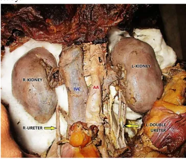
In our study we have observed one left sided ureter with complete duplication which originated from separate renal pelvis and accompanied its fellow ureter, running parallel to each other and finally drained to urinary bladder with separate opening. Fig 1 shows the course of duplicated ureter in the cadaver and table 1 shows the proportion of duplicated ureter observed in our study.
Fig 1: Duplicated left ureter
Table 1: Proportion of duplicated ureter
No of Renal
Specimens
No of normal ureter
% of Normal ureter
No of Duplicated
ureter % of duplicated
ureter
U/L B/L
T.C.Froebel Giftly et al JMSCR Volume 08 Issue 02 February 2020 Page 696 The proportion of duplication of ureter observed
in this study was 1.61% and percentage of normal ureter was seen in 98.4% of specimens.
Discussion
Two ureters connected with the single kidney open in the bladder either by a common orifice or by separate orifices-upper and lower. In the latter condition, a mesonephric duct gives rise to two ureteric buds, upper and lower, which invade the metanephric blastema independently and induce the development of the upper and lower poles of kidney respectively. As the mesonephric duct undergoes loop formation in the posterior wall of the bladder, the lower ureter opens in the bladder in normal position, whereas the upper ureter draining urine from the upper pole of the kidney migrates more caudally along with the caudal shift of the terminal part of the mesonephric duct and opens in ectopic position. Thus the normal and ectopic ureters cross each other[3]
Unilateral duplication has an incidence of 1 in 125 while bilateral duplication has an incidence of 1in 800 cases. Duplication is two to five times more common in females than in males. Moreover, incomplete duplication is three times more common than complete duplication. Incomplete type may remain asymptomatic or may cause complications like ureteric stenosis, urinary lithiasis and pyelonephritis.[10]
Table 2 Comparison of prevalence of duplicated ureter
AUTHORS YEAR NO OF SPECIMENS %
Prakash et al 2011 100 2
Parveen et al 2016 180 2.2
Umesh et al 2017 64 3
Roy M at al 2018 156 0.64
Anju et al 2017 72 4
Payal et al 2018 90 2.2
Present study 2020 62 1.61
Chart 1: Comparison of prevalence with various studies
Table 2 and chart 2 shows the prevalence of duplicated ureter in various studies which ranged from 0.64-4. In our study the prevalence observed was 1.61% which correlates with 2% in Prakash 20116
2 2,2
3
0,64
4
2,2
1,61
0 0,5 1 1,5 2 2,5 3 3,5 4 4,5
Prakash et al Parveen et al Umesh et al Roy M at al Anju et al Payal et al Present study
%
T.C.Froebel Giftly et al JMSCR Volume 08 Issue 02 February 2020 Page 697
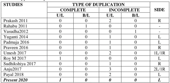
table 3: Comparison of types of duplicated ureter
STUDIES TYPE OF DUPLICATION
SIDE
COMPLETE INCOMPLETE
U/L B/L U/L B/L
Prakash 2011 0 0 2 0 R
Rahabu 2011 0 1 0 0 -
Vasudha2012 0 0 0 1 -
Yuganti 2014 0 0 1 0 L
Padmaja 2016 0 0 1 0 L
Praveen 2016 0 0 1 0 R
Umesh 2017 0 0 2 0 1L/1R
Roy M 2017 1 0 0 0 L
Sudhikshiya 2017 0 0 1 0 R
Anju2017 0 0 3 0 2L/1R
Payal 2018 0 0 2 0 L
Present 2020 1 0 0 0 L
Table 3 shows the types of duplication whether complete or incomplete as well as unilateral or bilateral observed by various studies. From the table it was observed that unilateral incomplete type was more commonly found and left sided duplication was more common. But in our study left sided complete duplication was found.
Rahabu et al 20117 observed a bilateral complete duplicated ureter associated with bilateral lower polar accessory renal artery and a right accessory renal vein. The lower polar accessory renal arteries took origin from the abdominal aorta to supply the respective poles of right and left kidneys. The right accessory renal vein opened directly into the inferior vena cava.
Padmaja vasi et al 2016 8 observed a left sided incomplete duplicated ureter associated with left accessory renal artery. One ureter was towards the upper pole and connected to one major calyx and five minor calyces. Other ureter was towards the lower pole and connected to one major calyx and six minor calyces
Rahabu et al 20117 observed a bilateral complete duplicated ureter associated with bilateral lower polar accessory renal artery and a right accessory renal vein. The lower polar accessory renal arteries took origin from the abdominal aorta to supply the respective poles of right and left kidneys. The right accessory renal vein opened directly into the inferior vena cava.
Yuganti 2014 9 in this case report, on the left side there were two separate renal pelves, one above
the other, each giving rise to a ureter. The relationship of both the pelves was maintained with the renal vein and artery as the sequence vein, artery and pelvis in before backward direction. The duplicated ureters were running parallel to each other, with the laterally placed ureter starting from the pelvis located below and the medially placed ureter starting from the pelvis located above
Praveen et al 201610 found out one incomplete duplicated left sided ureter in his study with no other associated abdominal or renal anomaly. Umesh 201717 observed two incomplete duplicated ureters in his study with prevalence of 3%.He also found out 6.25% of lobulation in left kidney and 3.12% bilateral lobulations
Payal Arvind 201815in his study on 90 specimens found out two incomplete left sided duplicated ureter associated with hypoplastic and lobulated kidneys. Bifid renal pelvis and ureter is often asymptomatic and incidentally diagnosed during radiographic imaging of abdomen and pelvicregion for other purposes. However, it may lead to clinical conditions like formation ofrenal calculi, ureterocele, hydronephrosis, and urinary tract infections.[13]
T.C.Froebel Giftly et al JMSCR Volume 08 Issue 02 February 2020 Page 698 ureteroureteric reflux. Thus, partial duplication is
associated with two problems .a) Ureteropelvic junction (UPJ) obstruction of the lower moiety. b) Retrograde yo-yo peristalsis of urine in ureter.[15]
Conclusion
According to our study,1 in 150 individuals may be having duplicated ureter. Most common type observed with comparison to previous studies was left sided unilateral incomplete duplication. Knowledge of developmental anomaly of the urinary system is of great importance as it can affect both the disease conditions and the interventional methods. In urogenital surgeries including renal transplant or gynaecological surgeries and in laparoscopic procedures, this information is also very useful for early detection through radiological examinations. Recognition of such variants before renal surgeries can decrease unnecessary complications and decrease the morbidity and mortality significantly.
References
1. Standring S; In Gray’s Anatomy. 38th ed. Edinburgh, London: Churchill Livingston; 2005:1285-1288
2. Sadler TW. Langman’s Medical Embryology. 9th Ed., Baltimore, Maryland, USA, Lippincott Williams &Wilkins, 2004: 329.
3. A.k. Dutta. Essentials of human embryology. 7th Ed., Kolkata, Current books international,2017:217
4. A.K. Dutta. Essentials of human Anatomy-Abdomen and Thorax.10th Ed., Kolkata, Current books international,2014:309-313 5. Vishram singh. Textbook of clinical
Embryology.2nd Ed.,New Delhi, Elseivier, 2017:260
6. Prakash et al. Double ureter and duplex system-A cadaveric and radiological study.
Urological journal 2011
DOI:10.22037/uj.v8i2.1027
7. Rahabu Varma. Bilateral double ureters and accessory renal vessels in a Tanzanian male cadaver: a rare urinary system variation. IJAV. 2011; 4: 164–166.
8. Padmaja Vasi. A case report on double ureter and accessory renal artery. Int J Anat Var (IJAV). 2014; 7: 48–50.
9. Yuganti et al. Unilateral isolated incompletely duplicated ureter-Case study. Journal of mahatmagandhi institute of medical sciences 2014:19(2);148-150. 10.Praveen et al.Unilateral incomplete
duplicated ureter – A clinical and embryological insight. international Journal of Medical Research &Health Sciences, 2016, 5, 8:68-70
11.Roy M, Singh BR, Gajbe UL, Thute P. Anatomical variations of ureter in central India: A cadaveric study. J Datta Meghe Inst Med Sci Univ 2017;12:277-9
12.Vasudha kulkarni. bilateral bifid ureter with accessory renal artery: a case report 2012:9:3(3);67-79
13.Sudikshiya. MED PHoEnix : An Official Journal of NMC, Birgunj, Nepal, Volume (3), Issue (1), July, 2018, 91-94
14.Dr.Anju et al. A cadaveric study on anatomical variations of kidney and ureter in a tertiary care teaching hospital Indian Journal of Basic and Applied Medical Research; June 2017: Vol.-6, Issue- 3, P. 300-305
15.Payal Arvind et al. A cadaveric study of variation in the urological system. Int J Anat Res 2018, Vol 6(3.3):5686-94
16.Privett JT, Jeans WD, Roylance J. The incidence and importance of renal duplication. Clin Radiol. 1976 Oct;27(4):521-30.