Predictors of exercise capacity in heart failure
Combined right ventricular dysfunction and raised left ventricular filling pressures predict
limited exercise capacity in heart failure with reduced ejection fraction (HFreF)
Pranvera Ibrahimi
1,2, Afrim Poniku
1, Violeta Hysenaj
1, Artan Ahmeti
1, Fisnik Jashari
2, edmond
Haliti
1and gani Bajraktari
1,2.
1Clinic of Cardiology and Angiology, University Clinical Centre of Kosova, Prishtina, Kosovo; 2Department of Public Health and Clinical Medicine and Heart Centre, Umeå University, Sweden
Abstract
Background and Aim:
Compromised exercise capacity is a major symptom in patients with heart failure (HF) and reduced left ventricular (LV) ejection fraction (eF). six-minute walk test (6-MWt) is popular for the objective assessment of exercise capacity in these patients but is largely confined to major heart centres. the aim of this study was to prospectively examine functional parameters that predict 6-MWt in patients with HF and reduced LVeF.
Methods: In 111 HF patients (mean age 60±12 years, 56% male), a 6-MWt and an echo-Doppler study were performed in the same day. In addition to conventional ventricular function measurements, global LV dyssynchrony was indirectly assessed by total isovolumic time - t-IVt [in s/min; calculated as: 60 – (total ejection time – total filling time)], and tei index (t-IVt/ejection time). Also, LV and right ventricular function were assessed by mitral and tricuspid annular plane systolic excursion (MAPse and tAPse, respectively). Based on the 6-MWt distance, patients were divided into 2 groups: group I: ≤300m and group II: >300m.
Results: the 6-MWt distance correlated with t-IVt and tei index (r=-0.37, p<0.001, for both), lateral and septal e’ velocities (r=0.41, p<0.001, and r=0.46, p<0.001, respectively), e/e’ ratio (r=-0.37, p<0.001) and tAPse (r=0.45, p<0.001), but not with the other clinical or echo parameters. group I patients had longer t-IVt, lower e/e’ratio, tAPse and lateral e’ (p<0.001 for all) compared with group II. In multivariate analysis, tAPse [0.076 (0.017-0.335), p=0.001], e/e’ [1.165 (1.017-1.334), p=0.027], t-IVt [1.178 (1.014-1.370), p=0.033] independently predicted poor 6-MWt performance (<300m). sensitivity and specificity for tAPse ≤1.9 cm were 66% and 77%, (AuC 0.78, p<0.001); e/e’ ≥10.7 were 66% and 62% (AuC 0.67, p=0.002) and t-IVt ≥13 s/min were 64% and 60% (AuC 0.68, p=0.002) in predicting poor 6-MWt. Combined tAPse and e/e’ had a sensitivity of 68% but specificity of 92% in predicting 6-MWt. Respective values for combined tAPse and t-IVt were 71% and 85%.
Conclusion: In patients with HF, the limited exercise capacity assessed by 6-MWt, is multifactorial being related both to the severity of right ventricular systolic dysfunction as well as to raised LV filling pressures and global dyssynchrony.
Key words: six-minute walk test; Doppler echocardiography; right ventricular function, heart failure, exercise capacity
Introduction
Heart failure (HF) is a major public health problem 1,2 with high morbidity and mortality 3. Left ventricular (LV) systolic dysfunction is the commonest cause of HF symptoms. Despite recent advances in pharmacological and device treatment, HF prognosis and quality of life remain poor 4,5. We and others have reported a number of clinical and echocardiographic predictors of prognosis in these patients 6-11. the main concern for professionals is the patient’s limiting symptoms and the mechanisms behind them. several LV function parameters have been shown to predict patient’s exercise capacity 12, 13 assessed by six-minute walk test – 6-MWt 14-17. Despite the fact that 6-MWt itself has been used to assess functional status of HF patients 18 and to predict prognosis 19,20 it is limited to a relatively few cardiac centres or specialized units. In contrast, Doppler echocardiography is the main-stay in the diagnosis, follow up and monitoring of HF patients, particularly with its ability to provide accurate assessment of cardiac physiology which can help explain exertional symptoms. the aim of this study was to identify objective right and left ventricular echocardiographic functional parameters that predict 6-MWt in a group of patients with HF and reduced LVeF.
Methods
Study population
We studied 111 consecutive patients with stable HF [New York Heart Association (NYHA) functional class I-III] secondary to ischemic heart disease or idiopathic dilated cardiomyopathy, between January 2007 and May 2012. Patients were on optimum cardiac medications at the time of the study, which included ACe-inhibitors, ß blockers and diuretics. Medications were optimized for individual patients at least 2 weeks prior to enrollment, based on symptoms and renal function. All patients were in sinus rhythm and had eF <45%, on baseline echocardiogram. Patients with clinical evidence for cardiac decompensation, limited physical activity due to factors other than cardiac (e.g. arthritis), more than mild renal failure, chronic obstructive pulmonary disease as well as recent acute coronary syndrome, stroke or anemia were excluded. Patients gave a written informed consent to participate in the study which was approved by the local ethics Committee.
Data collection
included haemoglobin, lipid profile, blood glucose level, and kidney function tests. estimation of body mass index (BMI) was made from weight and height measurements. Waist, hip measurements were also made and waist/hip ratio calculated.
Echocardiographic examination
Patients were examined by a Philips Ie-33 system with a multi-frequency transducer, and harmonic imaging. Images were obtained with the patient in the left lateral decubitus position and during quiet expiration. LV dimensions at end-systole and end-diastole were made from the left parasternal cross-sectional recording of the minor axis with the M-mode cursor positioned by the tips of the mitral valve leaflets. LV volumes and ejection fraction were calculated from the apical 2 and 4 chamber views using the modified simpson’s role. Ventricular long axis motion was studied by placing the M-mode cursor at the lateral and septal angles of the mitral ring (MAPse) and the lateral angle of the tricuspid ring (tAPse). total amplitude of long axis motion was measured as previously described 21. Ventricular long axis myocardial velocities were also studied using Doppler tissue imaging technique with the sample volume placed at the basal segment of LV lateral and septal segments as well as RV free wall. systolic (s’), as well as early and late (e’ and a’) diastolic myocardial velocities were measured. Mean value of the lateral and septal LV velocities were calculated. Left atrial diameter was measured from aortic root recordings with the M-mode cursor positioned at the level of the aortic valve leaflets.
Diastolic ventricular function was assessed from ventricular filling velocities using spectral Doppler with the pulsed wave sample volume positioned at the tips of the mitral and tricuspid valve leaflets. Peak LV and RV early (e wave), and late (A wave) diastolic filling velocities were measured and e/A and e/e’ ratios were calculated. LV filling pattern was considered ‘restrictive’ when e/A ratio was >2.0, e wave deceleration time < 140 ms and the left atrium dilated, > 40 mm in transverse diameter.
Measurements of LV dyssynchrony
Indirect assessment of LV dyssynchronous function was obtained by measuring total isovolumic time (t-IVt), tei Index and LV-RV pre-ejection time delay. total LV filling time was measured from the onset of the e wave to the end of the A wave and ejection time from the onset to the end of the aortic Doppler flow velocity. total isovolumic time (t-IVt) was calculated as 60 - (total ejection time + total filling time) and was expressed in s/min. tei index was calculated as the ratio between t-IVt and ejection time 22, 23.
Mitral and tricuspid regurgitation severity was assessed by colour and continuous wave Doppler and was graded as mild, moderate, or severe according to the relative jet area to that of the left and right atrium, respectively, as well as the flow velocity profile, in line with the recommendations of the American society of echocardiography 24. Retrograde transtricuspid pressure drop > 35 mmHg was taken as an evidence for pulmonary hypertension. All M-mode and Doppler recordings were made at a fast speed of 100 mm/s with a superimposed eCg (lead II).
Six minute walk test
Within 24 hours of the echocardiographic study a 6-MWt was performed on a level hallway surface, administered by a specialized nurse blinded to the results of the echocardiogram. According to the method of gyatt et al 25 patients were
informed of the purpose and protocol of the 6-MWt which was conducted in a standardized fashion while patients on their regular medications 25-27. A 15 meter flat, obstacle-free corridor was used and patients were instructed to walk as far as they can, turning 180º after they have reached the end of the corridor, during the allocated time of 6 minutes. Patients walked unaccompanied so as not to influence walking speed. At the end of the 6 min, the supervising nurse measured the total distance walked by the patient.
Statistical Analysis
Data are presented as mean ± sD or proportions (% of patients). Continuous data was compared with two-tailed unpaired student’s t test and discrete data with Chi-square test. Correlations were tested with Pearson coefficients. Predictors of 6-MWt distance were identified with univariate analysis and multivariate logistic regression was performed by the step-wise method, whereas multiple logistic regression analysis was used to identify the independent correlates of limited 6-MWt performance. A significant difference was defined as p < 0.05
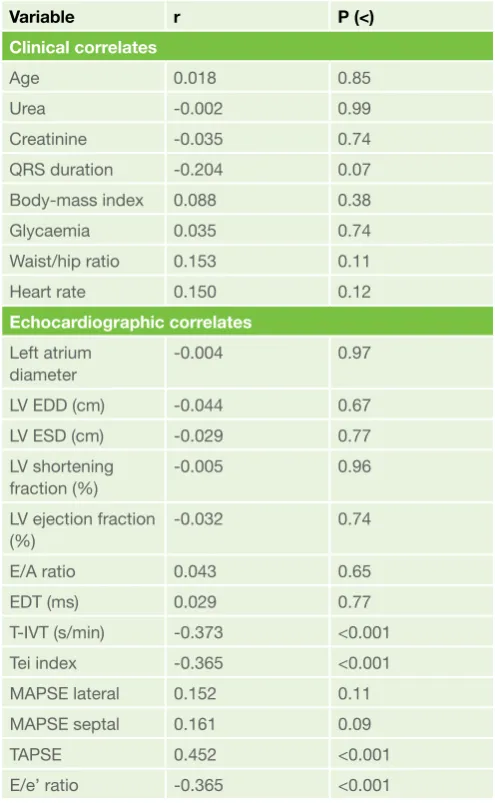
Table 1. Correlations of clinical and echocardiographic variables with 6 minute walk distance
Variable r P (<)
Clinical correlates
Age 0.018 0.85
urea -0.002 0.99
Creatinine -0.035 0.74
QRs duration -0.204 0.07
Body-mass index 0.088 0.38
glycaemia 0.035 0.74
Waist/hip ratio 0.153 0.11
Heart rate 0.150 0.12
Echocardiographic correlates
Left atrium diameter
-0.004 0.97
LV eDD (cm) -0.044 0.67
LV esD (cm) -0.029 0.77
LV shortening fraction (%)
-0.005 0.96
LV ejection fraction (%)
-0.032 0.74
e/A ratio 0.043 0.65
eDt (ms) 0.029 0.77
t-IVt (s/min) -0.373 <0.001
tei index -0.365 <0.001
MAPse lateral 0.152 0.11
MAPse septal 0.161 0.09
tAPse 0.452 <0.001
e/e’ ratio -0.365 <0.001
(2-tailed). Patients were divided according to their ability to walk >300m into good and Limited exercise performance groups, and were compared using unpaired student t-test.
Results
Patients mean age was 60±12 years, and 44.1% were female. the aetiology of heart failure was ischaemic cardiomyopathy in 65 patients and idiopathic dilated cardiomyopathy in 46 patients.
Clinical and echocardiographic correlates of 6-MWT
(Table 1)
Out of all Doppler echocardiographic measurements, LV lateral wall e’ velocity (r=0.41, p<0.001), e/e’ ratio (r=-0.37, p<0.001), t-IVt (r=-0.37, p<0.001) and tei index (r=-0.37, p<0.001) as well as tAPse (r=0.45, p<0.001) had the highest correlation with the 6-MWt distance (Table 1). Age, LV systolic function and dimensions, and kidney function correlated poorly with 6-MWt.
Patients with Limited vs. Good 6-MWT performance
Clinical and biochemical differences (Table 2)
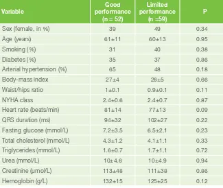
there were no significant differences in any of the clinical or biochemical measures.
Cardiac function differences (Table 3)
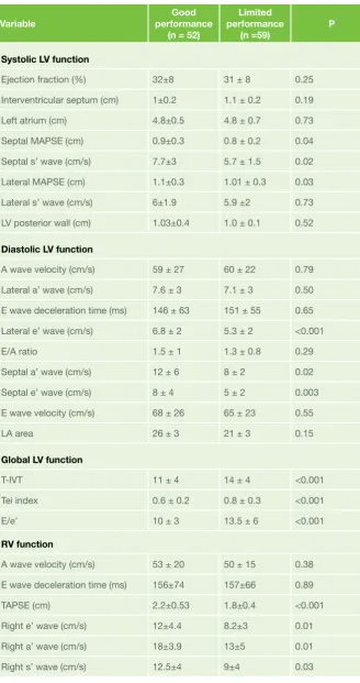
Patients with limited 6-MWt performance had lower septal and lateral MAPse (p=0.04 and p=0.03, respectively), lower septal s’ velocity (p=0.02) and a’ velocity (p=0.02), lower septal and lateral e’ velocities (p=0.003 and p<0.001, respectively), higher e/e’ ratio (p< 0.001), longer t-IVt (p< 0.001), higher tei index (p<0.001), lower tAPse (p< 0.001) and lower right ventricular s’, e’ and a’ velocities (p=0.03, p=0.01 and p=0.01, respectively) compared with those with good performance. the rest of the echo measurements were not different between the two patient groups.
Predictors of limited 6 minute walk
performance
Univariate predictors of limited 6-MWT performance (Table 4)
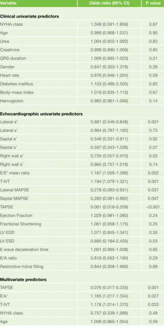
In univariate analysis [odds ratio 95% confidence interval], tAPse [0.061 (0.018-0.209)), p<0.001], e/e’ ratio [1.167 (1.059-1.286), p=0.002], t-IVt [1.194 (1.079-1.321), p=0.001], lateral e’ [0.681 (0.546-0.848), p=0.001], septal e’ [0.549 (0.331-0.911), p=0.02], right ventricular e’ [0.735 (0.557-0.970), p=0.03], lateral MAPse [0.278 (0.083-0.931), p=0.037, and septal MAPse [0.283 (0.081-0.982), p= 0.047], predicted limited 6-MWt.
Multivariate predictors of limited 6-MWT performance (Table 4)
In multivariate analysis [odds ratio 95% confidence interval], tAPse [0.076 (0.017-0.335)], e/e’ ratio [1.165 (1.017-1.334), p=0.027] and t-IVt [1.178 (1.014-1.370), p=0.033] independently predicted limited 6-MWt performance. A tAPse ≤1.9 cm was 66% sensitive and 77% specific (AuC 0.78, p<0.001), an e/e’ ≥10.7 was 66% sensitive and 62% specific (AuC 0.67, p=0.002) and a t-IVt ≥13 s/min was 64% sensitive and 60% specific (AuC 0.68, p=0.002) in predicting limited 6 MWt distance in these patients. Combined tAPse and e/e’ had a sensitivity of 68% but specificity of 92% in predicting poor exercise tolerance. Respective values for combined tAPse and t-IVt were 71% and 85%.
Discussion
Findings: Our findings show that conventional measurements of LV structure and function, i.e eF, do not correlate with exercise tolerance, as assessed by 6-MWt distance. In contrast, indirect measures of LV filling pressures, e/e’, and dyssynchrony, t-IVt and tei index, correlate with 6-MWt distance. the same parameters as well as systolic and diastolic MAPse and tAPse velocities were significantly worse in patients with limited 6-MWt compared to those with good performance. Finally, the only predictors of 6-MWt distance were tAPse, t-IVt and e/e’ with combined tAPse <1.9 cm and e/e’ >10.7 having a sensitivity of 68% and specificity of 92%, and a combined tAPse <1.9 cm and t-IVt ≥13 s/min showing a sensitivity of 71% and specificity of 85% in predicting poor 6-MWt performance.
Data interpretation: Our multivariate analysis shows that the combination right ventricular systolic function, measured by tAPse and raised LV filing pressure, shown by e/e’ or LV global dyssynchrony, t-IVt are good predictors of exercise capacity objectively assessed by 6-MWt distance in a group of HF patients with reduced ejection fraction. We and others have previously shown that tAPse and its systolic velocity correlate with peak oxygen consumption in heart failure 28-31. tAPse was also found to predict survival of heart failure patients with values <14 mm carrying poor prognosis compared to >14 mm 40. these findings highlight the crucial role of the right ventricle in maintaining optimum stroke volume required. the only way by which the body compensates for the compromised stroke volume is by increasing the heart rate which itself is an indication for pharmacological heart rate control and hence
Table 2. Comparison of clinical and biochemical data between patient’s groups
Variable
Good performance
(n = 52)
Limited performance
(n =59) P
sex (female, in %) 39 49 0.34
Age (years) 61±11 60±13 0.95
smoking (%) 31 40 0.38
Diabetes (%) 35 37 0.86
Arterial hypertension (%) 65 48 0.18
Body-mass index 27±4 28±5 0.66
Waist/hips ratio 1±0.1 0.9±0.1 0.11
NYHA class 2.4±0.6 2.4±0.7 0.87
Heart rate (beats/min) 81±14 77±13 0.09
QRs duration (ms) 94±32 102±27 0.22
Fasting glucose (mmol/L) 7.2±3.5 6.5±2.1 0.23
total cholesterol (mmol/L) 4.3±1.2 4.1±1.1 0.33
triglycerides (mmol/L) 1.6±0.7 1.7±1.1 0.72
urea (mmol/L) 10±4.8 10±4.9 0.94
Creatinine (μmol/L) 113±48 111±38 0.86
Hemoglobin (g/L) 132±15 125±25 0.12
the importance of resting and exercise reserve heart rate. Our results show that a tAPse <19 mm, in isolation, has a modest accuracy in predicting poor 6-MWt performance, possibly indicating an early stage of cardiac dysfunction before further irreversible falls in tAPse occur with an associated adverse effect on survival. the second observation was the combination of low tAPse and prolonged t-IVt, which reflects the degree of global LV dyssynchrony. the latter has been shown to predict 6-MWt and survival in a similar group of HF patients 8, 32, through its effect on compromising stroke volume entering the LV and consequently ejected out to the systemic circulation. Indeed, we found in the current paper that the combined disturbances have a specificity of 85% in predicting limited 6-MWt, again confirming the respective additive role in compromising stroke volume. Finally, our analysis has shown that the combined low tAPse and raised e/e’ has a specificity of 92% in predicting 6-MWt. e/e’ reflects raised LV filling pressures with the highest values consistent with poor LV filling during late diastole, due to high end-diastolic pressure. the complete loss of the LV filling component in late diastole is bound to compromise the stroke volume, raise left atrial pressure further resulting in pulmonary venous hypertension. Although our cut off value for e/e’ was only 10.7 it puts the patients in the grey zone with modest rise of filling pressures. In addition, the expected rise of left atrial pressure with exercise is likely to increase right ventricular systolic pressure and affect tAPse. Indeed, raised e/e’ has proved a sensitive predictor of peak oxygen consumption 33 and survival 8 in heart failure patients. And hereby, we are showing that its combination with reduced tAPse is highly specific in predicting limited 6-MWt distance.
Clinical implications: LV ejection fraction remains the most clinically used measure of cardiac function for diagnosis, monitoring and managing HF patients. eF failed to demonstrate any significant differences between our two groups of patients, with good and limited 6-MWt. In contrast, less used markers of right ventricular function (tAPse) and LV raised filling pressures (e/e’) and global dyssynchrony (t-IVt) discriminated between the two groups with high accuracy, and we suggest should therefore be implemented in routine HF practice.
Limitations: Despite 6-MWt objectively measuring the extent of exercise
Table 3. Comparison of echocardiographic data between patient’s groups
Variable
Good performance
(n = 52)
Limited performance
(n =59) P
Systolic LV function
ejection fraction (%) 32±8 31 ± 8 0.25
Interventricular septum (cm) 1±0.2 1.1 ± 0.2 0.19
Left atrium (cm) 4.8±0.5 4.8 ± 0.7 0.73
septal MAPse (cm) 0.9±0.3 0.8 ± 0.2 0.04
septal s’ wave (cm/s) 7.7±3 5.7 ± 1.5 0.02
Lateral MAPse (cm) 1.1±0.3 1.01 ± 0.3 0.03
Lateral s’ wave (cm/s) 6±1.9 5.9 ±2 0.73
LV posterior wall (cm) 1.03±0.4 1.0 ± 0.1 0.52
Diastolic LV function
A wave velocity (cm/s) 59 ± 27 60 ± 22 0.79
Lateral a’ wave (cm/s) 7.6 ± 3 7.1 ± 3 0.50
e wave deceleration time (ms) 146 ± 63 151 ± 55 0.65
Lateral e’ wave (cm/s) 6.8 ± 2 5.3 ± 2 <0.001
e/A ratio 1.5 ± 1 1.3 ± 0.8 0.29
septal a’ wave (cm/s) 12 ± 6 8 ± 2 0.02
septal e’ wave (cm/s) 8 ± 4 5 ± 2 0.003
e wave velocity (cm/s) 68 ± 26 65 ± 23 0.55
LA area 26 ± 3 21 ± 3 0.15
Global LV function
t-IVt 11 ± 4 14 ± 4 <0.001
tei index 0.6 ± 0.2 0.8 ± 0.3 <0.001
e/e’ 10 ± 3 13.5 ± 6 <0.001
RV function
A wave velocity (cm/s) 53 ± 20 50 ± 15 0.38
e wave deceleration time (ms) 156±74 157±66 0.89
tAPse (cm) 2.2±0.53 1.8±0.4 <0.001
Right e’ wave (cm/s) 12±4.4 8.2±3 0.01
Right a’ wave (cm/s) 18±3.9 13±5 0.01
Right s’ wave (cm/s) 12.5±4 9±4 0.03
capacity, it does not assess the exact underlying mechanism for its limitation. We relied in our data interpretation on Non-invasive Doppler and
echocardiographic measurements which have been used in numerous studies before and proved very reproducible. Although our resting Doppler echocardiographic results predicted mechanisms of exercise limitation, more accurate interpretation would have required exercise echo examination. Invasive pressure measurements could have added more value to the data interpretation.
Conclusion: Limited exercise tolerance assessed by 6-MWt distance is multifactorial involving reduced right ventricular systolic function, raised left ventricular filling pressures as well as significant global dyssynchrony.
Correspondence to: Dr. Pranvera Ibrahimi, MD
Clinic of Cardiology and Angiology, university Clinical Centre of Kosova, Prishtina, Kosovo, “Rrethi i spitalit”, p.n., Prishtina, Kosova
e-mail: [email protected] pranvera.ibrahimi@ medicin.umu.se
References
1. American Heart Association. Heart Disease and stroke statistics–2003 update. Dallas, tX:
American Heart Association, 2002.
2. Cowie MR, Mosterd A, Wood DA, Deckers JW, Poole-Wilson PA, sutton gC, grobbee De. the epidemiology of heart failure. Eur Heart J 1997; 18: 208–25.
3. White HD, Aylward Pe, Huang Z, Dalby AJ, Weaver WD, Barvik s, Marin-Neto JA, Murin J, Nordlander RO, van gilst WH, Zannad F, McMurray JJ, Califf RM, Pfeffer MA; VALIANt Investigators. Mortality and morbidity remain high despite captopril and/or Valsartan therapy in elderly patients with left ventricular systolic dysfunction, heart failure, or both after acute myocardial infarction: results from the Valsartan in Acute Myocardial Infarction trial (VALIANt). Circulation 2005; 112(22): 3391-9. 4. Davies M, Hobbs F, Davis R, Kenkre J, Roalfe
AK, Hare R, et al. Prevalence of left-ventricular systolic dysfunction and heart failure in the echocardiographic Heart of england screening study: a population based study. Lancet 2001; 358: 439-45.
5. Ahmed A. Quality and outcomes of heart failure care in older adults: role of multidisciplinary disease-management programs. J Am Geriatr Soc 2002; 50(9): 1590-3.
6. Bajraktari g, emini M, shabani X, Berisha V, selmani H, Rexhepaj N, elezi s, Ndrepepa g. Predictors of mortality in medically treated patients with congestive heart failure of nonrheumatic etiology and reduced systolic function. Eur J Intern Med 2009; 20(4): 362-5. 7. Dini FL, Buralli s, Bajraktari g, elezi s, Duranti
e, Metelli MR, Carpi A, taddei s. Plasma matrix metalloproteinase-9 better predicts outcome than N-terminal protype-B natriuretic peptide in patients with systolic heart failure and a high prevalence of coronary artery disease. Biomed Pharmacother 2010; 64(5): 339-42.
Table 4. Predictors of limited 6 minute walk test
Variable Odds ratio (95% CI) P value
Clinical univariate predictors
NYHA class 1.048 (0.591-1.859) 0.87
Age 0.999 (0.968-1.031) 0.95
urea 1.004 (0.922-1.092) 0.93
Creatinine 0.999 (0.990-1.009) 0.85
QRs duration 1.009 (0.995-1.023) 0.21
gender 0.647 (0.303-1.378) 0.26
Heart rate 0.976 (0.946-1.004) 0.09
Diebetes mellitus 1.103 (0.486-2.505) 0.82
Body-mass index 1.019 (0.935-1.110) 0.67
Hemoglobin 0.983 (0.961-1.006) 0.14
Echocardiographic univariate predictors
Lateral e’ 0.681 (0.546-0.848) 0.001
Lateral s’ 0.964 (0.787-1.182) 0.73
septal e’ 0.549 (0.331-0.911) 0.02
septal s’ 0.597 (0.343-1.038) 0.07
Right wall e’ 0.735 (0.557-0.970) 0.03
Right wall s’ 0.960 (0.757-1.219) 0.74
e/e’ mean ratio 1.167 (1.059-1.286) 0.002
t-IVt 1.194 (1.079-1.321) 0.001
Lateral MAPse 0.278 (0.083-0.931) 0.037
septal MAPse 0.283 (0.081-0.982) 0.047
tAPse 0.061 (0.018-0.209) <0.001
ejection Fraction 1.029 (0.981-1.080) 0.24
Fractional shortening 1.061 (0.958-1.175) 0.25
LV eDD 1.071 (0.855-1.341) 0.55
LV esD 0.665 (0.184-2.405) 0.53
e wave deceleration time 1.001 (0.995-1.008) 0.65
e/A ratio 0.818 (0.562-1.190) 0.29
Restrictive mitral filling 0.844 (0.358-1.990) 0.69
Multivariate predictors
tAPse 0.076 (0.017-0.335) 0.001
e/e’ 1.165 (1.017-1.334) 0.027
t-IVt 1.178 (1.014-1.370) 0.033
NYHA class 0.757 (0.339-1.689) 0.49
Age 1.008 (0.965-1.054) 0.59
8. Bajraktari g, Dini FL, Fontanive P, elezi s, Berisha V, Napoli AM, Ciuti M, Henein M. Independent and incremental prognostic value of Doppler-derived left ventricular total isovolumic time in patients with systolic heart failure. Int J Cardiol 2011; 148(3): 271-5.
9. Bajraktari g, Fontanive P, Qirko s, elezi s, simioniuc A, Huqi A, Berisha V, Dini FL. Independent and incremental value of severely enlarged left atrium in risk stratification of very elderly patients with chronic systolic heart failure. Congest Heart Fail 2012; 18(4): 222-8.
10. Ho KK, Anderson KM, Kannel WB, grossman W, Levy D. survival after onset of congestive heart failure in Framingham Heart study subjects.
Circulation 1993; 88: 107-15.
11. Packer M, Coats AJ, Fowler MD, et al. effect of carvedilol on survival in severe chronic heart failure. N Engl J Med 2001; 344: 1651-8. 12. smart N, Haluska B, Leano R, Case C, Mottram PM, Marwick tH.
Determinants of functional capacity in patients with chronic heart failure: role of filling pressure and systolic and diastolic function. Am Heart J 2005;149:152–158.
13. terzi s, Dayi su, Akbulut t, sayar N, Bilsel t, tangurek B, Akgoz H, Kose H, Yilmazer s, Yesilcimen K. Value of left atrial function in predicting exercise capacity in heart failure with moderate to severe left ventricular systolic dysfunction. Int Heart J 2005; 46:123–131.
14. Bajraktari g, elezi s, Berisha V, Lindqvist P, Rexhepaj N, Henein MY. Left ventricular asynchrony and raised filling pressure predict limited exercise performance assessed by 6 minute walk test. Int J Cardiol. 2011 Feb 3;146(3):385-9.
15. Ingle L, Rigby As, Carroll s, Butterly R, King RF, Cooke CB, et al. Prognostic value of the 6 min walk test and self-perceived symptom severity in older patients with chronic heart failure. Eur Heart J 2007; 28(5):560–8.
16. Rostagno C, Olivo g, Comeglio M, Boddi V, Banchelli M, galanti g, et al. Prognostic value of 6-minute walk corridor test in patients with mild to moderate heart failure: comparison with other methods of functional evaluation. Eur J Heart Fail 2003; 5(3): 247-52.
17. Bajraktari g, Batalli A, Poniku A, Ahmeti A, Olloni R, Hyseni V, Vela Z, Morina B, tafarshiku R, Vela D, Rashiti P, Haliti e, Henein MY. Left ventricular markers of global dyssynchrony predict limited exercise capacity in heart failure, but not in patients with preserved ejection fraction.
Cardiovasc Ultrasound 2012; 10(1): 36.
18. Crapo RO, Casaburi R, Coates AL, enright PL, MacIntyre NR, McKay Rt, Johnson D, Wanger Js, Zeballos RJ, Bittner V, Mottram C. Ats statement: guidelines for the six-minute walk test. Am J Respir Crit Care Med 2002; 166:1111–1117.
19. Ingle L, Rigby As, Carroll s, Butterly R, King RF, Cooke CB, et al. Prognostic value of the 6 min walk test and self-perceived symptom severity in older patients with chronic heart failure. Eur Heart J 2007; 28(5):560-8.
20. Rostagno C, Olivo g, Comeglio M, Boddi V, Banchelli M, galanti g, et al. Prognostic value of 6-minute walk corridor test in patients with mild to moderate heart failure: comparison with other methods of functional evaluation. Eur J Heart Fail 2003; 5(3): 247-52.
21. Henein MY, gibson Dg. Normal long axis function. Heart 1999; 81(2): 111-3.
22. Duncan AM, Francis DP, Henein MY, gibson Dg. Importance of left ventricular activation in determining myocardial performance (tei) index: comparison with total isovolumic time. Int J Cardiol 2004; 95: 211-7. 23. tei C, Ling LH, Hodge DO, Bailey KR, Oh JK, Rodeheffer RJ, et al. New
index of combined systolic and diastolic myocardial performance: a simple and reproducible measure of cardiac function - a study in normals and dilated cardiomyopathy. J Cardiol 1995; 26: 357–366.
24. Zoghbi WA, enriquez-sarano M, Foster e, grayburn PA, Kraft CD, Levine RA, et al. American society of echocardiography. Recommendations for evaluation of the severity of native valvular regurgitation with two-dimensional and Doppler echocardiography. J Am Soc Echocardiogr 2003; 16: 777-802.
25. gyatt gH, sullivan MJ, thompson PJ, Fallen eL, Pugsley sO, taylor DW, et al. the 6-minute walk test: a new measure of exercise capacity in patients with chronic heart failure. Can Med Assoc J 1985; 132: 919-23.
26. gyatt gH, thompson PJ, Berman LB, sullivan MJ, townsend M, Jones NL, et al. How should we measyre function in patients with chronic heart and lung disease? J Chronic Dis 1985; 28: 517-24.
27. Lipkin DP, scriven AJ, Crake t, Poole-Wilson PA. six minute walking test for assessing exercise capacity in chronic heart failure. BMJ 1988; 292: 653-5.
28. troisi F, greco s, Brunetti ND, Di Biase M. Right heart dysfunction assessed with echography, B-type natriuretic peptide and cardiopulmonary test in patients with chronic heart failure. J Cardiovasc Med (Hagerstown) 2008; 9(7):672-6
29. Webb-Peploe KM, Henein MY, Coats AJ, gibson Dg. echo derived variables predicting exercise tolerance in patients with dilated and poorly functioning left ventricle. echo derived variables predicting exercise tolerance in patients with dilated and poorly functioning left ventricle. Heart 1998; 80(6): 565-9.
30. D’Andrea A, gravino R, Riegler L, salerno g, scarafile R, Romano M, Cuomo s, Del Viscovo L, Ferrara I, De Rimini ML, Muto P, Limongelli g, Pacileo g,Bossone e, Russo Mg, Calabrò R. Right ventricular ejection fraction and left ventricular dyssynchrony by 3D echo correlate with functional impairment in patients with dilated cardiomyopathy. J Card Fail 2011; 17(4): 309-17.
31. ghio s, temporelli PL, Klersy C, simioniuc A, girardi B, scelsi L, Rossi A, Cicoira M, genta Ft, Dini FL. Prognostic relevance of a non-invasive evaluation of right ventricular function and pulmonary artery pressure in patients with chronic heart failure. Eur J Heart Fail 2013 Jan 10. [epub ahead of print].
32. Bajraktari g, Duncan A, Pepper J, Henein M. Prolonged total isovolumic time predicts cardiac events following coronary artery bypass surgery. Eur
J Echocardiogr 2008; 9(6): 779-83.
33. gardin JM, Leifer es, Fleg JL, Whellan D, Kokkinos P, Leblanc MH, Wolfel e, Kitzman DW; HF-ACtION Investigators. Relationship of Doppler-echocardiographic left ventricular diastolic function to exercise performance in systolic heart failure: the HF-ACtION study. Am Heart J 2009; 158 (4 suppl): s45-52.