R E S E A R C H
Open Access
Delaying the oocyte maturation trigger by one
day leads to a higher metaphase II oocyte yield in
IVF/ICSI: a randomised controlled trial
Frank Vandekerckhove
1*, Jan Gerris
1, Stijn Vansteelandt
2, An De Baerdemaeker
1, Kelly Tilleman
1and Petra De Sutter
1Abstract
Background:The negative impact of rising progesterone levels on pregnancy rates is well known, but data on mature oocyte yield are conflicting. We examined whether delaying the oocyte maturation trigger in IVF/ICSI affected the number of mature oocytes and investigated the potential influence of serum progesterone levels in this process.
Methods:Between January 31, 2011, and December 31, 2011, 262 consecutive patients were monitored using ultrasound plus hormonal evaluation. Those with > =3 follicles with a mean diameter of > =18 mm were divided into 2 groups depending on their serum progesterone levels. In cases with a progesterone level < = 1 ng/ml, which was observed in 59 patients, 30-50% of their total number of follicles (only counting those larger than 10 mm) were at least 18 mm in diameter. These patients were randomised into 2 groups: in one group, final oocyte maturation was triggered the same day; for the other, maturation was triggered 24 hours later. Seventy-two patients with progesterone levels > 1 ng/ml were randomised in the same manner, irrespective of the percentage of larger follicles (> = 18 mm). The number of metaphase II oocytes was our primary outcome variable. Because some patients were included more than once, correction for duplicate patients was performed.
Results:In the study arm with low progesterone (<= 1 ng/ml), the mean number of metaphase II oocytes (+/−SD) was 10.29 (+/−6.35) in the group with delayed administration of the oocyte maturation trigger versus 7.64 (+/−3.26) in the control group. After adjusting for age, the mean difference was 2.41 (95% CI: 0.22-4.61; p = 0.031). In the study arm with elevated progesterone (>1 ng/ml), the mean numbers of metaphase II oocytes (+/−SD) were 11.81 (+/−9.91) and 12.03 (+/−7.09) for the delayed and control groups, respectively. After adjusting for PCOS (polycystic ovary syndrome) and female pathology, the mean difference was−0.44 (95% CI:−3.65-2.78; p = 0.79).
Conclusions:Delaying oocyte maturation in patients with low progesterone levels yields greater numbers of mature oocytes.
Trial registration:B67020108975 (Belgian registration) and NCT01980563 (ClinicalTrials.gov).
* Correspondence:[email protected] 1
Centre for Reproductive Medicine, University Hospital Ghent, De Pintelaan 185, Gent 9000, Belgium
Full list of author information is available at the end of the article
Background
Various ultrasound and hormonal criteria have been used to determine the moment to trigger oocyte maturation in IVF/ICSI cycles. Historically, the moment for triggering oocyte maturation has been based on follicle diameters that were measured using ultrasound and levels of serum estradiol [1,2]. A Cochrane review [3] stated that the use of sonographic criteria alone might be sufficient but that the simultaneous determination of serum estradiol is still recommended as long as large randomised controlled tri-als have not shown that the incidence of ovarian hyper-stimulation syndrome is equal in both groups. Combined monitoring was recommended as "a precautionary good practice point". These findings were mainly gathered in agonist protocols.
The decision to advance trigger oocyte maturation by 24 hours did not seem to have a positive effect on the probability of pregnancy in an antagonist protocol [4].
Irrespective of the use of an agonist or an antagonist for suppression, we tested whether the protocol of Garcia-Velasco et al. [5], in which oocyte maturation is triggered as soon as 3 follicles reach diameters of 18 mm, could be adapted further. These authors used it to compare ovarian steroid production when either an agonist or an antagonist was used. We also applied a uniform protocol for monitoring and hypothesised that delaying the administration of the trigger for 24 hours would result in a higher yield of mature oocytes, which served as our primary outcome variable. To evaluate whether this modification had any consequences on pregnancy rates or pregnancy outcomes, these variables were further evaluated as secondary outcomes. This ran-domised controlled trial was performed in patients with normal serum progesterone levels.
It is known that high progesterone levels (>1.5 ng/ml) have a deleterious effect on the endometrium and, thus, on pregnancy rates [6]. When progesterone levels are slightly elevated (>1 ng/ml), it can be difficult to decide whether to continue the stimulation procedure for one more day. A randomised controlled trial was carried out on this group of patients to evaluate the number of ma-ture oocytes at retrieval as the primary outcome variable. Pregnancy rates and outcomes were important second-ary variables.
Methods
This study was approved by the Ethical Committee of the Ghent University Hospital (B67020108975) and as a clinical trial internationally (NCT01980563 at Clinical-Trials.gov). It was part of a larger prospective trial in a single university hospital (Gent, Belgium) that compared cycle monitoring for IVF/ICSI in two parallel control groups: those with ultrasound monitoring versus those with combined monitoring (ultrasound plus hormonal
monitoring). Between January 31, 2011, and December 31, 2011, 262 consecutive patients from the latter group were eligible for the present trial.
Inclusion criteria were the presence of both ovaries and being a female less than 45 years of age. Patients with ovarian cysts at the start of the ovarian stimulation pro-cedure were excluded. Ovarian reserve was determined by measuring anti-Müllerian hormone (AMH) before starting treatment (Immunotech, Beckman Coulter Company, Brea, CA, USA).
Various protocols for controlled ovarian hyperstimulation were applied. Either recombinant FSH (Gonal F®, Merck Serono, Geneva, Switzerland) or urinary FSH (Menopur®, Ferring Pharmaceuticals, Saint-Prex, Switzerland) was used with daily doses between 150 and 300 U, dependent on age, anti-Müllerian hormone (AMH) levels and previous re-sponse, if applicable. In the agonist group, 0.1 mg triptore-lin (Decapeptyl®, Ipsen, Paris, France) was administered subcutaneously for 7 days starting on cycle day 1, and go-nadotrophins were started on cycle day 3. In the antagonist group, a fixed protocol was used: gonadotrophins were started on cycle day 3, and 0.25 mg cetrorelix (Cetrotide®, Merck Serono, Geneva, Switzerland) was injected subcuta-neously as a daily dose from the 6th day of stimulation until the day of oocyte maturation triggering. After 1 week of stimulation with gonadotrophins, a first ultrasound moni-toring session was planned. Serum levels of estradiol, LH and progesterone were determined simultaneously. All samples were analysed with ECLIA (Modular E170, Roche, Vilvoorde, Belgium). The inter- and intra-assay coefficients of variability for the progesterone assay were 3.46–6.71% and 1.1–7.0%, respectively. The cut-off for the sensitivity of the test (minimal detectable level) was 0.15 ng/ml. Depend-ing on the findDepend-ings, patients were scheduled for additional monitoring every 1 or 2 days. As soon as three follicles reached a diameter of at least 18 mm, patients were divided into two groups: those with serum progesterone > 1 ng/ml and those with a low progesterone level, defined as≤1 ng/ ml. The results of the individual monitoring of the patients were centralised and discussed at a daily staff meeting. All 6 staff members who were performing monitoring enrolled patients equally.
a double lumen needle was performed in all other cases. All laboratory procedures were carried out as previously described [7]. In cases of ICSI treatment, the number of mature oocytes was dependent on their morphological appearance after denudation. In IVF cycles, all oocytes that were inseminated were classified as mature, as is ac-ceptable convention per the literature. A maximum of 2 embryos were transferred 3 days after oocyte retrieval. Luteal support consisted of 600 mg micronised progester-one (Utrogestan®, Besins Healthcare, Bangkok, Thailand) that was administered vaginally in three daily doses, start-ing after oocyte collection, and continued until 2 weeks after transfer if not pregnant or until a clinical pregnancy was confirmed by ultrasound.
For patients with a low progesterone level (<1 ng/ml), a different approach was used. When > 50% of the follicles were larger than 18 mm, 5000 U of hCG was injected on the same day. Only follicles of at least 10 mm were counted to obtain this ratio. If the number of follicles with a diameter of at least 18 mm was between 30 and 50% of the total number counted, the patient was randomised. They received 5000 U of hCG on the same day (low gesterone early [LPE] group) or 24 hours later (low pro-gesterone late [LPL] group), and no extra monitoring procedure was performed the day after randomisation in the LPL group. Allocation was performed as previously described. Further treatment was completed as described above for patients with a progesterone level > 1 ng/ml.
The results of the trial were processed anonymously by a single observer (FV). The number of metaphase II oocytes (MII) was our primary outcome variable. Sec-ondary variables that demonstrate the oocyte yield were the number of oocytes retrieved, the number of fertilised oocytes (2PN) and the number of good quality embryos (GQE) on day 3. Other secondary outcome variables were defined according to the literature [8]: pregnancy rates (PR), clinical pregnancy rates (CPR), ongoing pregnancy rates (OPR) and live birth rates (LBR) as expressed per cycle; clinical implantation ratios (CIR/E), ongoing im-plantation ratios (OIR/E) and live birth ratios (LBR/E) were calculated for each individual embryo that was transferred.
The sample size calculation for the group with proges-terone levels≤1 ng/ml was based on a mean yield of 6 mature oocytes (SD = 3) in the LPE group versus 11 (SD = 6) in the LPL group [4,9], resulting in a required sample size of 15 in each group (Welch’s t-test, 5% signifi-cance level, 80% power). For patients with high progester-one levels (>1 ng/ml), we found no comparable data in the literature as a reference. We therefore decided to in-clude patients in the HPE and HPL groups concomitantly with the LPE and LPL groups.
The descriptive analyses in Table 1 were based on Fisher's exact tests for proportions and Student’s t-tests for continuous outcomes; when the data were skewed or contained outliers, the non-parametric Mann–Whitney
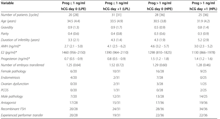
Table 1 Descriptive statistics
Variable Prog≤1 ng/ml Prog≤1 ng/ml Prog > 1 ng/ml Prog > 1 ng/ml hCG day 0 (LPE) hCG day +1 (LPL) hCG day 0 (HPE) hCG day +1 (HPL)
Number of patients [cycles] 20 [28] 31 [31] 28 [36] 25 [36]
Age (years) 34.5 (4.4) 33.5 (4.9) 30.5 (3.8) 31.9 (4.2)
Gravidity 0.9 (1.3) 0.9 (1.7) 0.5 (0.9) 0.8 (1.4)
Parity 0.4 (0.6) 0.4 (0.8) 0.3 (0.6) 0.3 (0.9)
Duration of infertility (years) 3.3 (2.1) 4.3 (1.4) 4.3 (1.9) 5.2 (2.9)
AMH (ng/ml)* 2.7 (2.1 - 5.0) 4.1 (2.5 - 6.2) 4.6 (3.2 - 5.7) 3.0 (2.3 - 5.2)
E2 (pg/ml)* 1460 (956–2150) 1390 (964–2110) 1298 (810–1825) 1130 (866–1978)
Progesteron (ng/ml)* 0.7 (0.5 - 0.9) 0.8 (0.5 - 0.9) 1.5 (1.2 - 1.8) 1.4 (1.2 - 1.6)
Number of embryos transferred 1.25 (0.64) 1.52 (0.72) 1.29 (0.60) 1.28 (0.46)
Female pathology 6/20 10/31 16/28 9/25
Endometriosis 4/20 2/31 7/28 0/25
Ovarian dysfunction 0/20 2/31 3/28 1/25
PCOS 0/20 1/31 0/28 2/25
Male pathology 7/20 12/31 13/28 14/25
Antagonist 17/28 15/31 17/36 19/36
Recombinant FSH 20/28 24/31 28/36 34/36
Experienced performer transfer 20/28 19/31 22/36 22/36
Numbers are expressed as numbers or as means (SD) unless explained differently. *Numbers are expressed as median (first and third quartile).
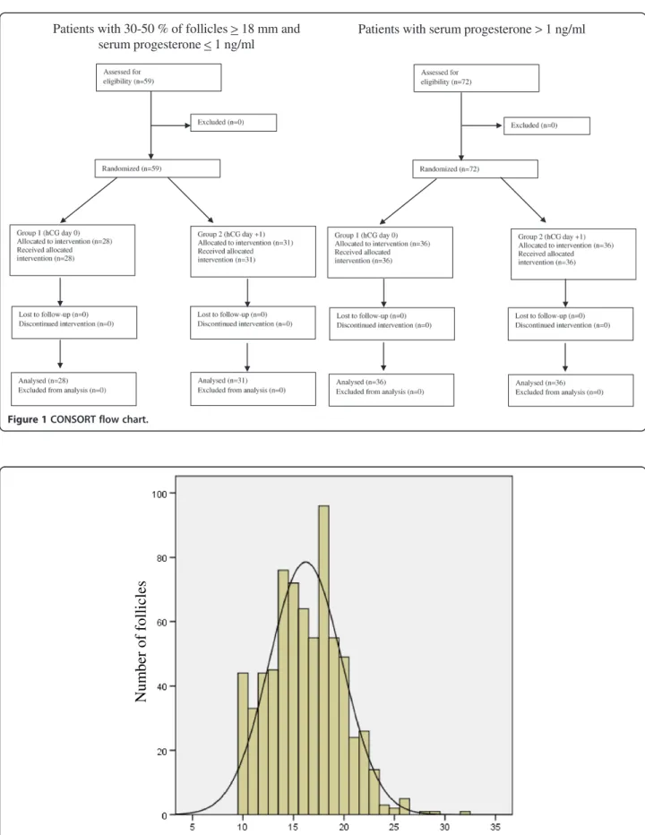
Patients with 30-50 % of follicles >18 mm and serum progesterone <1 ng/ml
Patients with serum progesterone > 1 ng/ml
Figure 1CONSORT flow chart.
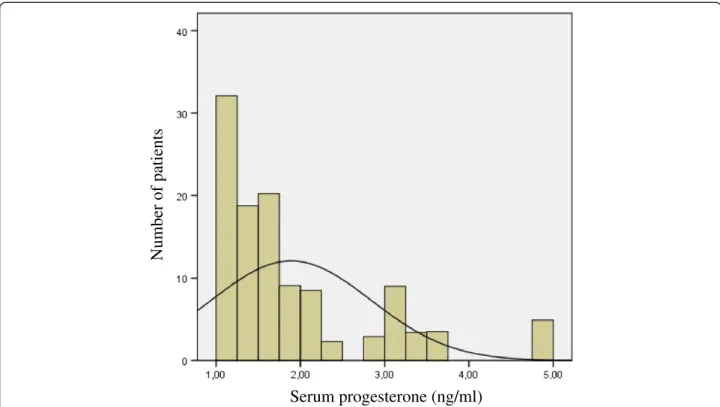
Follicle diameter (mm)
Num
b
er of
follicles
U-test was used. To account for correlations between measurements for women with repeated cycles, all fur-ther analyses were based on linear and logistic marginal regression models that were fitted using generalised esti-mating equations with exchangeable working correla-tions. Although adjustment for baseline covariates was not required in view of the randomised study design, ad-justments for age, PCOS and female pathology were used to improve precision in some of the linear models. All tests were performed at the 5% significance level. Statistical analyses were performed using SPSS, version 21, and R Studio, version 0.97.320.
Subsequent to ending the trial, additional evidence came to suggest that serum progesterone levels > 1.5 ng/ ml at the moment of triggering oocyte maturation might lower pregnancy rates. Therefore, we performed an add-itional comparison of 2 subgroups of patients: those with low progesterone levels (<1.0 ng/ml; group A) and those with highly elevated levels (>1.5 ng/ml; group B). Fisher's exact test and Student's t-test were again used as de-scribed above.
Results
Seventy-two patients with at least three follicles≥18 mm had serum progesterone levels > 1 ng/ml. They were ran-domised into 2 groups (HPE and HPL) of 36 individuals each. In the HPE group, the oocyte maturation trig-ger was administered on the same day. In the HPL group, hCG was injected 24 hours later. In the remaining cases with low serum progesterone levels
(<1 ng/ml), 59 patients had 30 to 50% of their folli-cles measuring equal to or greater than 18 mm. After randomisation, 28 patients were allocated to the LPE group and received hCG on the same day. The remaining 31 patients (LPL group) were trig-gered 24 hours later. All eligible patients were ran-domised and could be allocated (Figure 1). The data were analysed after correcting for duplicate patients in each group.
No important differences were observed between the LPE and LPL groups and between the HPE and HPL groups for all controlled variables (Table 1): age of the female, gravidity, parity, duration of infertility, ovarian reserve determined by AMH, peak estradiol level, num-ber of transferred embryos, diagnostic criteria (such as female pathology, endometriosis, ovarian dysfunction and PCOS), male pathology, stimulation protocol, num-ber of cancellations, numnum-ber of failed fertilisations and the experience of the physician performing the embryo transfer.
What is the effect of delaying the oocyte maturation trigger by 24 hours in patients with low serum progesterone (<1 ng/ml)?
In Figure 2, the distribution of the follicle diameters in these patients is illustrated. By waiting 24 hours (LPL group), we obtained more oocytes and predominantly more mature ones compared with the LPE group, where hCG was administered on the same day (Table 2). Multivariate analysis with correction for the female’s age showed a
Table 2 The influence of delaying oocyte maturation in patients with a normal progesterone level on the yield of (mature) oocytes, fertilised oocytes and good quality embryos
Day hCG Number included (cycles/[patients]) Oocytes MII 2PN GQE
0 (LPE) 28 [20] 9.36 (3.21) 7.64 (3.26) 5.29 (3.26) 4.32 (2.62)
+1 (LPL) 31 [31] 12.58 (7.62) 10.29 (6.35) 7.48 (5.25) 5.74 (4.61)
Mean difference 2.97 2.41 1.80 1.18
95% CI 0.45 to 5.49 0.22 to 4.61 −0.15 to 3.76 −0.53 to 2.88
P 0.021 0.031 0.071 0.18
Numbers are expressed as means (SD). Adjusted for age.
Table 3 The influence of delaying oocyte maturation in patients with a normal progesterone level on pregnancy and pregnancy outcome
Day hCG PR/cycle CPR/cycle OPR/cycle LBR/cycle CIR/E* OIR/E* LBR/E*
0 (LPE) 31.3% 31.3% 27.1% 27.1% 0.27 (0.42) 0.22 (0.40) 0.22 (0.40)
+1 (LPL) 35.5% 29.0% 25.8% 25.8% 0.26 (0.43) 0.24 (0.43) 0.24 (0.43)
Odds ratio 1.24 0.92 1.00 1.00 Mean difference −0.017 0.017 0.017
95% CI 0.40 to 3.86 0.29 to 2.92 0.30 to 3.37 0.30 to 3.37 95% CI −0.23 to 0.20 −0.20 to 0.23 −0.20 to 0.23
P 0.71 0.88 1.00 1.00 P 0.88 0.87 0.87
significant difference in the number of oocytes (p = 0.021) and in the number of mature oocytes (p = 0.031) in favour of the LPL group (Table 2). No statistically significant dif-ferences were found in the number of fertilised oocytes and the number of good-quality embryos.
Several secondary variables were evaluated to compare the pregnancy rates and pregnancy outcomes. No signifi-cant differences were found between groups LPE and LPL (Table 3). Multivariate analysis did not influence the final results.
What is the effect of delaying the oocyte maturation trigger by 24 hours in patients with high serum progesterone (>1 ng/ml)?
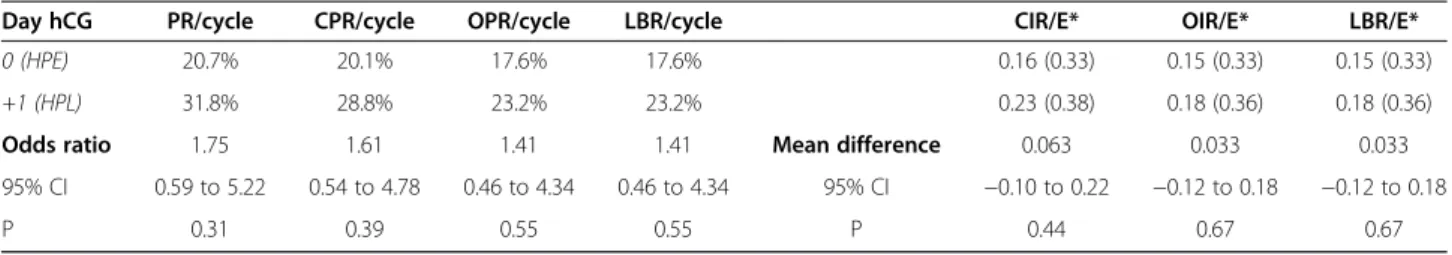
Figure 3 reveals a non-normal distribution of elevated progesterone levels. Thirty patients (48 cycles) had moderately elevated serum progesterone levels (>1 ng/ml
and≤1.5 ng/ml). Levels higher than 1.5 ng/ml were found in 23 patients (24 cycles). Cases with very high proges-terone levels (>3 ng/ml) were very scarce (3 patients).
The yield of mature oocytes was not different be-tween the HPE (hCG administered the same day) and HPL (hCG administered 24 hours later) groups. Multi-variate analysis did not affect the results (Table 4). The other variables (viz., number of oocytes recovered, ferti-lised oocytes, good quality embryos and all variables describing implantation and pregnancy rate) were also comparable (Table 5).
What is the effect of highly elevated serum progesterone (>1.5 ng/ml)?
We also compared the findings between patients with low progesterone levels (group A) and patients with very high progesterone levels (>1.5 ng/ml) (group B) who were
Serum progesterone (ng/ml)
Number of patients
Figure 3The distribution of progesterone levels in patients with elevated serum progesterone (>1 ng/ml).
Table 4 The influence of delaying oocyte maturation in patients with an elevated progesterone level on the yield of (mature) oocytes, fertilised oocytes and good quality embryos
Day hCG Number included (cycles/[patients]) Oocytes MII 2PN GQE
0 (HPE) 36 [28] 14.23 (8.44) 12.03 (7.09) 8.22 (5.84) 6.5 (5.02)
+1 (HPL) 36 [25] 13.06 (10.00) 11.81 (9.91) 7.78 (5.86) 6.2 (4.80)
Mean difference −1.52 −0.44 −0.97 −0.75
95% CI −5.30 to 2.27 −3.65 to 2.78 −3.49 to 1.55 −2.98 to 1.48
P 0.43 0.79 0.45 0.51
triggered on the day of the final monitoring (i.e., no delay of 24 hours). The numbers of (mature) oocytes, fertilised oocytes and good quality embryos were all significantly higher in group B. The variables describing implantation and pregnancy outcome were lower in group B, although statistical significance was lacking. More data are required to confirm our findings (Table 6).
Discussion
The effect of increasing progesterone levels
Increasing serum progesterone during stimulation for IVF/ICSI may have a negative impact on pregnancy rates. Venetis [10] reviewed this phenomenon and in-cluded studies with progesterone levels≥0.9 ng/ml. Ini-tially, this effect was described in only a fraction of the reports. In a recent systemic review [11], he expanded and further confirmed his findings. It seemed that a ratio
of progesterone-to-estradiol > 0.48 reduced pregnancy rates in antagonist cycles and that this ratio was found to be an independent predictor of pregnancy [12]. The effect of elevated progesterone levels was higher in pa-tients with a rather low ovarian response [11]. An emer-ging progesterone increase could be predicted by the number of follicles and an increase in serum estradiol [13]. This possible negative effect was the consequence of endometrial changes, because it was not described in oocyte donation programs [14] or when embryos that were obtained in a cycle with high progesterone were cryopreserved and subsequently thawed and transferred [15]. Van Vaerenbergh [16] demonstrated that gene ex-pression in the endometrium thoroughly changed when serum progesterone levels were higher than 1.5 ng/ml. This threshold of 1.5 ng/ml was further used by Bosch [6], who clearly demonstrated a negative effect on preg-nancy rates in both agonist and antagonist cycles. Although our study was not powered to compare implantation and pregnancy rates, the additional data in Table 6 confirmed these findings.
The yield of mature oocytes was either not mentioned or only calculated as a secondary variable in most stud-ies. Mio [17], Bustillo [18] and Venetis [11] demon-strated a higher number of retrieved oocytes in cycles with elevated progesterone levels. The cut off values for positive progesterone levels differed between studies, and uniform conclusions were not formulated. In our own study (Table 4), there was no significant difference in the number of (mature) oocytes.
From these observations, we may conclude that there is no evidence that increasing progesterone levels have a negative effect in cases where eggs are recruited for do-nation or for further cryopreservation, be it for medical or non-medical reasons.
Another possibility is that, if rising progesterone levels are encountered during the stimulation procedure, we can delay for a few days more to yield a maximum num-ber of good-quality eggs. They can be fertilised and cryo-preserved and be used for transfer later on, the so-called “segmented procedure”[19].
Table 5 The influence of delaying oocyte maturation in patients with an elevated progesterone level on pregnancy and pregnancy outcome
Day hCG PR/cycle CPR/cycle OPR/cycle LBR/cycle CIR/E* OIR/E* LBR/E*
0 (HPE) 20.7% 20.1% 17.6% 17.6% 0.16 (0.33) 0.15 (0.33) 0.15 (0.33)
+1 (HPL) 31.8% 28.8% 23.2% 23.2% 0.23 (0.38) 0.18 (0.36) 0.18 (0.36)
Odds ratio 1.75 1.61 1.41 1.41 Mean difference 0.063 0.033 0.033
95% CI 0.59 to 5.22 0.54 to 4.78 0.46 to 4.34 0.46 to 4.34 95% CI −0.10 to 0.22 −0.12 to 0.18 −0.12 to 0.18
P 0.31 0.39 0.55 0.55 P 0.44 0.67 0.67
*Numbers are expressed as means (SD). Unadjusted.
Table 6 The oocyte yield and pregnancy outcome in subgroups categorized by highly different progesterone levels
Group A Group B P Prog≤1
ng/ml D0
Prog > 1,5 ng/ml D0 Number of patients included 28 14
Number of oocytes 9,36 (3,21)* 16,71 (9,93)* 0.02
Number of mature oocytes 7,64 (3,26)* 13,43 (8,36)* 0.03
Number of fertilised oocytes 5,29 (3,26)* 10,00 (6,50)* 0.02
Number of good quality embryos 4,32 (2,62)* 7,93 (5,14)* 0.03
PR/cycle 31.3% 7,14% 0.07
CPR/cycle 31.3% 7,14% 0.07
OPR/cycle 27.1% 7,14% 0.11
LBR/cycle 27.1% 7,14% 0.11
CIR/embryo 0.27 (0.42) 0.07 (0.27) 0.07
OIR/embryo 0.22 (0.40) 0.07 (0.27) 0.16
LBR/embryo 0.22 (0.40) 0.07 (0.27) 0.16
The importance of follicle diameters
The ultrasound criteria to decide the best moment for triggering oocyte maturation have always been a point of discussion.
When no GnRH agonist/antagonist has been used, a leading follicle diameter of 16 mm or more and a serum estradiol of at least 600 pg/ml have served as guidelines for administering 10000 IU hCG [20]. Using a GnRH agonist—either in a long or short protocol—multiple cri-teria have been suggested. This can be explained by the different stimulation protocols that have been used and the variations in study designs. The leading follicles have had to reach diameters of 16 to 20 mm in most cases [1,2,21-28].
In antagonist cycles, most studies have proposed leading follicle diameters of 16 to 17 mm [4,9,29-31]. It seems that, in cycles where an antagonist has been used, the decision has been made somewhat earlier than in agonist cycles. In 2006, the Brussels GnRH an-tagonist Consensus Workshop Group stated that the optimal timing for triggering oocyte maturation when using a GnRH antagonist protocol needed to be ex-plored further [32].
In our study, we focused on follicle diameters in se-lected patients with low progesterone levels (<1 ng/ml). They all reached the threshold of having a least 3 folli-cles≥18 mm [5] with 30–50% of them being large enough. When waiting 24 hours to inject hCG, a larger number of (mature) oocytes were obtained (Table 2), as has already been mentioned by others [27].
In our series, we could not confirm a higher pregnancy rate in the group in which we triggered oocyte matur-ation 1 day later (Table 3). This confirms the findings by Tremmelen and Lane [33], who found that advancing or delaying hCG administration by 1 day from ‘ideal’ had no adverse impact on IVF treatment outcomes in non-programmed GnRH antagonist cycles. Again, we must notice that our study was not powered to compare preg-nancy rates, so conclusions on cycle outcomes must be interpreted with caution.
In spite of this, our findings support the idea that a higher yield of mature oocytes indirectly contributes to a higher overall productivity rate, as mentioned by Stanger and Yovich [34].
Conclusions
As soon as three follicles have a diameter≥18 mm, fur-ther decisions to pinpoint the moment for administering hCG depend on the progesterone level. If the progester-one level is higher than 1 ng/ml, delaying the adminis-tration of hCG by 24 hours has no effect on the number of mature oocytes. If the progesterone level is≤1 ng/ml and 30–50% of the follicles have diameters≥18 mm, delaying oocyte maturation by 24 hours is advised.
Abbreviations
2PN:2 pro-nuclear; AMH: Anti-Müllerian hormone; CIR: Clinical implantation rate; CPR: Clinical pregnancy rate; GnRH: Gonadotrophin releasing hormone; GQE: Good quality embryos; hCG: Human chorionic gonadotrophin; HPE: High progesterone early group; HPL: High progesterone late group; LBR: Live birth rate; LH: Luteinizing hormone; LPE: Low progesterone early group; LPL: Low progesterone late group; MII: Metaphase 2 oocytes; OIR: Ongoing implantation rate; OPR: Ongoing pregnancy rate;
PCOS: Polycystic ovary syndrome; PR: Pregnancy rate; SD: Standard deviation.
Competing interests
The authors declare that they have no competing interests.
Authors’contributions
FV designed the study protocol, included patients, analysed the data and wrote the manuscript. JG included patients and helped in analysing the data. SV performed the statistical analysis. AD included patients and helped in putting the data into the database. KT helped in structuring the database. PD included patients and all authors critically revised the article and approved it to be published.
Acknowledgements
PD is holder of a clinical research mandate by the Flemish Foundation for Scientific Research (FWO-Vlaanderen).
Author details
1Centre for Reproductive Medicine, University Hospital Ghent, De Pintelaan
185, Gent 9000, Belgium.2Department of Applied Mathematics, Computer Science and Statistics, Ghent University, Krijgslaan 281 S9, Gent 9000, Belgium.
Received: 1 February 2014 Accepted: 17 April 2014 Published: 23 April 2014
References
1. Ectors FJ, Vanderzwalmen P, VanHoeck J, Nijs M, Verhaegen G, Delvigne A, Schoysman R, Leroy F:Relationship of human follicular diameter with oocyte fertilization and development after in-vitro fertilization or intracytoplasmic sperm injection.Hum Reprod1997,12:2002–2005. 2. Inaudi P, Germond M, Senn A, Degrandi P:Timing of Hcg administration
in cycles stimulated for in-vitro fertilization - specific impact of heterogeneous follicle sizes and steroid concentrations in plasma and follicle fluid on decision procedures.Gynecol Endocrinol1995,9:201–208. 3. Kwan I, Bhattacharya S, McNeil A, Van Rumste MME:Monitoring of stimulated
cycles in assisted reproduction IVF and ICSI.Cochrane Database Syst Rev
2008 [http://onlinelibrary.wiley.com/doi/10.1002/14651858.CD005289.pub2/ abstract].
4. Kyrou D, Kolibianakis EM, Fatemi HM, Tarlatzis BC, Tournaye H, Devroey P:Is earlier administration of human chorionic gonadotrophin (hCG) associated with the probability of pregnancy in cycles stimulated with recombinant follicle-stimulating hormone and gonadotrophin-releasing hormone (GnRH) antagonists? A prospective randomized trial.Fertil Steril
2011,96:1112–1115.
5. Garcia-Velasco JA, Isaza V, Vidal C, Landazabal A, Remohi J, Simon C, Pellicer A:Human ovarian steroid secretion in vivo: effects of GnRH agonist versus antagonist (cetrorelix).Hum Reprod2001,16:2533–2539. 6. Bosch E, Labarta E, Crespo J, Simon C, Remohi J, Jenkins J, Pellicer A:
Circulating progesterone levels and ongoing pregnancy rates in controlled ovarian stimulation cycles for in vitro fertilization: analysis of over 4000 cycles.Hum Reprod2010,25:2092–2100.
7. Dirckx K, Cabri P, Merien A, Galajdova L, Gerris J, Dhont M, De Sutter P:
Does low-dose aspirin improve pregnancy rate in IVF/ICSI? A randomized double-blind placebo controlled trial.Hum Reprod2009,24:856–860. 8. Zegers-Hochschild F, Adamson GD, De Mouzon J, Ishihara O, Mansour R,
Nygren K, Sullivan E, van der Poel S:International Committee for Monitoring Assisted Reproductive T, World Health O: The International Committee for Monitoring Assisted Reproductive Technology (ICMART) and the World Health Organization (WHO) Revised Glossary on ART Terminology, 2009.Hum Reprod2009,24:2683–2687.
hCG administration results in a higher incidence of endometrial advancement on the day of oocyte retrieval in GnRH antagonist cycles. Hum Reprod2005,20:2453–2456.
10. Venetis CA, Kolibianakis EM, Papanikolaou E, Bontis J, Devroey P, Tarlatzis BC:
Is progesterone elevation on the day of human chorionic gonadotrophin administration associated with the probability of pregnancy in in vitro fertilization? A systematic review and meta-analysis.Hum Reprod Update
2007,13:343–355.
11. Venetis CA, Kolibianakis EM, Bosdou JK, Tarlatzis BC:Progesterone elevation and probability of pregnancy after IVF: a systematic review and meta-analysis of over 60 000 cycles.Hum Reprod Update2013,
19:433–457.
12. Cetinkaya ES, Berker B, Aytac R, Atabekoglu C, Sonmezer M, Ozmen B:The value of the progesterone-to-estradiol ratio on the day of hCG administration in predicting ongoing pregnancy and live birth rates in normoresponders undergoing GnRH antagonist cycles.Eur J Obstet Gynecol Reprod Biol2013,
170:452–457.
13. Kyrou D, Al-Azemi M, Papanikolaou EG, Donoso P, Tziomalos K, Devroey P, Fatemi HM:The relationship of premature progesterone rise with serum estradiol levels and number of follicles in GnRH antagonist/recombinant FSH-stimulated cycles.Eur J Obstet Gynecol Reprod Biol2012,162:165–168. 14. Al-Azemi M, Kyrou D, Kolibianakis EM, Humaidan P, Van Vaerenbergh I,
Devroey P, Fatemi HM:Elevated progesterone during ovarian stimulation for IVF.Reprod Biomed Online2012,24:381–388.
15. Silverberg KM, Martin M, Olive DL, Burns WN, Schenken RS:Elevated serum progesterone levels on the day of human chorionic-gonadotrophin administration in in-vitro fertilization cycles do not adversely affect embryo quality.Fertil Steril1994,61:508–513.
16. Van Vaerenbergh I, Fatemi HM, Blockeel C, Van Lommel L, In't Veld P, Schuit F, Kolibianakis EM, Devroey P, Bourgain C:Progesterone rise on HCG day in GnRH antagonist/rFSH stimulated cycles affects endometrial gene expression.Reprod Biomed Online2011,22:263–271.
17. Mio Y, Sekijima A, Iwabe T, Onohara Y, Harada T, Terakawa N:Subtle rise in serum progesterone during the follicular phase as a predictor of the outcome of invitro fertilization.Fertil Steril1992,58:159–166. 18. Bustillo M, Stern JJ, Coulam CB:Serum progesterone at the time of
human chorionic gonadotrophin does not predict pregnancy in in-vitro fertilization and embryo transfer.Hum Reprod1995,10:2862–2867. 19. Fatemi HM, Blockeel C, Devroey P:Ovarian stimulation: today and
tomorrow.Curr Pharm Biotechnol2012,13:392–397.
20. Murad NM:Ultrasound or ultrasound and hormonal determinations for in vitro fertilization monitoring.Int J Gynaecol Obstet1998,63:271–276. 21. De Sutter P, Dhont M:Poor response after hormonal stimulation for
in vitro fertilization is not related to ovarian aging.Fertil Steril2003,
79:1294–1298.
22. Elgindy EA:Progesterone level and progesterone/estradiol ratio on the day of hCG administration: detrimental cutoff levels and new treatment strategy.Fertil Steril2011,95:1639–1644.
23. Golan A, Herman A, Soffer Y, Bukovsky I, Ronel R:Ultrasonic control without hormone determination for ovulation induction in in-vitro fertilization-embryo transfer with gonadotrophin-releasing-hormone analog and human menopausal gonadotrophin.Hum Reprod1994,
9:1631–1633.
24. Kovacs P, Kovats T, Bernard A, Zadori J, Szmatona G, Kaali SG:Comparison of serum and follicular fluid hormone levels with recombinant and urinary human chorionic gonadotrophin during in vitro fertilization. Fertil Steril2008,90:2133–2137.
25. Lass A:Group UKToh: Monitoring of in vitro fertilization-embryo transfer cycles by ultrasound versus by ultrasound and hormonal levels: a prospective, multicenter, randomized study.Fertil Steril2003,80:80–85. 26. Miller KF, Goldberg JM, Falcone T:Follicle size and implantation of
embryos from in vitro fertilization.Obstet Gynecol1996,88:583–586. 27. Mochtar MH, Custers IM, Koks CAM, Bernardus RE, Verhoeve HR, Mol BW,
Van Wely M, van der Veen F:Timing oocyte collection in GnRH agonists down-regulated IVF and ICSI cycles: a randomized clinical trial. Hum Reprod2011,26:1091–1096.
28. Tan SL, Balen A, El Hussein E, Mills C, Campbell S, Yovich J, Jacobs HS:
A prospective randomized study of the optimum timing of human chorionic gonadotrophin administration after pituitary desensitization in in vitro fertilization.Fertil Steril1992,57:1259–1264.
29. Hauzman EE, Bodri D, Guillen JJ, Vidal R, Coll O, Vernaeve V:Exploring different criteria by 2D or 3D ultrasound for triggering final oocyte maturation in an oocyte donation program: a randomized pilot study. Hum Reprod2011,26:I95–I96.
30. Kolibianakis EM, Albano C, Camus M, Tournaye H, Van Steirteghem AC, Devroey P:Prolongation of the follicular phase in in vitro fertilization results in a lower ongoing pregnancy rate in cycles stimulated with recombinant follicle-stimulating hormone and gonadotrophin-releasing hormone antagonists.Fertil Steril2004,82:102–107.
31. Kolibianakis EM, Papanikolaou EG, Tournaye H, Camus M, Van Steirteghem AC, Devroey P:Triggering final oocyte maturation using different doses of human chorionic gonadotrophin: a randomized pilot study in patients with polycystic ovary syndrome treated with gonadotrophin-releasing hormone antagonists and recombinant follicle-stimulating hormone. Fertil Steril2007,88:1382–1388.
32. Tarlatzis BC, Fauser BC, Kolibianakis EM, Diedrich K, Devroey P:GnRH antagonists in ovarian stimulation for IVF.Hum Reprod Update2006,
12:333–340.
33. Tremellen KP, Lane M:Avoidance of weekend oocyte retrievals during GnRH antagonist treatment by simple advancement or delay of hCG administration does not adversely affect IVF live birth outcomes. Hum Reprod2010,25:1219–1224.
34. Stanger JD, Yovich JL:Follicle recruitment determines IVF productivity rate via the number of embryos frozen and subsequent transfers. Reprod Biomed Online2013,27:286–296.
doi:10.1186/1477-7827-12-31
Cite this article as:Vandekerckhoveet al.:Delaying the oocyte maturation trigger by one day leads to a higher metaphase II oocyte yield in IVF/ICSI: a randomised controlled trial.Reproductive Biology and Endocrinology201412:31.
Submit your next manuscript to BioMed Central and take full advantage of:
• Convenient online submission
• Thorough peer review
• No space constraints or color figure charges
• Immediate publication on acceptance
• Inclusion in PubMed, CAS, Scopus and Google Scholar
• Research which is freely available for redistribution