SHORT COMMUNICATION
Beyond the passive interactions
at the nano-bio interface: evidence of Cu
metalloprotein-driven oxidative dissolution
of silver nanoparticles
Daniel N. Freitas
1, Andrew J. Martinolich
1,2, Zoe N. Amaris
1and Korin E. Wheeler
1*Abstract
Background: In a biological system, an engineered nanomaterial (ENM) surface is altered by adsorbed proteins that modify ENM fate and toxicity. Thus far, protein corona characterizations have focused on protein adsorption, interac-tion strength, and downstream impacts on cell interacinterac-tions. Given previous reports of Ag ENM disrupinterac-tion of Cu traf-ficking, this study focuses on Ag ENM interactions with a model Cu metalloprotein, Cu(II) azurin. The study provides evidence of otherwise overlooked ENM-protein chemical reactivity within the corona: redox activity.
Results: Citrate-coated Ag ENMs of various sizes (10–40 nm) reacted with Cu(II) azurin resulted in an order of magni-tude more dissolved ionic silver (Ag(I)(aq)) than samples of Ag ENMs only, ENMs mixed Cu(II) ions, or control proteins such as cytochrome c and horse radish peroxidase. This dramatic increase in ENM oxidative dissolution was observed even when Cu(II) azurin was combined with a diverse mixture of Escherchia coli proteins to mimic the complexity of the cellular conona. SDS PAGE results confirm that the multiprotein ENM corona includes azurin. A Cu(I)(aq) colorimet-ric indicator confirms Cu(II) azurin reduction upon interaction with Ag ENMs, but not with the addition of ionic silver, Ag(I)(aq).
Conclusions: Cu(II) azurin and 10–40 nm Ag ENMs react to catalyze Ag ENM oxidative dissolution and reduction of the model Cu metalloprotein. Results push the current evaluation of protein-ENM characterization beyond passive binding interactions and enable the proposal of a mechanism for reactivity between a model Cu metalloprotein and Ag ENMs.
Keywords: Nanomaterials, Nanoparticles, Silver nanoparticles, Protein corona, Redox chemistry, Cu metalloprotein, Azurin
© 2016 Freitas et al. This article is distributed under the terms of the Creative Commons Attribution 4.0 International License (http://creativecommons.org/licenses/by/4.0/), which permits unrestricted use, distribution, and reproduction in any medium, provided you give appropriate credit to the original author(s) and the source, provide a link to the Creative Commons license, and indicate if changes were made. The Creative Commons Public Domain Dedication waiver (http://creativecommons.org/ publicdomain/zero/1.0/) applies to the data made available in this article, unless otherwise stated.
Findings Background
The design of metal and metal oxide engineered nanoma-terials (ENMs) are complicated by chemical and physical changes in a physiological system. Protein adsorption, for example, forms a “corona” and leaves ENM surfaces with little resemblance to the original material [1–3] to permanently alter ENM reactivity [2, 4–8]. Current ENM
protein corona studies focus on characterization of pro-tein adsorptions [9–11] and mediation of downstream biological uptake and toxicity [5, 6, 12, 13]. Proteins and metal ENMs are, however, likely to undergo chemical reactions and alter biological and environmental reactiv-ity of ENMs. Despite the evidence that ENMs are bio-chemically reactive [14–16] and some even demonstrate enzyme-like activity [16], characterization of ENMs chemically reacting with proteins (beyond protein bind-ing and unfoldbind-ing) has been mostly overlooked.
With this in mind, Cu metalloproteins were chosen as a case study characterization of protein-ENM biochemical
Open Access
*Correspondence: [email protected]
1 Department of Chemistry and Biochemistry, Santa Clara University,
Santa Clara, CA 95053, USA
reactivity because recent studies by Armstrong et al. [17] and others [18] have demonstrated that Ag ENMs can disrupt copper trafficking. Importantly, free silver ions (Ag(I)(aq)), by contrast, were found to have no impact on Cu trafficking. Results suggest that Cu-metalloenzymes react uniquely with Ag ENMs within the organisms stud-ied and establish the need to specifically evaluate the bio-chemical interactions between Cu metalloproteins and Ag ENMs.
Cu(II) azurin, a model Cu metalloprotein, is extensively characterized, redox active, and structurally simple with one metal center [19]. Importantly, previous studies of Cu(II) azurin–Ag ENM interactions align with findings by Armstrong et al. [17]; when directly interacting with the Ag ENM surface Cu(II) azurin forms biologically inactive, but fully folded apo- and Ag(I) azurin [20]. Ag(I) (aq), however, cannot displace the tightly bound Cu(II) within azurin, a result consistent with previous work [21]. The reactivity of Cu(II) azurin with Ag ENMs, but not with Ag(I)(aq), justifies further study in the elucida-tion of biochemical reactivity between Cu metallopro-teins and Ag ENMs.
Here, we evaluate the hypothesis that Cu(II) azurin– Ag ENM are a redox pair. Evidence is presented for Cu(II) azurin binding to Ag ENMs and increasing oxida-tive dissolution to form Ag(I)(aq), even in the presence of a complex mixture of other bacterial proteins. Evi-dence is also presented for reduction of Cu(II) azurin. Taken together, these experiments explain the unique behavior of Ag ENMs with Cu(II) azurin and, by exten-sion, provide biochemical reactivity as a foundation for Ag ENM modification of Cu homeostasis. More broadly, these data introduce the importance of considering the biochemical reactivity of ENMs beyond passive binding interactions.
Methods
Sample preparation
Citrate-coated ENMs were purchased from Nanocom-posix Inc (San Diego, CA). Cu(II) azurin was overex-pressed and purified as previously described [20, 22]. Other chemicals were purchased from Fisher Scientific, unless otherwise noted. Eppendorf Centrifuge 5424 and Molecular Devices SpectraMax M2 were used for centrif-ugation and UV–Vis spectra, respectively.
Unless otherwise stated, all samples included 50 μM Cu(II) azurin, horseradish peroxidase (HRP, Sigma), or equine cytochrome c (cyt c, US Biologicals) reacted with Ag ENMs in nanopure water (18 mΩ). ENM con-centrations ensured equal surface area and protein bind-ing sites [23] at 3.73, 0.955, 0.416 and 0.233 nM for 10,
20, 30, and 40 nm ENMs, respectively. Soluble protein extract (SPE) was taken from E. coli Migula Castellani
and Chalmers (ATCC) and used at 0.7 and 0.07 mg/ml, which is equivalent to 50 and 5 µM azurin. Procedures for SPE extraction and purification are provided in sup-plemental materials.
ICP‑MS quantification of Ag ENM dissolution
Samples were centrifuged (30 min, 21 K RCF) to remove ENMs from solution after 6 h incubation. 85 % of the supernatant was removed and re-centrifuged. Again, 85 % of the supernatant was removed for preparation and analysis of Ag(I) concentration using an Agilent 7500CE ICP-MS (Agilent Technologies, Palo Alto, CA, USA) by the Interdisciplinary Center for Plasma Mass Spectrom-etry (University of California at Davis, CA, USA). The samples were introduced using a MicroMist Nebulizer (Glass Expansion, Pocasset, MA, USA) into a temper-ature-controlled spray chamber. Instrument standards diluted from Certiprep 2A (SPEX CertiPrep, Metuchen, NJ, USA) encompassed the range 0, 0.5, 1, 10, 50, 100, 200, 500, 1000 parts per billion (ppb) in 3 % trace element grade HNO3 (Fisher Scientific, Fair Lawn, NJ, USA) in
18.2-MΩ water. A separate 100 ppb Certiprep 2A stand-ard was analyzed as every tenth sample as a quality con-trol. Sc, Y and Bi Certiprep standards (SPEX CertiPrep) were diluted to 100 ppb in 3 % HNO3 and introduced by
peripump as an internal standard.
BCA analysis for Cu(I) detection
The reactions for Cu(I) detection were executed as described above. After 6 h, 100 µL of each sample was combined with 1 mM bicinchoninic acid (BCA, MP Bio-medicals, LLC) and analyzed immediately via UV–Vis spectrophotometry using a Shimadzu UV-1800 for Cu(I) detection at 562 nm (ε562 = 14,150 M−1 cm−1). To enable
peak analysis, ENMs were centrifuged out of solution (30 min, 15 K RPM) and the supernatant was analyzed for Cu(I) using BCA. To ensure BCA was not reacting with Cu(II) from Cu(II) azurin, Cu(II)(aq), or Ag(I)(aq), an array of controls were also run as described in supple-mental materials.
Results and discussion
of Ag ENM oxidative dissolution within a complex pro-tein mixture like that in the cell. Independent evidence of redox activity was measured through quantification of Cu(II) azurin reduction and Cu(I) release. With multi-ple sources of evidence for Ag ENM- Cu(II) azurin redox activity, a mechanism of Ag ENM-Cu(II) azurin reactivity is proposed.
Cu(II) azurin catalyzes Ag ENM dissolution
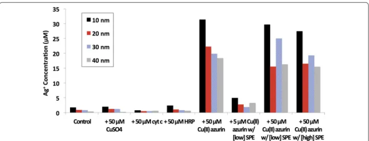
Formation of Ag(I)(aq) by oxidative dissolution of 10–40 nm Ag ENMs was quantified by ICP-MS (Fig. 1). Oxidative dissolution was small at 2 µM Ag(I)(aq) or less for Ag ENMs alone, or with CuSO4, cyt c, and HRP.
Addition of Cu(II) azurin, however, increased Ag(I)(aq) concentrations by roughly an order of magnitude. Protein driven oxidative dissolution has been previously reported for Ag ENMs, as well as other metal and metal oxide ENMs.
When azurin was mixed with varying concentrations of SPE, Cu(II) azurin still catalyzed Ag ENM dissolution. When the contribution of SPE is removed, Ag(I)(aq) con-centrations are similar to those found in samples of ENM and azurin alone (Fig. 1, see Additional file 1: Figure S2 for raw concentrations). Even at lower, 5 μM azurin con-centrations, where azurin does not dominate the protein corona (SDS PAGE results, Additional file 1: Figure S3) oxidative dissolution is two to four times that measured in control samples. SDS PAGE gels indicate that Cu(II) azurin is within the hard corona even when mixed with SPE (Additional file 1: Figure S3).
Notably, although Ag ENM reduction potentials are size dependent, there is not a clear size dependence in Ag ENM oxidative dissolution within this sample set; the sole exception is that highest dissolved Ag(I)(aq) con-centrations were consistently observed in the smallest, 10 nm ENM samples.
Ag ENMs reduce Cu(II) azurin
Reduction of Cu(II) azurin was assessed with bicin-choninic acid (BCA) as a colorimetric indicator of Cu(I)(aq) (Fig. 2). The Cu(I)-BCA absorption peak appears as a shoulder on the Cu(II)-thiol ligand to metal charge transfer (LMCT) band of Cu(II) azurin. As expected, samples with the strongest Cu(I)-BCA shoulder have smaller LMCT bands from Cu(II) azurin, indicative of reduction or loss of Cu(II) from azurin. Control studies demonstrate that the BCA was reactive only with Cu(I)(aq), but not with azurin or Ag(I)(aq) alone (Additional file 1: Figure S3). In addi-tion, 10-nm citrate-coated Au ENMs reacted with Cu(II) azurin show no evidence of Cu(I) and ESI–MS analysis of the resulting azurin did not reveal any apo- or Au- azurin (data not shown). Consistent with previ-ous reports [21] that the very strong Cu(II)-thiol bond must be reduced before copper release, these results confirm both ENM and redox activity are necessary for Cu(II) azurin-ENM reactivity.
Electronic absorbance spectra were deconvo-luted (Additional file 1: Figure S3.b) and spectral contributions were used to calculate the respective
concentrations of Cu(I)(aq) and Cu(II) azurin (Fig. 2b). Monovalent copper concentrations were uniformly low at 5 µM or less. Consistent with oxidative disso-lution results to form Ag(I)(aq), no size-dependence was observed in formation of Cu(I)(aq), but 10 nm Ag ENMs did react to form the most Cu(I). Concen-trations of Cu(II) azurin decreased in accordance with Cu(I)(aq) formation; 10 nm Ag ENMs resulted in the lowest amount of Cu(II) in azurin.
Notably, measured total concentrations of cop-per from Cu(I)(aq) and Cu(II) azurin were consistent across samples, at 21.1 ± 1.2 µM. This concentration is 25–30 % lower in than the starting concentration of copper in azurin and has been confirmed by ICP-MS. The remaining copper may be bound to Ag ENMs and Ag ENM-azurin complexes pelleted out during sample processing. These results suggest a small percentage of released copper or Cu azurin may be adsorbing to the ENM surface; a conclusion consistent with evidence of strong azurin–Ag ENM complex formation previously reported [20] and SDS PAGE results indicating azurin is in the hard corona. The calculated Cu(I)(aq) concen-trations from deconvoluted electronic spectra do not follow an Ag ENM size trend. This may be due to the loss of copper during sample processing, but it may also be explained by variations in Ag ENM curvature with size that alter protein-Ag ENM interactions.
Conclusions
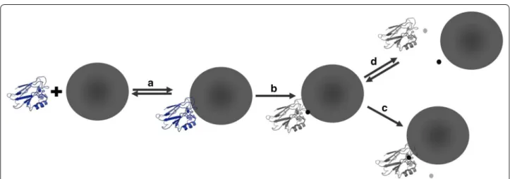
Evidence of a protein-Ag ENM redox reaction is two-fold. First, Cu(II) azurin increases Ag ENM oxida-tive dissolution by an order of magnitude over other model redox proteins, even in the presence of a com-plex mixture of proteins within the corona. Second, Ag ENMs, not Ag(I)(aq) or Au ENMs, reduce and dis-place Cu(II) from azurin. We propose a mechanism of Cu(II) azurin–Ag ENM interactions wherein complex formation results in oxidation of surface Ag on the ENM and reduction of Cu(II) in azurin (Fig. 3). After redox, the Cu(I) is displaced from azurin to either form apo-azurin, or to bind the Ag(I) and form Ag(I) azurin. Either way, the fate of both reactants is altered by increasing Ag ENM dissolution and disrupting the physiological function of the Cu metalloprotein. This mechanism provides a biochemical explanation for Cu(II) azurin reactivity with Ag ENMs, but not with Ag(I)(aq) or when Ag ENM-protein complex forma-tion is prevented [20].
This work provides an example of an adventitious pro-tein-ENM redox reaction that alters both metal ENM and protein reactivity. Although many consider dissolu-tion the main mechanism of silver ENM toxicity [14, 24, 25], few have considered the role of the protein corona in redox activity of ENMs and potential role in metal homeostasis [17]. More broadly, this work emphasizes
the need to further study adventitious protein redox reactivity with ENMs, especially considering the enzyme-like reactivity of some ENMs, and broadly demonstrates the chemical reactivity of the protein corona at the ENM surface.
Abbreviations
ENMs: engineered nanomaterials; BCA: bicinchoninic acid; ICP-MS: inductively coupled plasma mass spectrometry; SPE: soluble protein extract from E. coli; SDS PAGE: sodium dodecyl sulfate polyacrylamide gel electrophoresis; LMCT: ligand metal charge transfer; cyt c: cytochrome c; HRP: horse radish peroxidase.
Authors’ contributions
DNF designed and performed all ICP-MS sample preparation and analysis and made all figures. AJM carried out the BCA analysis of ENM-azurin reactions, and participated in the design of the study; ZA carried out experimental controls; DNF and AJM worked with KEW to design experiments and in manu-script preparation; KEW conceived of, designed, and coordinated the study, and drafted the manuscript. All authors gave final approval for publication. All authors read and approved the final manuscript.
Author details
1 Department of Chemistry and Biochemistry, Santa Clara University, Santa
Clara, CA 95053, USA. 2 Present Address: Department of Chemistry, Colorado
State University, Fort Collins, CO 80523-1872, USA. Acknowledgements
We would like to thank Paul Abbyad for providing assistance in fitting the electronic spectra; we also thank Kyle Lancaster and Harry Gray for fruitful Additional file
Additional file 1: Additional methods. SPE extraction and SDS PAGE gels. Figure S1. ICP-MS quantification of Ag(I)(aq) concentrations from oxidative dissolution of Ag ENMs. Figure S2.a. Control UV–Vis spectra for BCA detection of Cu(I)(aq). Figure S2.b. Sample spectra showing deconvolution of UV–Vis spectra. Figure S3. SDS PAGE gels of the protein corona of Ag ENMs.
discussions on azurin purification and reactivity. We thank Joel Commisso and Austin M. Cole (UC Davis Interdisciplinary Center for Plasma Mass Spectrom-etry) for technical assistance and ICP-MS data.
Competing interests
The authors declares that they have no competing interests. Funding
This research was supported by an award from Research Corporation for Science Advancement, with additional financial assistance from Santa Clara University.
Received: 5 August 2015 Accepted: 14 January 2016
References
1. Monopoli MP, Aberg C, Salvati A, Dawson K. A. Nat Nanotechnol. 2012;7(12):779.
2. Docter D, Westmeier D, Markiewicz M, Stolte S, Knauer SK, Stauber RH. Chem Soc Rev. 2015;44(17):6094.
3. Hellstrand E, Lynch I, Andersson A, Drakenberg T, Dahlback B, Dawson KA, et al. Complete high-density lipoprotiens in nanoparticle corona. FEBS J. 2009;276:3372.
4. Arvizo RR, Giri K, Moyano D, Miranda OR, Madden B, McCormick DJ, et al. Identifying new therapeutic targets via modulation of protien corona formation by engineered nanoparticles. PLoS One. 2012;7(3):e33650. 5. Walkey CD, Olsen JB, Song F, Liu R, Guo H, Olsen DWH, et al. Protein
corona fingerprinting predicts the cellular interaction of gold and silver nanoparticles. ACS Nano. 2014;8(6):5515.
6. Walkey CD, Chan WCW. Understanding and controlling the interaction of nanomaterials with proteins in a physiological environment. Chem Soc Rev. 2012;41(7):2780.
7. Setyawati MI, Tay CY, Docter D, Stauber RH, Leong DT. Understanding and exploiting nanoparticles’ intimacy with the blood vessel and blood. Chem Soc Rev. 2015;44(22):8174.
8. Walczyk D, Bombelli FB, Monopoli MP, Lynch I, Dawson KA. What the cell “sees” in bionanoscience. J Am Chem Soc. 2010;132:5761.
9. Tenzer S, Docter D, Kuharev J, Musyanovych A, Fetz V, Hecht R, et al. Rapid formation of plasma protein corona critically affects nanoparticle patho-physiology. Nat Nanotechnol. 2013;8(10):772.
• We accept pre-submission inquiries
• Our selector tool helps you to find the most relevant journal • We provide round the clock customer support
• Convenient online submission • Thorough peer review
• Inclusion in PubMed and all major indexing services • Maximum visibility for your research
Submit your manuscript at www.biomedcentral.com/submit
Submit your next manuscript to BioMed Central
and we will help you at every step:
10. Eigenheer R, Castellanos ER, Nakamoto MY, Gerner KT, Lampe AM, Wheeler KE. Environ Sci Nano. 2014; 1(3):238.
11. Zhou JD, Mortimer G, Martin D, Minchin RF. Differential plasma protein binding to metal oxide nanoparticles. Nanotechnology. 2009;20(45):455101.
12. Fleischer CC, Kumar U, Payne CK. Biomater. Sci. 2013;1(9):975. 13. Durán N, Silveira CP, Durán M, Martinez DST. Silver nanoparticle protein
corona and toxicity: a mini-review. J Nanobiotechnology. 2015;13:55. 14. Behra R, Sigg L, Clift MJD, Herzog F, Minghetti M, Johnston B, et al.
Bioavailability of silver nanoparticles and ions: from a chemical and biochemical perspective. Soc Interface. 2013;10(87):20130396.
15. Yokel RA, Hussain S, Garantziotis S, Demokritou P, Castranova V, Cassee FR. The Yin: An adverse health perspective of nanoceria: uptake, distribu-tion, accumuladistribu-tion, and mechanisms of its toxicity. Environ Sci Nano. 2014;1(5):406.
16. Hirst SM, Karakoti AS, Tyler RD, Sriranganathan N, Seal S, Reilly CM. Anti-inflammatory properties of cerium oxide nanoparticles. Small. 2009;5(24):2848.
17. Armstrong N, Ramamoorthy M, Lyon D, Jones K, Duttaroy A. Mechanism of silver nanoparticles action on insect pigmentation reveals intervention of copper homeostasis. PLoS One. 2013;8(1):e53186.
18. Wang B, Feng W, Zhao Y, Chai Z. Metallomics insights for in vivo studies of metal based nanomaterials. Metallomics. 2013;5(7):793.
19. Gray HB, Malmström BG, Williams RJP. Copper coordination in blue pro-teins. J Biol Inorg Chem. 2000;5(5):551.
20. Martinolich AJ, Park G, Nakamoto MY, Gate RE, Wheeler KE. Structural and functional effects of Cu metalloprotein-driven silver nanoparticle dissolu-tion. Environ Sci Technol. 2012;46(11):6355.
21. Tordi MG, Naro F, Giordano R, Silvestrini MC. Silver binding to Pseu-domonas aeruginosa azurin. Biometals. 1990;3(2):73.
22. Piccioli M, Luchinat C, Mizoguchi TJ, Ramirez BE, Gray HB, Richards JH. Inorg Chem. 2002;34(3):737.
23. Mattoussi H, Mauro JM, Goldman ER, Anderson GP, Sundar VC, Mikulec FV, et al. J Am Chem Soc. 2000;122(49):12142.
24. Liu J, Sonshine DA, Shervani S, Hurt RH. Controlled release of biologically active silver from nanosilver surfaces. ACS Nano. 2010;4(11):6903. 25. Yang X, Gondikas AP, Marinakos SM, Auffan M, Liu J, Hsu-Kim H, et al.