Genome Sequencing and Phylogenetic Analyses as a Basis for Molecular
Subtyping of Male-Specific (FRNA) Coliphages
by
Stephanie Dianne Friedman
A dissertation submitted to the faculty of the University of North Carolina at Chapel Hill in partial fulfillment of the requirements for the degree of Doctor of Philosophy in the
Department of Environmental Sciences and Engineering, School of Public Health
Chapel Hill 2008
Approved by:
Mark D. Sobsey, PhD
Jan Vinjé, PhD
Fred J. Genthner, PhD
Lola V. Stamm, PhD
Frederick K. Pfaender, PhD
Louise M. Ball, PhD
©2008
Abstract
Stephanie Dianne Friedman
Genome Sequencing and Phylogenetic Analyses as a Basis for Molecular Subtyping of Male-Specific (FRNA) Coliphages
(Under the direction of Mark D. Sobsey)
Monitoring programs for recreational waters utilize indicator bacteria concentrations as predictors of sewage-exposure related illness risks. However, most illnesses contracted through exposure to recreational waters may be of viral etiology. Identifying the fecal sources (non-human vs human) is also valuable information for risk management and source mitigation. Male-specific (FRNA) coliphages are proposed as sensitive enteric viral
indicators for source-tracking fecal pollution in environmental waters. Classified as family
Leviviridae of two genera, Levivirus and Allolevivirus, and four genogroups (I, II, III, IV) the
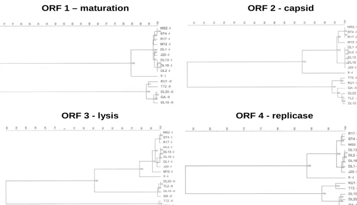
genogroup, nucleotide sequence similarities ranged from 75-99%, 83-93%, 69-95% and 74-95% for genogroups I, II, III and IV, respectively. Genogroup II lysis protein tree formed a unique branch that was not observed in the full-length nucleotide tree. Thus, both full-length nucleotide and individual protein sequences need to be evaluated when genotyping or
phylogenetically clustering these FRNA coliphages. From conserved regions within each genogroup, four genogroup-specific primer sets were designed for a reverse-transcription polymerase chain reaction (RT-PCR) assay. The assay was then evaluated successfully on a panel of environmental FRNA strains demonstrating their usefulness to assess the sanitary quality of recreational waters and provide data identifying and subsequently eliminating the contamination source.
Acknowledgments
It has been my privilege to be a member of the UNC Environmental Virology laboratory and to study under the direction of Drs. Mark Sobsey, Jan Vinjé and Fred Genthner. A generous thank you is extended to each of you for your guidance and assistance. A special gratitude to Dr. Genthner for going above and beyond to secure funding for the project and for dealing with the never ending layers of administration. I would also like to thank Drs. Ball, Stamm, Pfaender and Noble for serving on my committee.
vi
Table of Contents
Table of Contents... vii
List of Tables...ix
List of Figures...x
I. Introduction...1
II. Background...5
III. Research Objectives...9
IV. Literature Review...10
References... 49
V. Gene Mapping and Phylogenetic Analysis of the Complete Genome of 30 ssRNA Male-Specific Coliphages of the Family Leviviridae...56
Introduction... 57
Purpose... 59
Materials and Methods... 60
Results... 69
Discussion... 88
Summary... 94
References... 95
vii
Introduction... 100
Purpose... 103
Materials and Methods... 104
Results... 112
Discussion... 124
Summary... 129
References... 130
VII. A Reverse Transcription-PCR Assay to Distinguish the Four Genogroups of Male-Specific (F+) RNA Coliphages...133
Introduction... 134
Purpose... 136
Materials and methods... 137
Results... 148
Summary... 153
References... 154
VIII. Overall Discussion...157
IX. Summary and Conclusions...171
X. Recommendations for Future Research...173
References... 179
Appendix A1...182
Group I... 182
Group II ... 201
viii
Group III QB-like ... 235
Group IV... 253
Appendix B1...271
Group I... 271
Group II ... 279
Group III... 285
Group IV... 294
Appendix C...303
Group I and JS Nucleotides... 303
JS Amino Acids ... 325
Group I and JS Amino Acids... 329
ix
List of Tables
Table Page
Table 4.1 Bacteriophage classification. ... 14
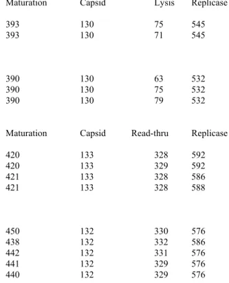
Table 5.1 Open Reading Frames positions for Leviviridae.... 70
Table 5.2 Leviviridae nucleotide percent similarity ... 72
Table 5.3 Start condons and Shine-Dalgarno sequences ... 76
Table 5.4 Leviviridae number of amino acids per gene... 79
Table 5.5 Comparison of genomic traits. Allolevivirus and Levivirus... 85
Table 6.1 JS strains and genogroup I... 115
Table 6.2 Pairwise comparison JS strains and genogroup I ... 116
Table 6.3 Pairwise comparison of amino acids for JS and genogroup I... 118
Table 6.4 Pairwise comparison of replicase protein JS and genogroup I. ... 119
Table 7.1 Sources of Leviviridae strains... 138
Table 7.2 Leviviridae accession numbers ... 142
x
List of Figures
Figure Page
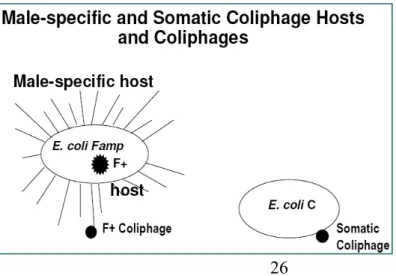
Figure 4.1 Somatic and male-specific coliphage hosts... 26
Figure 4.2. Schematic of male-specific coliphage classification... 29
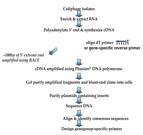
Figure 5.1. Flow-chart of sequencing methods... 62
Figure 5.2 Phylogenetic analysis of Leviviridae nucleotide sequences... 73
Figure 5.3 SimPlot nucleotide similarity and genome organization... 73
Figure 5.4 (A) Phylogenetic analysis of each protein: Levivirus I, II. ... 86
Figure 5.4 (B) Phylogenetic analysis of each protein: Allolevivirus III, IV. ... 87
Figure 6.1 SimPlot analysis of genome nucleotide profile strain MS2 and DL52. ... 120
Figure 6.2 SimPlot analysis of genome nucleotide profile strain MS2 and fr... 120
Figure 6.3 SimPlot analysis of replicase amino acid profile strain MS2 and DL52... 121
Figure 6.4 Phylogenetic analysis of replicase amino acids groups I, II and JS... 122
Figure 6.5 JS strains replicase frame-shift... 123
Figure 7.1 Gel electrophoresis of FRNA phages using one-step RT-PCR... 143
I. Introduction
A link between waterborne transmission of disease and sewage was first observed in 1854 by the historical achievements of Dr. John Snow. The observations by Dr. Snow pioneered the sciences of epidemiology, preventative medicine and public health
intervention. His observations of the cholera outbreak led to the removal of the Broad Street pump handle in London followed by a rapid, subsequent decline in cholera-related deaths (Snow, 1855).
Sewage disposal into marine and freshwater systems has occurred since the
municipal wastewater discharges, inputs such as stormwater runoff, septic-tank seepage from on-site systems, agricultural runoff, urban runoff and other fecal waste sources can enter aquatic environments. Other naturally occurring, non-pathogenic and potentially pathogenic viruses are already present in aquatic environments, thus adding to the complexity of the viral ecology of aquatic systems.
To minimize adverse impacts and to protect the public health and aquatic environments, management systems based on fecal indicators of microbial origin were implemented as a “warning flag” and a means by which to estimate fecal contamination based on direct microbial measurements. The World Health Organization (WHO; Ashbolt et al, 2001) defines microbial indicators of public health concern by one of three groups:
“(1) process indicator - group of organisms that demonstrates the efficacy of a process, such as total heterotrophic bacteria or total coliforms for chlorine disinfection
(2) fecal indicator - a group of organisms that indicates the presence of a fecal contamination, such as thermotolerant coliforms or E. coli. Hence, they only infer
that pathogens may be present (3) index and model organisms - a group or species indicative of pathogen presence and behavior, respectively. For example, Ecoli as an
index for Salmonella; F-RNA coliphages as models of human-enteric viruses.”
Criteria for an ideal fecal indicator selection are as follows: (1) consistently present in feces at higher concentration than those of pathogens; (2) cannot/should not replicate outside the intestinal tract; (3) easily detected and quantified; (4) non-pathogenic; (5) at least as resistant as pathogens to disinfection treatments and environmental conditions/inactivation rates; (6) indicator concentration in water is quantitatively associated with potential risks to human populations, typically from enteric illness; and (7) applicable to all water types (marine, freshwater, estuarine).
provide quantitative estimates of E. coli and enterococci. These methods are neither
real-time nor do theyprovideinformationregardingsource. The best thatcurrent methods can do is indicate that possible fecal contamination occurred within the last 24 hours. To minimize risks to human health, resource managers and human health advisors need an early-warning indicator that will enable them to assess the sanitary condition of waters in real-time or at least shortly after the sample is collected for analysis. An additional limitation ofthe current indicators, enterococci and E.coli, is that they do not correlate with the presence and
concentrations of allpotential water-borne pathogens (Griffin et al., 2003). Most
importantly, current EPA recreational water-quality criteria using two bacterial indicators have little or no correlation to the presence and concentration of human pathogenic viruses. To date, no viral indicator has been mandated for regulatory purposes in recreational waters in the USA.
Male-specific coliphages have been suggested as a viral indicator for: (1) fecal contamination (Osawa, 1981; Furuse, 1983), (2) enteric bacterial contamination (Gerba, 1987), (3) enteric viral contamination (Grabow, 2001; Leclerc et al, 2000) and (4) risks of gastro-intestinal illness from recreational water exposures (Colford et al., 2007). Male-specific coliphages, Male-specifically the ssRNA Leviviridae family, are superficially
indistinguishable from most human enteric viruses (Grabow, 2001), occur in higher numbers in sewage and wastewater effluents than viral enteric pathogens (Grabow, 2001), their presence implies the presence of pathogenic viruses (Grabow, 2001), and, in a majority of cases, they provide human/animal fecal-source specificity (Vinjé et al, 2004; Cole et al, 2003; Furuse, 1987; Schaper et al, 2002; Scott et al, 2002; Stewart, 2002; Long et al, 2005).
sequencing at least three to five strains from each FRNA coliphage genogroup (I, II, III and IV), (2) develop and analyze for identification of preferred targeted genomic regions the sequence database representing environmental and prototype strains and (3) based on the primers identified in step 2, design and validate a molecular assay to detect and ultimately subtype the different genogroups. To develop a genetic database, 19 FRNA strains were sequenced. In addition, two new undescribed Levivirus strains were sequenced. In addition,
II. Background
To develop valid fecal-indicator criteria based on credible epidemiology design, US EPA undertook a series of marine and freshwater public-beaches studies (EPA-600/1-84-004; EPA-600/1-80-031). Objectives were to assess the mathematical relationship between microbiological indicator concentrations in bathing water and illness rates resulting from recreational water exposure (swimming), in order to correct for perceived deficiencies in US Public Health Service studies conducted before 1972. Additional goals were to provide a statistically sound study outcome correlating the concentration of the best bacterial indicator with magnitude of health effects in bathers, resulting in specific bacterial concentration-health risk outcome relationships associated with swimming in sewage-contaminated waters. Two key US EPA documents were published from these epidemiological studies “Health Effects Criteria for Fresh Recreational Waters” (EPA-600/1-84-004) and “Health Effects Criteria for Marine Recreational Waters” (EPA-600/1-80-031). Epidemiological data supported the use of E. coli and enterococci as primary fecal indicators because they were
associated with statistically significant increased gastrointestinal illness rates to
swimmers/bathers at increasing concentration in water. Subsequently, EPA guidelines for recreational waters were established in the 1986 “Ambient Water Quality Criteria for Bacteria in Recreational Waters.”
Act of 2000. The BEACH Act was an amended Section 303 of the Clean Water Act. The focus was to improve public health and recreational water quality programs by
1) strengthening beach testing and standards, 2) providing faster testing methods, 3) predicting fecal pollution, 4) better defining the criteria as to which fecal indicators and water quality standards are based, 5) investing in health and methods research and 6) informing the public (EPA, 2003).
There are limitations associated with the two current bacterial fecal indicators for recreational water quality, enterococci and E.coli. They do not provide a complete
assessment of the sanitaryqualityofwater. Their presence and densities do not correlate with allpotential water-borne pathogens causing adverse environmental effects (Griffin et al., 2003). For example, most illnesses contracted by swimmers appear to be of viral etiology. Environmental and health risks associated with microbes can be as much a
consequence of viral contamination as it can of contamination by any other microorganisms. Both microbial and epidemiological evidence suggests that the current EPA recreational water-quality criteria using two bacterial indicators have little or no correlation to the
presence of pathogenic viruses or to health-risks from non-point source fecal contamination. In microbial water quality studies conducted in Florida, indicator bacteria counts satisfied EPA criteria limits in the presence of detectable human pathogenic viruses (Griffin et al., 2003; Griffin et al., 1999). In other studies, waters satisfying E. coli standards for shellfish
2000; Sinton et al., 1999; Sinton et al., 2002). Even when current bacteriological standards are met in recreational waters, risks to human health may be posed by viruses.
In other studies, E. coli and enterococci were detected in environmental niches of
tropical or temperate climates (Hawaii, Guam and Florida) where there was no evidence of human fecal contamination. Such environmental reservoirs of these bacteria led to high indicator counts that were not associated with fecal contamination (Hardina and Fujioka, 1991; Roll and Fujioka, 1997; Byappanahalli and Fujioka, 1998; Fujioka and
Byappanahalli, 2000; Solo-Gabriele HM et al., 2000; Genthner et al., 2005). In some cases, elevated bacterial indicator counts exceeding EPA water-quality criteria were influenced by soil run-off and not a result of sewage input (Byappanahalli and Fujioka, 2004).
The apparent weaknesses in current bacterial indicators and the need to develop better, faster methods to accurately measure fecal contamination and determine source
al., 2003; Furuse, 1987; Schaper et al., 2002; Scott et al., 2002; Stewart, 2002; Long et al., 2005) and the potential exists to develop and use rapid nucleic-acid based molecular detection for real-time public-health risk management.
To design and develop a robust molecular genogroup-specific assay, the analytical system must be based on a genetically-representative sequence database. To identify
appropriate genomic regions to target for primer design and amplification the use of only the few FRNA phages that have been fully sequenced over the past few decades is inadequate. As of 2007, eleven full-length genome sequences were available in GenBank
III. Research Objectives
Specific Goal:
1. To develop and validate a rapid genetic typing assay for the detection and characterization of FRNA coliphages from various geographical locations and sources representing each genogroup (I, II, III, IV).
Study Design:
1. Increase the full-genomic FRNA sequence database by selecting and sequencing representative strains from each genogroup.
2. Conduct phylogenetic analyses, identify nucleotide similarities, locate Open Reading Frame (ORF) positions and gene locations from these sequences.
3. Perform bioinformatics analysis to reveal protein domains, family conservations and discrete amino acid sequence features, or motifs, by Pfam and PROSITE patterns. 4. Identify optimal target regions for the development of a one-step reverse
transcription-PCR assay specific for each genogroup.
5. Design and validate the one-step RT-PCR assay specific for each genogroup.
6. Identify optimal target regions to design primers and probes for the development of a real-time PCR assay specific for each genogroup.
7. Design the real-time assays to detect and discriminate the four Leviviridae
IV. Literature Review Coliphage History
Between 1915-1917, bacterial viruses or bacteriophages, were first noted
independently by Frederick William Twort and Felix Hubert d’Hérelle. Approximately 20 years earlier, plant viruses (1892 by Ivanowski) and animal viruses (1902 by Loeffler and Froesch) had been described (Duckworth, 1987). During the late 1930's, Emory L. Ellis isolated phages from Pasadena, CA, sewage by using E. coli as a host bacterium. In 1942,
bacteriophages were first observed using electron microscopy (Ackermann, 2006) by Tom Anderson (www.asm.org). Bacteriophage lambda was discovered in 1951 by Esther Lederberg while observing a lysogenic phage isolate from E. coli (Ackermann, 2006).
Throughout the 20th century, the culmination of phage research laid the foundation of modern molecular biology.
(Fiers et al., 1976). Genomic sequences could now be compared to serological and physicochemical typing.
Taxonomy
Early taxonomic classification of phages, between 1920s and 1930s, was based on bacterial host specificity. In the 1940s and 1950s, the advent of electron microscopy propelled phage taxonomy to morphological descriptions (Nelson, 2004). Techniques to isolate nucleic acids and determine the genome composition, i.e., dsDNA, dsRNA, ssRNA, ssDNA, provided a system of viral taxonomy based on nucleic acid type and morphology published by Lwoff, Horne and Tournier in 1962 (Ackermann, 2006). Bradley proposed a classification scheme based on basic phage features, i.e., nucleic acid type (DNA or RNA, ss or ds, circular or linear), capsid morphology, tailed or filamentous phages, enveloped or non-enveloped, etc. By the late 1960s to early 1970s, the International Committee on Taxonomy of Viruses (ICTV) formalized phage classification into six genera based on Bradley’s
proposed scheme. As new phage types are discovered, they are added to the ICTV classification of one order and 13 families. The largest viral group is the bacteriophages, with the predominant phage type having dsDNA and a smaller number of phage types having ssRNA (Ackermann, 2006).
characteristics. The more individual characteristics that were shared such as protein sequences, nucleic acid form, etc, the stronger the relationship. Protein-distance programs calculated the number of amino acid changes from protein-to-protein and were a resourceful tool in determining the fine parameters of the phage proteomic tree. The resulting proteomic tree was compatible with the ICTV arrangement (Rohwer and Edwards, 2002).
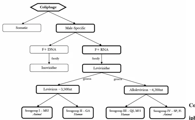
Male-specific phages belong to either the Leviviridae, single-strand RNA, or
Inoviridae, a single-stranded DNA family (Sobsey et al., 2005) (Fig 4.1, Fig 4.2).
Male-specific RNA phages (FRNA) are non-enveloped, positive sense, single-strand RNA (ssRNA) contained within a 26 nm diameter icosahedral-shaped capsid (Buchen-Osmond, 2003). The Leviviridae family (Table 4.1) comprises two genera, Levivirus and
Allolevivirus. These genera are further divided into four genogroups (I, II, III and IV). Levivirus are genetically divided into genogroups I and II, and Allolevivirus are subdivided
into genogroups III and IV.
Leviviridae FRNA phages were initially grouped primarily according to their
group IV were assigned to FRNA phages based on template specificity of RNA replicase (Miyake et al., 1971; Miyake et al., 1973).
Based on a limited number of complete sequences four FRNA genes could be
identified (reviewed by Bollback & Huelsenbeck, 2001). These genes code for an assembly or maturation protein, capsid protein, lysis protein and replicase protein in the Leviviruses
whereas in Alloleviviruses, the lysis protein is replaced by a read-through protein. Each
levivirus virion contains one molecule of positive sense ssRNA, 180 copies of the capsid or coat protein, one copy of the maturation protein and, in alloleviviruses, approximately 15
copies of the read-through protein (Weber & Konigsberg, 1975; van Duin, 2000; van Duin and Tsareva, 2006). Read-through protein synthesis occurs at a rate of about 6% and,
although the exact function is unknown, the combination of the read-through protein with the maturation protein is required for an infectious viral stage (van Duin and Tsareva, 2006). The Leviviridae maturation protein is needed for viral infection and virus particle maturation
(Olsthoorn et al., 1995) whereas the numerous coat proteins in the virion are used for assembly of phage progeny (Klovins et al. 1997; Weber and Konigsberg, 1975). A single, small (< 100 amino acids), hydrophobic peptide, the lysis peptide, is responsible for cell lysis at the end of the infection cycle in the Levivirus genogroups (van Duin, 1988). No distinct
lysis protein is present in Alloleviviruses; lysis is mediated by the maturation protein (Karnik
and Billeter, 1983). The replicase in both genera, also known as viral RNA-dependent RNA
polymerase, is required in small amounts early in the infection process. Twenty minutes after infection, synthesis of this protein ceases (van Duin, 1988).
Family Number of Genera Morphology Nucleic Acid Enveloped
Cystoviridae 1 isometric ssRNA, L, S Yes
Leviviridae 2 icosahedral ssRNA, L
Corticoviridae 1 isometric dsDNA, C, T
Microviridae 4 icosahedral ssDNA, C
Tectiviridae 1 icosahedral dsDNA, L
Inoviridae 2 rod ssDNA, C
Myoviridae 6 contractile tail dsDNA, L
Siphoviridae 6 noncontractile tail dsDNA, L
Podoviridae 3 short tail dsDNA, L
Lipothrixviridae 1 rod dsDNA, L Yes
Rudiviridae 1 rod dsDNA, L
Plasmaviridae 1 pleomorphic dsDNA, C, T Yes
Fuselloviridae 1 lemon-shaped dsDNA, C, T Yes
___________________________________________________________________________ ss - single stranded; L - linear; S - segmented; C - circular; T - superhelical; ds - double stranded
Ecology and Source Specificity
predominant in the southwest islands of Japan, the Philippines, Indonesia, Taiwan and Singapore and group II phages were most abundant in mainland Japan (Furuse et al., 1978; Miyake et al., 1971). Domestic drains located in Asian countries had very few FRNA from groups I and IV. Phages isolated from domestic drainage in Korea were from groups II and III whereas southeast Asia phages were predominantly group III (Furuse, 1987). FRNA phages isolated from sewage water in Australia were from group II and, in the USA, groups II and III FRNA phages were collected (Furuse et al., 1975).
FRNA phage isolation frequencies from various countries varied greatly. Only 2.5% of phages collected from sewage in a study in Peru were FRNA, 5% were FRNA in Brazil and by comparison, FRNA were 38% in Taiwan and 30% in Japan (Furuse et al., 1975). The FRNA phages from sewage samples in Brazil and West Germany belonged to group I exclusively. It was unknown whether or not these sewage treatment plants received
slaughterhouse waste (Furuse, 1987). Furuse states “it can be reasonably assumed that group I phage observed in raw sewage from treatment plants are most likely introduced from animal sources, and group II and group III phage from human sources.” A separate study in Brazil found that thirteen out of 353 sewage and/or fecal samples contained FRNA phages, or approximately 4% and of these thirteen, five were typed as group I, two as group II and six as group III (Miyake et al., 1973).
and III, respectively (Aoi et al., 1972).
Domestic sewage samples in Korea had 56% RNA phages belonging primarily to groups II and III along with 4 group I phages (Osawa et al., 1981). Taiwan sewage and fecal samples included 20% FRNA with 2 group IV, 22 group III, 8 group II and 6 group I isolates (Miyake et al., 1971).
Furuse and colleagues continued to further explore the distribution patterns of the four genogroups of FRNA phages in samples from the following sources: 1) gastrointestinal contents of cows and pigs, 2) feces of domesticated animals (pigs, horses, cattle and fowl), 3) human feces 4) animal feces and 5) sewage obtained from slaughterhouse treatment plants. FRNA groups II and III were isolated in almost equal proportions from human feces whereas the gastrointestinal tract of swine harbored groups I and II. Group I was isolated from feces and gastrointestinal contents from all other animals. Slaughterhouse samples were
predominately group I along with a few isolates from group II. In theses studies, group III was only isolated from human subjects and from no other host organism. Group IV
slaughterhouse sewage. However, their results conflict with the fact that group I strains MS2 and R17 were isolated from municipal sewage systems located in the USA.
FRNA phages isolated in The Netherlands from fecal samples obtained from humans and domesticated animals were serologically typed. As in the Furuse studies, groups I and IV phages were isolated exclusively from animals whereas human sources harbored groups II and III (Furuse, 1987)
Groups II and III were isolated from human feces in FRNA distribution studies from South Africa and Spain (Schaper et al., 2002). In contrast to earlier reports of groups II and III occurring only in human feces and I and IV in animals, the Schaper study isolated group II from cattle and swine and group II and III from poultry. This study casts doubt on the absolute association of phage genotype and source-specificity, but emphasized the
observation that group I has not been isolated from human feces. Nonetheless, group I has been isolated in domestic sewage and whether or not animal waste was present in these municipal treatment facilities is a question that cannot be easily answered.
The lack of consistent numbers of FRNA in human feces and the discrepancies of phage types harbored in various animals does not minimize the fact that FRNA are isolated from sewage in numbers ranging from 102 - 104 PFU/ml (Leclerc et al., 2000). From
multiple samples over time, FRNA were isolated from raw sewage at an average of 4.2 x 10⁴ PFU/L (Brion et al., 2002).
FRNA Coliphages as Indicators of Fecal Pollution
FRNA coliphages have been recommended as a possible indicator of enteric viruses as their presence indicates fecal pollution from either humans or animals. A highly
enterovirus virus concentrations in river water, coagulated effluent, chlorinated and UV-irradiation effluents, coagulated river water and lake water but not in raw sewage or
biologically-treated sewage (Havelaar et al., 1993). FRNA was proposed as a viral indicator in recreational waters due to the strong correlation between FRNA and enteroviruses. Limitations to this study were that only freshwater, not marine or other environmental water bodies were studied, and only a small geographical area was evaluated (Havelaar et al., 1993).
Marine, freshwater and estuarine waters were selected to study the relationship between coliphages, their E. coli host and a few pathogenic bacteria. Multiple sampling
stations were located near domestic and industrial sewage discharges. A correlation between male-specific coliphage concentration and E. coli concentration were found to be
dependent upon the direction and distance from the effluent plume. A greater statistical relationship was noted for male-specific coliphages and the pathogens Salmonella, P.
areuginosa and C. albicans when compared to fecal and total coliforms (Borrego et al.,
1987). In this study, FRNA and FDNA phages were not resolved separately.
Drinking water sources were analyzed for the presence of male-specific phages, somatic phages and Bacteroides fragilis phages for 30 months. Bacterial indicator assays,
total coliforms and fecal coliforms, were also evaluated to determine if a relationship existed between the bacterial and viral indicators. B. fragilis HSP40 host was used to select the
Phage B. fragilis was isolated less frequently two out of three years. Male-specific phages
were suggested as an indicator similar to total coliforms and phage B. fragilis was similar to
fecal coliforms as a measure of fecal pollution (Armon and Kott, 1995).
For a period of two years, raw sewage and surface water samples were surveyed once or twice per week from sites reflective of different land use and agricultural areas. Following double-agar overlay and RNase sensitivity, plaques were genotyped by hybridization using Hsu’s method (1995). FRNA phages were the most abundant F+ phage collected in surface waters (67% out of 105 samples) and sewage (87.5% out of 288) samples. Surface waters were positive for type I (81%) and only one sample had type III, suggesting these surface waters were not influenced by human-impacted effluent. However, type III was the
predominant genotype as 57% were isolated from raw sewage (Brion et al., 2002). During this study, type III FRNA were recovered more frequently than the other genotypes when a group of male campers were staying near the sampling site. Following their departure, the presence of type III declined and was no longer detectable after a week. Source-tracking with type III may indicate sewage contamination occurred within the past 7 days (Brion et al., 2002).
A ratio of FRNA to FDNA densities were compared from samples collected in animal feces, municipal wastewater facilities, in potentially impaired surface waters and in
isolation frequencies were bovine waste (82%), swine wastewaters (50%), gull (4%), goose (0) and human wastewater (77%) FDNA. The remaining animals, horse, buffalo, cat, cormorant, rooster, dog, llama, donkey and pig did not yield F+ coliphages. In animals, goose had the highest percentage of group I (approximately 98%) followed by swine (51%) and cattle (30%). Groups II and III were only isolated from human (50% group II, 15% III), swine (5% II, 22% III) and bovine (15% II, 0 III). Both FDNA and FRNA phages were isolated from surface waters impaired by human, swine, bovine and background sources. Group I FRNA were predominant in background surface waters (97%) whereas bovine-impacted surface waters had 82% group I followed by 75% group I from human-impaired waters. Human land use sites had the greatest percentage of group II isolates (12%) with a low group II recovery from background (2%) and bovine-waste sites. A correlation of FRNA source-associated genogroups was confounded by differential survival rates for each genogroup and/or strain. The authors concluded that there was a statistically significant link of group II FRNA associated with human-land use sites (Cole et al., 2003).
The US EPA sponsored a field validation of Methods 1601 and 1602 to detect coliphages, both male-specific and somatic, in groundwater systems. In addition, fecal indicators E coli, enterococci, total coliforms, Clostridium perfringens spores and the
Association., 2004).
Distinct geographical locations (New Mexico, Massachusetts, Connecticut, Michigan, the Carolinas and Southern California) across the USA were surveyed in an ecological study of FDNA, FRNA and somatic phages. Phage concentrations were compared to fecal
coliform counts. Viruses were isolated and enumerated on double-agar overlay and the appropriate phage-specific E. coli host. Further separation of phage types were determined
by RNase sensitivity followed by serotyping. RT-PCR methods followed by Reverse Line Blot (RLB) methods were used to genotype the phages. Direct fecal samples did not have detectable levels (<3 PFU/gram feces) of male-specific phages in feces other than chicken litter, gull and goose. Only FRNA groups I and IV were detected in these avian species. Cow lagoon isolates typed to group I, whereas hog lagoon predominantly typed group I (32%), 3% group IV along with 18% group III. Hog lagoon had the highest percent of group III FRNA. Wastewater influent and effluent were predominately group II with 12 and 15%, respectively. In addition, wastewater influent had 6% group III, 2% I and 1% IV of FRNA phages. Interestingly, septic water samples did not contain FRNA, but had 100% FDNA strain M13. Grazing animal feces contained large numbers of somatic phages. It was concluded that FDNA M13-like was most prevalent in wastewaters, FDNA were detectable in fecal samples and the link between FRNA group III as a human-associated effluent was not absolute (Long et al., 2005).
transferred and fixed onto a membrane and hybridized with the Beekwilder et al., (1996) designed probes. Hybridization probes detected groups II and III in hospital wastewaters, groups III and IV from swine waters, groups I, III and IV from poultry and groups I and IV from cattle wastes. As a whole, river water samples contained all four genogroups.
However, seven of the individual river samples were positive for only genogroup II. Analysis of FRNA from an assortment of sources led the authors to conclude that group III was not necessarily specific for human excreta, but the trend on the specificity of groups I and IV for animal sources and II and III for human sources supported previous findings (Sundram et al., 2006).
evidence from human and swine hepatitis E virus populations being consistent with their finding for FRNA phages (Stewart et al., 2006).
Ninety-six surface water stations on the State of South Carolina’s impaired list were selected as study sites to quantify FRNA and somatic coliphages. Typing FRNA occurred by serological and/or nucleic acid hybridization methods. Fourteen of these sites identified FRNA genogroups II (5%) and III (1%) while the majority of isolates typed as group I (94%). Direct wastewater samples typed 73% group III, 14% II and 11% group I. Isolates collected and typed from a swine lagoon had 70% group I, 19% group III and 6% group IV. Chicken litters contained approximately equal amounts of group I and IV FRNA. The
presence of groups II and III from the 14 surface water sites were mainly located downstream of wastewater effluent discharges and considered to be contaminated by human fecal
pollution (Stewart-Pullaro et al., 2006).
Sewage-polluted tropical river waters and animal fecal samples were examined for male-specific RNA, male-specific DNA and somatic coliphages. Male-specific DNA and FRNA occurred at similar quantities per gram feces or per ml of water at 7% and 6.5%, respectively. F-specific phages were isolated by plaque assay from 50% of the river samples and 4.4% from the animal fecal material. The study concluded the presence of FRNA phages in the tropical waters of Klang Valley, Malaysia, and proposed FRNA as a tool for
monitoring fecal-polluted waters in their country (Yee et al., 2006).
A small-scale study using male-specific, somatic phages and F+ E. coli as tracers of
clinic with manholes specific to each location. Phages were not isolated from the manholes that served the laundry area and were found to occur in very low numbers (<10 PFU/ml) from the elderly home, the day-care center that used pampers, the greenhouse and the factory. The dairy farm was prevalent in somatic phages (102 - 103 PFU/ml) but had low counts of male-specific phages (1- 10 PFU/ml). Direct fecal material from newborn infants contained male-specific phages at concentrations of 10 - 105 PFU/g feces with one child excreting phages for almost 8 months. Absence or presence of F+ E. coli in the same water
sample(s) correlated with low or high male-specific phage counts. Throughout the year-long study, there were higher numbers and more positive samples of male-specific phages than somatic phages with approximately 96-98.5% of typed phages being FRNA (Gino et al., 2007). Phages were not genogrouped or typed in this study.
FRNA Host Specificity
E. coli K-12 strain was studied in terms its ability or inability to transfer a sex factor,
termed F. If cells transferred F to other cells by means of chromosomal markers, this was termed Hfr strains. If the F factor was transferred independent of the chromosome but
through an extra-chromosomal state, or plasmid, the strains were known as F+ (Clark, 1963). Electron microscopy shed light onto the aggregate nature of FRNA phages adsorbed to the host cell’s fimbriae, the F pili (Bradley, 1964) (Fig 4.1). The conjugative pili serve to transfer genetic information by horizontal gene transfer in gram-negative bacteria. Both Hfr and F+ strains are derived from E. coli K-12 (Havelaar and Hogeboom, 1984).
The male-specific DNA and RNA coliphages adsorb to these conjugative pili (Daehnel et al., 2005), a fertility (F+) sex-pili, coded in E. coli by the F-plasmid
DNA phages bind to the tip of the F-pili and RNA phages attach to the sides (Daehnel et al., 2005).
The Furuse studies enumerated FRNA phages by incorporating male strains of E. coli
(F+, F’ or Hfr) into the media plates followed by RNase treatment (Furuse, 1987).
Salmonella typhimurium strain WG 49 that express F+ by the presence of an F-plasmid have
also been used as a selective host to isolate male-specific phages (Havelaar et al., 1984). S.
typhimurium detected somatic and FDNA phages, however, the host selected 90-95% FRNA
phages (Sundram et al., 2006). E. coli strain Famp (ATCC 700891) is commonly used in
host-selection procedures for F+ coliphages.
Phage replication is restricted to environments such as the intestinal tract of warm-blooded animals as the sex-pili are only expressed at temperatures greater than 30 o C (Grabow, 2001). Interesting to note, Zinder (1963) and Bradley (1964) theorized that in order for male-specific phages to be plentiful in nature, then subsequently, the E. coli male
strains must either be equally prevalent in nature or they are not the natural environmental host. However, bacteria from animal intestines harbor hosts with pili at 104 CFU/ml in
wastewater, thereby possibly allowing phage replication (Long et al., 2005) in municipal wastewater systems.
the host-pili expression, it seems plausible that FRNA phage environmental replication would be restricted to > 30 o C. Optimal phage growth temperatures were found to be 30 o C for genogroup II and 37 o C for genogroups I, III and IV when three strains per genogroup were evaluated (Furuse, 1987). Other studies also reported different survival temperatures in water temperatures > 15 o C (Sobsey, ftp.sccwrp.org). In addition, phage replication
requires a host density of approximately 104 bacteria/ml to be successful (Goyal et al., 1987). The need for concomitant events of optimal temperatures and the presence of an F+ host in log phase would restrict environmental replication of FRNA coliphages.
Additional Potential Viral Indicators of Fecal Pollution
Bacteriophages are ubiquitous in nature and as a whole, are not suitable as an
environmental water quality fecal indicator. However, unique bacteriophages are associated with sewage. Various phages have been proposed as viral indicators. Somatic coliphages (Fig 4.1), FRNA coliphages, FDNA coliphages and the Bacteriodes phage have been
recommended. Advantages and limitations of each indicator will be addressed. Somatic coliphages are present among four bacteriophage taxonomic groups,
Myoviridae, Styloviridae, Podoviridae and Microviridae, hence the morphologic
heterogeneity. Three families of somatic phages contain dsDNA and one family,
Microviridae, contain ssDNA. The somatic phages are non-enveloped and are present in
sewage, often in high abundance in untreated sewage, ranging from 104 - 105 PFU/ml (Gerba, 2006). These coliphages have been detected in humans, chickens, pigs, other animals
(Gerba, 2006) and are easily cultured using E. coli strain CN-13 (ATCC 700609). The
concentrations of somatic phages are similar to FRNA, 102 - 104 infectious units per liter (Sobsey, ftp.sccwrp.org) The phage adsorb to the E. coli host and other enterobacteria
species via the cell wall with a basic receptor site. A limitation of somatic phages is the possibility that the bacteria host origin, especially from environmental reservoirs, may not have originated from a fecal source. Some somatic phages may replicate in environmental waters in the absence of fecal pollution (Ashbolt et al., 2001; Gerba, 2006). If the presence of somatic phages were unrelated to fecal contamination, then it would not serve to predict human health risk (Leclerc et al., 2000).
are filamentous, non-enveloped with a genome size of 6 kb to 9 kb and a virion length of approximately 700 nm to 2000 nm (www.virustaxonomyonline.com). Male-specific DNA coliphages are not morphologically similar to human enteric viruses and their ecology has not been extensively studied (Leclerc et al., 2000). Contradictions in the literature exist on whether FDNA or FRNA are more abundant in nature. Leclerc et al., (2000) noted FDNA were less abundant when compared to FRNA phages. Different distributions of FDNA and FRNA have been reported ranging from 52% FRNA and 48% FDNA (Vinjé et al., 2004) to 77% FDNA and 23% FRNA (Long et al., 2005). Cole et al., (2003) found higher numbers of FDNA in bovine wastes. Using Reverse-Line Blot hybridization and reverse-transcription-PCR followed by sequence analyses, one study found no link between FDNA type and source (Vinjé et al., 2004). However, a second study observed FDNA strain M13 as the dominant phage in septic system samples (Long et al., 2005). Thus, a definitive association between FDNA and human fecal pollution has not yet been established.
Certain phages belonging to the Siphoviridae family are approx 60 nm virion size,
contain dsDNA with long non-contractile tails and infect via cell wall attachment to the host bacteria, strain Bacteroides fragilis. Host strain B. fragilis HSP 40 selects for a
human-specific phage. The B. fragilis human-specific phage has been isolated in sewage, human
fecal samples, polluted groundwater, seawater, river water and sediments but not detected in animal feces (Bitton, 2005; Cornax et al., 1990; Tartera and Jofre, 1987). Unlike other viral or bacterial indicators, the Bacteroides phage is considered to be an exclusive viral indicator
of human fecal pollution and unlikely to replicate in the environment as the host is a strict anaerobe (Tartera et al., 1989). Human-specific phages can be rapidly detected with
are available (Bernhard et al., 2003). A limiting factor lies in the observations of a low isolation frequency, 0-15%, of B. fragilis phages were isolated from human feces (Gantzer et
al., 2002). B. fragilis host are anaerobic and die-off rapidly under ambient water
environmental conditions. As with other viruses, molecular detection only reveals the
nucleic acid presence or persistence of the phage, not necessarily the infective or biologically active form. Another Bacteroides host, strain RYC2056, is more abundant but not human
specific (Ashbolt et al., 2001). B. fragilis strain RYC2056 occurs in domestic sewage and
was used to select phage from 30% of swine and 28% of human fecal samples (Puig et al., 1999).
Figure 4.2. Schematic of male-specific coliphage classification.
As sewage and its accompanying microbial population are dispersed from point or non-point sources, microbes entering marine and freshwaters are subjected to the
surrounding ecological conditions. Water-quality parameters such as pH, hardness, salinity, degree and intensity of sunlight exposure and temperature (climate) strongly influence microbe survival. Survival differs among microbial types, even from virus to virus. Virus types, enveloped or non-enveloped, vary greatly in environmental survival, as enveloped viruses tend to survive poorly outside of their host compared to the survival of non-enveloped viruses. F+ phages and enteric virus groups such as adenovirus, enterovirus, norovirus (calicivirus) and rotavirus are all non-enveloped viruses.
Survival was evaluated for the indicator organisms E. coli, C. perfringens, fecal
coliforms and F+ phages in estuarine conditions. A series of parameters were selected such as temperature, salinity, dissolved oxygen, solar radiation, season and geographic location to study inactivation rates. The highest decay rates were influenced by sunlight and/or
temperature. F+ phages exhibited the least decay (83%) whereas fecal coliforms had the highest decline at 99%. Phages and C. perfringens were least affected by temperature. This
study provides corroborative evidence that F+ phage inactivation rates differ from bacterial indicators (Burkhardt III et al., 2000).
Representative prototype and genotyped field isolated strains of FRNA were spiked at concentrations of 105 - 107 infectious units into FRNA-free, untreated surface waters (freshwater). Samples were incubated at 25 o C in the dark and titered over time to
At day 4, rates of inactivation for prototype strains were as follows: genogroup I MS2 4.7 log10, genogroup II GA 2.6 log10, genogroup III QB 1.7 log10 and genogroup IV FI 2.7 log10
and SP (inactivation log data not provided). Prototype strain SP had the poorest survival and was not detectable after 4 days. Neither strain SP or QB could be detected by the two-step enrichment at day 36. Strains MS2, GA and FI were detected by enrichment at day 36. GA had the longest survival duration and was detectable by DAL throughout the experiment (day 36) (Brion et al., 2002). Survival of genotyped environmental isolates for FRNA coliphages was compared to their respective prototype strain. Of four, group I field strains, two
survived similar to MS2 whereas the remaining two isolates were detectable by DAL to the end of the experiment, day 36. In group II, GA and one field II strain survived the longest (day 36) and the remaining three, group II field isolates declined by day 15. Type III isolates were below detection limits by day 13. Survivability was strain-specific and not necessarily influenced by genotype or prototype vs field isolates in freshwater systems (Brion et al., 2002).
A non-water matrix was used to compare the inactivation of MS2 to other pathogenic viruses, norovirus and poliovirus type 1, and E. coli. Different microcosms were filled with
representative soils, i.e., organic muck, clay and sand. For one month, viruses seeded in a groundwater matrix at approx 106 were dosed into the soil microcosm twice/week. The following month, the columns were dosed with a simulation of rainwater. Microcosm effluent was drained and viral strains were detected by RT-PCR or infectivity assays. With the exception of E coli, the viruses were detected and shown to pass through the column.
The results suggested that E. coli was not a reliable viral indicator as the bacteria did not pass
transport would not mimic virus transport. None of the tested indicators passed through the clay column (Meschke and Sobsey, 2003).
Freshwater, specifically lake waters, were used in a microcosm format to evaluate survival rates on the four FRNA genogroups for 110 days. Strains MS2, two environmental group I, a group II, a group III and two group IV phages were inoculated (2 x 10⁴ PFU/ml) into the microcosms. Incubation temperatures were 4 and 20 o C. Survival time was greater at lower temperature (4 o C). Group IV had the fastest decay rate (5 log10 within 10 days, 4 o
C) followed by group III (5 log 10 at 3 weeks, 4 o C), where these isolates reached the limits
of detection at 10 days and 3 weeks, respectively. Rates of inactivation for groups I and II at 110 days, 4 o C were 1 log10 and 3 log10, respectively. The more persistent genogroups,
groups I and II, were detected at 110 days (Long and Sobsey, 2004).
Prototype strains MS2, GA, Qβ and FI were spiked into seawater at concentrations of approximately 106 PFU/ml and incubated, in the dark, at 21-23 o C. One ml aliquots were removed daily and titers observed by double-agar overlay (DAL) and real-time PCR, concurrently. Within 7 days, all four subgroups in seawater were no longer detected by DAL. In contrast, the real-time PCR detected the four groups after 20 days (Kirs and Smith, 2007). PCR detection of RNA, as demonstrated in these experiments, does not predict or indicate the presence of infectious phage but only the presence of RNA.
significantly increase at 25 o C compared to 4 o C. Infectivity reduction rates were not observed at 4 o C (Bae and Schwab, 2008).
FRNA inactivation rates are influenced by sunlight intensity, fresh vs saltwater matrices and soil composition. For example, increasing salinity and temperature
demonstrated faster rates of inactivation in FRNA strains. In contrast, lower temperatures (4
o C) and freshwater environments decreased inactivation rates on FRNA coliphages.
FRNA Coliphage Detection Methods - Plaque Assays
To quantify and accurately detect FRNA phages, initial purification methods are imperative. Depending upon the application, detection methods include Single Agar Overlay,
Double-Agar Overlay, enrichment, Most Probable Number, RNase sensitivity, serotyping and genotyping.
To prepare a Single Agar Overlay (SAL) for enumeration of FRNA, collect 250 ml environmental water sample and place on ice up to 6 hrs. Beforehand, begin a log-phase of the E. coli host FAMP (ATCC # 700891) by adding a 1ml inoculum from an overnight (O/N)
culture into 50 ml Trypticase Soy Broth. Gently shake at 37 o C for 4 hr. Host culture will be in log-phase at 4 hr. Divide the environmental sample by dispensing two aliquots of 100 ml into sterile bottles. To the 2X TSA (trypticase soy agar) add 1 ml of 100 X
streptomycin/ampicillin, 0.5 ml sterile 4M MgCl2. Add 10 ml log-phase F+ host to the 100
ml sample. Quickly combine the sample/E. coli host with the 2X TSA/antibiotics flask.
PFU/100 ml. Questionable plaques can be verified by spot plate assay (EPA Method 1602, 2001).
A presence/absence method involves a two-step enrichment. Using a sterile bottle, divide the 1 L environmental water sample into two, 500 ml aliquots. To each 500 ml, add the following: 50 ml of 10X cold TSB per L (media is cold to diminish phage growth), 12.5 ml 4M MgCl2, 10 ml of 100 X streptomycin/ampicillin and 5 ml log-phase F+ host. Cap and
invert to mix. Incubate overnight (16-24 hr) at 36 o C + 1 o C. The following day, spot 10 ul from each enrichment onto a grid spot plate (TSA) containing the appropriate host and incubate overnight. Score as positive or negative lysis zones in spots (EPA Method 1601).
An MPN (Most Probable Number) can be estimated from a 1L environmental water sample. Enrich the 1L sample with 2X TSB, antibiotics, 4M MgCl2 and E. coli host as stated
Applications to detect somatic phages are the same as those for male-specific except the selection host is one that is F-minus (F-) such as E. coli strain CN-13 (ATCC 700609).
For this host a 1% stock of antibiotic nalidixic acid is used in the media instead of strep/amp as used for E. coli Famp (EPA Method 1601, 2001). E. coli host C-3000 (ATCC 15597)
selects for both F+ and somatic phages but findings from field studies reveal strain C-3000 recovers lower numbers of somatics and male-specific phages when compared to the sum of phages recovered by their respective host-specific strain. In a recent study, E. coli strain
CB390 was effective at recovering the sum of both phage types (Guzman et al., 2008). Coliphage antisera are obtained by inoculating experimental animals to elicit an immune response that results in polyclonal antibodies against the desired phage strain. For serotyping, isolated plaques are spotted (10 ul) onto an agar plate containing a specific antisera, either to MS2, GA, Qβ, SP or FI. The agar plate contains the antisera and log phase host. Once the plaque is applied, the plate is incubated O/N at 37 o C. Phage growth
suppression on one of the plates, which contains homologous neutralizing antibodies to the phage group, is scored as positive. Serotyping is not 100% reliable and can produce ambiguous results (Beekwilder et al., 1996). For example, two isolates in the UNC collection were first serotyped to group II but later sequenced and genotyped to group I.
RNase sensitivity is determined by re-plating phage isolates on plates containing or lacking Ribonuclease A. On the plates with RNase, FRNA phages do not form lysis zones, however, FDNA phages will form zones of lysis.
FRNA Coliphage Detection Methods - Genotyping
FDNA and FRNA phages. An FRNA assay applicable for microbial source-tracking must first and foremost precisely identify FRNA. Genogroup-targeted methods solve two considerations, (i) FRNA selection and (ii) genotype-associated source tracking.
To distinguish the individual FRNA groups I, II, III, IV and a combination of groups I and II, III and IV, a molecular hybridization assay was developed. Genogroup-specific hybridization probes were designed using multiple alignment software to strains MS2, GA, Qβ, and SP. Environmental isolates of FRNA phage as lysis zones on agar plates were adsorbed to a membrane, denatured to unfold RNA secondary structures and linked to the membrane using UV light. Digoxigenin (DIG-dUTP) labeled probes were incubated overnight with the fixed membranes and visualized with alkaline phosphatase-conjugated anti-DIG antibody. Assay development involved optimization of various hybridization solutions, selection of the most efficient membrane and denaturation solutions. To test the hybridization assay, 203 FRNA field isolates from sewage, oysters, surface waters and feces were first grouped by serotyping. Serotyping and hybridization classified 79 and 109
isolates, respectively, from surface waters, adult and piglet swine feces, treated and untreated sewage and oysters as genogroup II. Almost half of the piglet isolates, 12 out of 26, were type IV. Isolates from chickens were grouped almost equally into I and IV. Serotyping cross-reactivity was observed for 37 isolates neutralized by anti-sera GA and partial neutralization to anti-sera MS2. However, these 37 serotyped strains were eventually genotyped by hybridization to be in group II. Similar classification results were obtained by both serotyping and hybridization genotyping (Hsu et al., 1995).
was designed to MS2 nucleotide region 1248 (Hsu) and 1260 (Beekwilder), group II was designed to GA region 431 (Hsu) and 2100 (Beekwilder), group III was designed to Qβ
region 27 (Hsu) and 660 (Beekwilder) and group IV probe was designed to SP region 35 (Hsu) and 40 (Beekwilder). Probes were designed to each genogroup by Beekwilder and colleagues by alignment of 3-5 strains/group. If the completed sequenced genome was not available, they proceeded to sequence partial regions to provide adequate sequence
representation for each genogroup. In addition, a probe A for Levivirus (groups I and II) and
probe B for Allolevivirus (groups III and IV) was developed. To validate the hybridization
probes, a combination of “blinded” but previously serotyped samples and field samples were analyzed. Approximately 78% of the samples were correctly identified by both the
genogroup and genus-specific probes. Isolates collected from human impacted areas in the form of hospital waste and domestic wastewater identified 1 group I, 1 group II, 4 group III and 1 group IV. The two human feces samples had ambiguous classifications in that both samples were weakly positive for group I and positive for group IV but hybridized to both genus-specific probes, Levivirus and Allolevivirus. Twenty phages isolated from animal
sources hybridized to groups I or IV in all cases except two. Of those two, one isolate was positive to groups I and II, and the second isolate (porcine slaughterhouse) was positive to group III (Beekwilder et al., 1996). These hybridization studies lend supporting evidence to the trends that genogroups II and III are associated with human waste and groups I and IV occur more often in animal sources.
cluster-specific oligonucleotide probes for hybridization were designed to FRNA strains, 1 group I probe for MS2-like, 1 probe for GA-like, 2 group III probes, Qβ and M11, and 2 group IV probes, SP and FI-like. Three cluster-specific FDNA probes were also designed, M13, fd and CH and one generic FDNA consensus-specific probe, termed “con.” In order to initially divide FRNA from FDNA isolates, a generic RT-PCR assay was performed.
Broadly reactive primer sets yield different PCR amplicon sizes for Allolevivirus vs.
Levivirus. Allolevivirus was amplified by primers MJV82 forward, JV41 reverse and Levivirus was detected by primers MJV82 forward and JV81 reverse. Primer pair SL2
forward and SL3 reverse amplified FDNA phages. Assay validation began by collecting a total of 557 environmental samples. Phages were isolated by single or double-agar overlay plating, followed by RNase sensitivity testing and serotyped by anti-sera neutralization or genotyped by hybridization (Hsu et al., 1995). RT-PCR identified 100% of the FRNA and FDNA strains. Identified strains were used to validate the reverse-line blot hybridization (RLB), resulting in 98% agreement of the FRNA strains and 100% confirmation of the FDNA phages (Vinjé et al., 2004).
When comparing the basic hybridization assays, the RLB involves a two-step process since the RT-PCR step occurs prior to hybridization. Nonetheless, RLB typing had a higher predictability, 100% FDNA, 98% FRNA, when compared to 38% by serotyping (Hsu et al., 1995), 54% by genotyping (Hsu et al., 1995) and 78% by genotyping (Beekwilder et al., 1996).
Prior to 2007, 10 full-length or nearly full-length FRNA phage genomes were
A universal forward and reverse primer set was designed (Kelly Reynolds, Univ of AZ) to a consensus sequence in the replicase gene to detect all FRNA strains. A two-step reverse transcription polymerase chain reaction (RT-PCR) was performed. Prior to RT-PCR, environmental samples were column filtered to remove inhibitors. RT-PCR sensitivity was improved when the samples were column filtered and detection limits were 0.10 PFU of laboratory control MS2. RT-PCR amplified FRNA from 2 samples that were
plaque-negative by soft agar overlay and was in agreement with the plaque-positive overlay methods (Rose et al., 1997). According to the authors, the primers amplified all four FRNA coliphage groups, but specific strains were not mentioned.
Phage MS2 is routinely used as a surrogate for pathogenic and environmental studies. Five sets of real-time primers and probes were designed to strain MS2. Two sets targeted the assembly gene, 1 for the coat region, 1 targeted the lysis gene and 1 primer and probe set targeted the replicase gene. Cross-reactivity of the real-time PCR was tested against non-targeted organisms, specifically pathogenic bacteria. MS2 primer sets did not cross-react with bacteria (O’Connell et al., 2006). Detection or cross-reactivity to other FRNA phages was not discussed.
Gentzer, 2006) and MS2, GA, Qβ, SP and FI (Kirs and Smith, 2007). Although the authors state their assay was template specific and lacked cross-hybridization, assay validation was limited to four or five FRNA strains. Raw sewage (Ogorzaly and Gentzer, 2006) and raw sewage and chicken litter (Kirs and Smith, 2007) samples were collected for QRT-PCR field validation. Ogorzaly and Gentzer detected 100% of groups I, II and 85% group III by QRT-PCR of double-agar overlay plaques. Group IV was not detected. Twenty plaques from sewage and 20 plaques from chicken litter were isolated, RNA purified and subjected to QRT-PCR (Kirs and Smith, 2007). Sewage isolates were group III and chicken stool isolates were group IV. Three genogroup-specific primer pairs were developed for a two-step RT-PCR assay and validated with strains MS2, GA and SP. Primer design for genogroups I, II and IV were based on GenBank genomic sequences for MS2, GA and SP, respectively. Phages isolated and enumerated from individual septic systems, poultry farm, municipal sewage and a background site were enumerated by SAL after filtration of the water sample. An MPN was also conducted. Purified phage RNA, from October 2004 environmental samples, tested positive for groups I and IV with RT-PCR and was in agreement with plaque-positive samples. The septic system tested negative with RT-PCR but plaque-positive by SAL and MPN. In January, 2005, 12 positive SAL and MPN samples tested positive for group I by RT-PCR. The May, 2005, sampling season had 3 out of 7 samples positive by SAL and MPN detection. In those 3 samples, RT-PCR identified group II upstream from the
wastewater treatment plant sample but the 3 remaining samples taken downstream of the sewage plant, upstream of a different sewage plant and lake waters near the poultry farm were negative by RT-PCR. The authors concluded FRNA indicators were not useful to distinguish between human and non-human sources and suggested the presence of either somatic or FDNA phages in their positive samples (Dryden et al., 2006). Limitations to their study design are as follows. Primers were designed to three individual strains and not an alignment of multiple strains per genogroup. These primers would not detect all four genogroups or environmental strains as reflected in the results. A group III primer set was not designed and yet, the authors concluded FRNA could not distinguish between human and non-human sources. SAL and MPN host selection was E. coli strain C-3000 (ATCC 15597),
a host that selects for both somatic and male-specific phages.
An antibody-based agglutination assay, termed “latex agglutination”, was developed to rapidly detect, <24 hr, genogroups of FRNA coliphages. Environmental strains were collected from bird droppings, shellfish and water bodies from diverse geographical locations across the USA. A two-step enrichment protocol was modified to a culture time of 180 min based on preliminary sampling and phage measurement at 0, 30, 60, 90, 120, 180 and 360 min on TSA plates containing E. coli host FAMP . Male-specific plaques were confirmed as
agglutination and typing (CLAT) assay, polyclonal antibodies were first generated against MS2, GA, Qβ, SP and FI. Phage strain-specific antisera was bound to polystyrene particles and used in the agglutination step of the CLAT. Coliphage enriched samples showed agglutination within 30-60 sec when 2.5 ul enriched phage cultures were mixed with 2.5 ul strain-specific antibody particle on an agglutination card. Out of 192 FRNA field isolates, CLAT correctly identified 185 (96%) and RLB identified 92%. When the two methods were compared, some ambiguity was noted in that CLAT identified more group II and RLB identified more groups III and IV. Twenty-four strains yielded inconsistent typing. Sequencing of the capsid region and phylogenetic clustering of these 24 strains yielded 19 group I and 5 group II. CLAT clustered 17 of the 19 group I strains as I and II, but matched
100% of the 5 sequenced group II isolates. A total of 164 FDNA isolates were also identified by CLAT at a rate of 97.7% (Love and Sobsey, 2007).
FRNA Relationship to Enteric Viruses and as Predictors of Health Risk
An important attribute of an ideal indicator is the relationship between the indicator density in polluted waters to human-health risks. Few epidemiology studies exists correlating health risks with F+ coliphage densities. A European cohort study (Lee et al., 1997) showed a statistically significant relative risk (RR) with increased F+ coliphage exposures.
However, a threshold value was not extrapolated and the slight increased RR (2.6-2.8) is minimal compared to the large coliphage density range of 26 - 32 and 69-308 PFU/10 ml, respectively.
and fever. The authors noted that when F+ phages were detected, a low number of beach-goers were exposed (Colford et al., 2007).
Meta-analysis of epidemiological studies revealed no correlation between bacteriophage in marine waters to GI illness, whereas the freshwater studies reported an elevated GI risk with elevated bacteriophage exposure (Wade et el., 2003). The meta-analysis did not clarify which type of bacteriophage showed associations with human health risks.
Six wastewater treatment facilities from FL, AZ and CA producing and distributing reclaimed water were monitored for indicator and pathogen load. Although reclaimed water is routinely assessed for fecal or total coliforms, the degree of microbial indicator and pathogen removal has not been evaluated. Results using male-specific coliphages did not show a correlation with enteric viral load. However, the coliphage predicted an absence of enteric viruses at levels less than 10 coliphage/100 ml (Rose et al., 2004).
Adenovirus presence was statistically correlated to FRNA (r = 0.99) in a brackish (salinity from 9-34 ppt) but no correlation to FDNA phages were observed in coastal waters (Jiang et al., 2001).
Future Applications
Molecular detection methods, i.e., PCR, real-time PCR and microarrays, allow a more timely assessment of the microbial quality of recreational waters. A prospective study at two Great Lakes beaches using real-time PCR detection found that enterococci was statistically associated with increased gastro-intestinal (GI) illness at both beach sites. A strong positive trend was noted with the presence of Bacteroides at one of the beaches (Wade
Microarrays have been constructed with genomic DNA purified from raw wastewaters (Lee and el., 2006) and 16S rRNA and cpn60 genes extracted from several specific pathogens (Maynard et al., 2005). Primers and oligonucleotide microarray probes were designed from sequences derived from specific pathogenic bacteria strains. Validation of the microarrays generated a positive hybridization signal in raw sewage samples for the E
.coli gene uidA (Lee et al., 2006).
Commercially-available field PCR instruments could potentially be applicable to molecular detection of FRNA genogroups. For example, a hand-held fluorogenic real-time PCR instrument, the Advanced Nucleic Acid Analyzer (ANAA), was used to detect bacteria spores and MS2 virions. The microbes were analyzed using a micro-chip in the ANAA and a positive signal was viewed within 18-26 min (Belgrader et al., 1998).
Portable real-time thermocyclers (Cepheid, Inc) and nucleic acid sequence based amplification (NASBA) instruments (BioMereux, Inc) can be transported to provide on-the-spot analysis of environmental samples. NASBA, unlike PCR, does not require a
thermocycler. NASBA relies on an isothermal process (41 o C), three RNA-associated enzymes and a molecular beacon. Single-stranded RNA is generated in a single-tube, emitting fluorescence by the molecular beacon upon hybridization with ssRNA and a target-specific oligonucleotide. NASBA technology targets RNA viruses and would be applicable as a portable instrument to detect FRNA coliphages.
Summary and Conclusion
whereby decisions must be made to select and validate the selection of such tools. For example, (1) define the problem (nearby source discharge or runoff, history of monitoring data, weather patterns, hydrology, etc), (2) formulate objectives as to the suspected source and category (human vs. non-human), (3) presence/absence vs. quantification of loading values (4) consider if the application linked to regulatory values for microbial indicator concentration or if fecal presence or absence is an acceptable result for decision making, (5) consider if fecal presence is linked to public health risk, (6) consider if there are legal ramifications, (7) select the most appropriate source-tracking protocol (8) define a sampling strategy or study design, (9) define a method of data collection and quality control, (10) define data analyses and (11) have a plan for data interpretation (Stoeckel, 2005).
Currently, the toolbox consists of DNA fingerprinting, antibiotic resistance, ribotyping, pulse-field electrophoresis and carbon utilization profiles of E. coli and/or
enterococci (library-dependent), host-specific Bacteroides and Prevotella bacteria markers
independent) and serotyping or genotyping of FRNA or FDNA coliphages (library-independent). The “library” per se, is a collection of bacterial or viral isolates from which the source of collection is known as well as the fingerprint, marker, genotype or serotype. One aspect of method selection depends upon the analytical question, library-dependent
(epidemiological matching or clustering) vs. library-independent (source could be traced in any water body type or geographical location).