The K1 Protein of Kaposi’s Sarcoma-Associated Herpesvirus
Augments Viral Lytic Replication
Zhigang Zhang, Wuguo Chen, Marcia K. Sanders, Kevin F. Brulois, Dirk P. Dittmer, Blossom Damania
Lineberger Comprehensive Cancer Center and Department of Microbiology and Immunology, University of North Carolina at Chapel Hill, Chapel Hill, North Carolina, USA
ABSTRACT
The K1 gene product of Kaposi’s sarcoma-associated herpesvirus (KSHV) is encoded by the first open reading frame (ORF) of
the viral genome. To investigate the role of the K1 gene during the KSHV life cycle, we constructed a set of recombinant viruses
that contained either wild-type (WT) K1, a deleted K1 ORF (KSHV
⌬
K1), stop codons within the K1 ORF (KSHV-K15
ⴛSTOP), or arevertant K1 virus (KSHV-K1
REV). We report that the recombinant viruses KSHV
⌬
K1 and KSHV-K1
5ⴛSTOPdisplayed
signifi-cantly reduced lytic replication compared to WT KSHV and KSHV-K1
REVupon reactivation from latency. Additionally, cells
infected with the recombinant viruses KSHV
⌬
K1 and KSHV-K1
5ⴛSTOPalso yielded smaller amounts of infectious progeny upon
reactivation than did WT and K1REV-infected cells. Upon reactivation from latency, WT and
KSHV-K1
REV-infected cells displayed activated Akt kinase, as evidenced by its phosphorylation, while cells infected with viruses deleted
for K1 showed reduced phosphorylation and activation of Akt kinase. Overall, our results suggest that K1 plays an important
role during the KSHV life cycle.
IMPORTANCE
Kaposi’s sarcoma-associated herpesvirus (KSHV) is the etiological agent of three human malignancies, and KSHV K1 is a
signal-ing protein that has been shown to be involved in cellular transformation and to activate the phosphatidylinositol 3-kinase
(PI3K)/Akt/mTOR pathway. In order to investigate the role of the K1 protein in the life cycle of KSHV, we constructed
recombi-nant viruses that were deficient for K1. We found that K1 deletion viruses displayed reduced lytic replication compared to the
WT virus and also yielded smaller numbers of infectious progeny. We report that K1 plays an important role in the life cycle of
KSHV.
K
aposi’s sarcoma (KS)-associated herpesvirus (KSHV), also
known as human herpesvirus 8, is the causative agent of KS, a
vascular neoplasm of endothelial cell origin (
1
). KSHV infection is
linked to two B cell lymphoproliferative disorders: primary
effu-sion lymphoma (PEL) and the plasmablastic variant of
multicen-tric Castleman’s disease (MCD) (
2–4
). KSHV predominantly
dis-plays a latent state in infected cells and in KSHV-associated
tumors, but a small percentage of KSHV-infected cells undergo
reactivation, which is thought to be important for KS
tumorigen-esis (
5
). KSHV reactivation can occur through multiple events (
6
,
7
), and the KSHV replication and transcription activator (RTA)
protein is the only viral protein that is both necessary and
suffi-cient to reactivate KSHV. The expression of RTA leads to the
activation of downstream lytic genes and ultimately the
produc-tion of progeny virions (
8–12
).
K1 is a transmembrane glycoprotein encoded by the first open
reading frame (ORF) in the KSHV genome (
13
). Although K1 is
highly upregulated during the lytic cycle, it has also been shown to
be expressed at lower levels during latency (
14
). K1 is expressed in
KS lesions and primary effusion lymphoma cell lines (
13–17
). The
K1 protein contains an immunoreceptor tyrosine-based
activa-tion motif (ITAM) in its cytoplasmic tail, which is involved in
activating signal transduction pathways (
4
,
17–20
). K1 transforms
mouse fibroblasts (
21
) and immortalizes primary human
endo-thelial cells (
22
). The transforming activity of K1 is thought to be
conferred through phosphatidylinositol 3-kinase (PI3K)/Akt/
mTOR signaling. The PI3K/Akt/mTOR pathway is activated by
K1 in B and endothelial cells (
18
,
22
), and this provides a survival
advantage to K1-expressing cells (
4
,
18
,
22
,
23
). K1-expressing
cells also secrete increased amounts of vascular endothelial growth
factor (VEGF), which is an angiogenic factor that promotes
vas-cularization (
24
). The K1 ITAM is required for signal
transduc-tion in B and endothelial cells (
18
,
21
,
22
,
25
,
26
).
The role of K1-mediated activation of signaling pathways
dur-ing KSHV reactivation from latency has been studied in the
con-text of exogenous expression of K1 (
26
,
27
). However, thus far, a
K1 mutant virus has not been made and tested for its ability to
affect KSHV lytic replication in the context of the whole virus.
In this study, we employed bacterial artificial chromosome
(BAC) technology to construct a set of recombinant KSHVs:
wild-type (WT) K1 (WT KSHV K1), wild-wild-type FLAG-tagged K1
(KSHV-K1
FLAG), a K1 deletion virus removing the entire K1 ORF
(KSHV
⌬
K1), and a K1 mutant virus (KSHV-K1
5⫻STOP) in which
stop codons were inserted into the K1 ORF to prevent the
expres-sion of the protein. A K1 revertant virus (KSHV-K1
REV), in which
a FLAG-tagged K1 coding sequence was restored, was also
con-structed. These different viruses were used to assess the effect of K1
on the KSHV life cycle. We found that deletion of the K1 gene or
Received9 December 2015 Accepted20 April 2016
Accepted manuscript posted online15 June 2016
CitationZhang Z, Chen W, Sanders MK, Brulois KF, Dittmer DP, Damania B. 2016. The K1 protein of Kaposi’s sarcoma-associated herpesvirus augments viral lytic replication. J Virol 90:7657–7666.doi:10.1128/JVI.03102-15.
Editor:R. M. Longnecker, Northwestern University
prevention of K1 expression in KSHV
⌬
K1 or KSHV-K1
5⫻STOP,
respectively, resulted in decreased production of infectious virus
following reactivation of the virus from latency. Moreover, these
mutant viruses exhibited less phosphorylation of Akt in infected
cells following reactivation. Akt phosphorylation and virus
pro-duction were restored in K1 revertant virus-infected cells
com-pared to K1 mutant virus-infected cells. Our results suggest that
K1 plays an important role in lytic reactivation and replication as
well as the activation of the Akt pathway in infected cells.
MATERIALS AND METHODS
Cell culture and recombinant virus construction.BAC16 and iSLK-RTA cells were kindly provided by Jae U. Jung (28). Vero and HEK293T cells harboring WT KSHV, KSHV-K1FLAG, KSHV-K15⫻STOP, KSHV-K1REV, and KSHV⌬K1 as well as iSLK-RTA and iSLK cells harboring these viruses were grown in Dulbecco’s modified Eagle’s medium (DMEM) supple-mented with 10% fetal bovine serum (FBS) and 1% penicillin-streptomy-cin. iSLK-RTA cells were cultured in the presence of 1g/ml puromycin and 250 g/ml G418. HEK293T cells harboring WT KSHV, KSHV-K1FLAG, KSHV-K15⫻STOP, KSHV-K1REV, and KSHV⌬K1 were con-structed and maintained in the presence of 100g/ml hygromycin. iSLK cells harboring WT KSHV, and the recombinant viruses KSHV-K1FLAG, KSHV-K15⫻STOP, KSHV-K1REV, and KSHV⌬K1 were established and maintained in the presence of 1g/ml puromycin, 250g/ml G418, and 1,200g/ml hygromycin B.
Construction of KSHV-K1FLAG, KSHV-K15ⴛSTOP, KSHV-K1REV,
and KSHV⌬K1. BAC16 was kindly provided by Jae U. Jung (28). pcDNA3-WT K1FLAGand pcDNA3-K15⫻STOPwere used as the templates for WT K1 and mutant K1 genes for the construction of recombinant viruses. pcDNA3 WT K1FLAG, which has a flag epitope tag at the N termi-nus of WT K1 from the BCBL1 genome and was constructed as previously described (24). pcDNA3-K15⫻STOPwas constructed by the insertion of the K15⫻STOPfragment into the BamHI and EcoRI sites of pcDNA3 WT K1FLAG. The pcDNA3-K15⫻STOPconstruct contains 3 stop codons fol-lowing the start codon of WT K1FLAGand also has 2 TGA stop codons instead of ATG codons at positions 481 and 763 to prevent the expression of WT K1FLAG. The following primers were used: forward 1 (5=-GCAGG
ATCCATGTAAATAGGTGAGCCCTGCCCGTGACCGCCCTG),
re-verse 1 (5=-CGATGGAATTCTCAGTACCAATCCACTGGTTGC),
for-ward 2 (5=-GCAGGATCCATGTAAATAGGTGAGCCCTGCCCGTGAC
CGCCCTG), reverse 2 (5=-GTATAACACCCCTCAGTTTGGTTTCACAG
CGTAAAATTATAGTATTTAG), forward 3 (5=-CACTCGTAGCTCTGAT
AGGAACCTGATGTGGTATCTTAGGAACTATTATC), and reverse 3
(5=-GATAATAGTTCCTAAGATACCACATCAGGTTCCTATCAGAGC
TACGAGTG).
DH10B harboring BAC16 was a gift from Jae U. Jung (28). KSHV-K1FLAG, KSHV-K15⫻STOP, KSHV-K1REV, and KSHV⌬K1 recombinant viruses were made by using BAC16. The original K1 gene is located at positions 105 to 959 in the KSHV BAC16 genome (28) (GenBank acces-sion number GQ994935). The recombinant viruses KSHV-K1FLAG, KSHV-K15⫻STOP, KSHV-K1REV, and KSHV⌬K1 were constructed with the Red/ET recombination system, purchased from Gene Bridges Inc., according to the manufacturer’s instruction. This system utilizes a dual-antibiotic cassette for the positive selection of kanamycin resistance and negative selection of streptomycin sensitivity rendered by the bacterial ribosomal protein S12 (rpsL⫹) gene. In brief, a Red/ET expression plas-mid was transformed into DH10B cells harboring BAC16 with electropo-ration. Next, an RpsL-neomycin cassette flanked by homologous nucleo-tides upstream of the original K1 gene and other homologous nucleonucleo-tides downstream of K1 was generated by PCR with forward primer
5=-GTGTAAACCTGTCTTTCAGACCTTGTTGGACATCCCGTAC
AATCAAGGGCCTGGTGATGATGGCGGGATCG and reverse primer
5=-CACATTACAATTATGTTACAGAGAATATTTAGATTATC
TTACCTGAATGTCAGAAGAACTCGTCAAGAAGGCG, and the cas-sette was then electroporated into DH10B-Red/ET cells harboring
BAC16. To replace the K1 gene with the linear RpsL-Neo cassette flanked by homologous arms, we usedL-arabinose in the growth medium to in-duce the expression of the Red/ET plasmid and allow for recombination. The transformed bacteria were plated onto LB agar plates and selected with chloramphenicol and kanamycin. Positive colonies growing under chloramphenicol and kanamycin selection were isolated and analyzed by PCR and pulsed-field gel electrophoresis (PFGE). Next, colonies harbor-ing BAC16 containharbor-ing the RpsL-Neo cassette in place of the K1 gene were subjected to a second recombination step. DNA fragments with homolo-gous arms, including K1FLAGor K15⫻STOP, were amplified by PCR with f o r w a r d p r i m e r 5=- G T G T A A A C C T G T C T T T C A G A C C T T G T TGGACATCCCGTACAATCAAGATGGCCCTGCCCGTGACCGC-CCTGC and reverse primer CACATTACAATTATGTTACAGAGAAT ATTTAGATTATCTTACCTGAATGTCAGAAGAACTCGTCAAGAAG
GCG for K1FLAG or forward primer GTGTAAACCTGTCTTTCAGAC
CTTGTTGGACATCCCGTACAATCAAGATGTAAATAGGTGAGCC CTGC and reverse primer ACACATTACAATTATGTTACAGAGAATAT TTAGATTATCTTACCTGAATGTCAGTACCAATCCACTGGTTGC for K15⫻STOPand were transformed into DH10B cells harboring BAC16 with the RpsL-Neo cassette in the K1 location. The bacteria were plated onto plates containing streptomycin and chloramphenicol, and colonies were analyzed by using PCR and PFGE to check for correct clones con-taining full-length K1FLAGor K15⫻STOP, etc., in the original K1 location and in place of the RpsL-Neo cassette. To make KSHV-K1REV, we re-peated the above-described procedure to replace the K1 mutant gene first with the RpsL-Neo cassette and then replaced the RpsL-Neo cassette with WT K1FLAG. All BAC plasmid DNAs were isolated by the alkaline lysis method for analysis of restriction digestion or with the NucleoBond BAC 100 kit (Clontech) for sequencing.
Establishment of stably infected HEK293T cells and their deriva-tives. WT or recombinant virus BAC16 DNA was transfected into HEK293T cells with Lipofectamine 2000 (Invitrogen) according to the manufacturer’s instructions. Briefly, 5g of DNA was transfected into HEK293T cells in one well of a 6-well plate. A total of 50g/ml of hygro-mycin B was added to the medium for selection at 48 h posttransfection. The concentration of hygromycin B was increased up to 100g/ml at 96 h posttransfection. Stable infected HEK293T cells were maintained with 100g/ml hygromycin B.
Establishment of stably infected iSLK cells and their derivatives.
iSLK cells were cocultured with recombinant virus-infected HEK293T cells. Briefly, a 2:1 ratio of stable recombinant virus-infected HEK293T cells was plated with iSLK-RTA cells so that the total number of cells was 2.5⫻105cells per well of a 6-well plate. Three milliliters of 2% FBS– DMEM with final concentrations of 25 ng/ml of tetradecanoyl phorbol acetate (TPA) and 0.5 mM sodium butyrate was added to the cells at 24 h postseeding to induce reactivation. Four days later, cells were selected with 250g/ml G418, 1g/ml puromycin, and 1.2 mg/ml hygromycin B. Next, recombinant virus-infected stable iSLK cells, which were 100% green fluorescent protein (GFP) positive, were reactivated with 3g/ml doxycycline and 1 mM sodium butyrate for 3 days. The supernatant was collected, cleared by centrifugation at 950⫻gfor 10 min, and filtered through a 0.45-m filter. A new batch of uninfected iSLK-RTA cells was infected with this filtered viral supernatant in the presence of 8g/ml of Polybrene and centrifuged for 2 h at 2,500 rpm at 30°C. The cells were then placed into an incubator with 5% CO2at 37°C. At 48 h postinfection, 1 g/ml puromycin, 250g/ml G418, and 1.2 mg/ml hygromycin were added to the medium to select for stable iSLK-WT KSHV, iSLK-KSHV-K1FLAG, iSLK-KSHV-K15⫻STOP, KSHV-K1REV, and KSHV⌬K1 cell lines. These cell lines were maintained in the presence of 1g/ml puromycin, 250g/ml G418, and 1.2 mg/ml hygromycin.
alkaline lysis. Purified BAC16 DNA was digested with KpnI and separated on 1% pulsed-field-certified agarose by using PFGE (Chef-Mapper; Bio-Rad) under the following conditions: 6 V/cm for 15 h at 14°C and initial and final switch times of 0.1 and 11.15 s, respectively. Recombinant KSHV DNA purified with the NucleoBond BAC 100 kit was used for sequencing with the 454 GS Junior sequencer. Finished genomic data were submitted to GenBank.
Preparation of viruses.A total of 5⫻105iSLK-WT and recombinant virus-infected cells were plated into one well of a 6-well plate overnight, after which cells were reactivated with 3g/ml doxycycline in DMEM with 2% FBS for 72 h. The supernatant was collected and centrifuged at 950⫻gfor 10 min. The supernatant was used for infection studies. Genomic DNA of recombinant KSHVs was isolated from the supernatant or infected cells with the DNeasy blood and tissue kit (Qiagen) for the detection of KSHV genomic copies by quantitative PCR (qPCR).
Infectivity assay.A total of 1.9⫻104Vero cells or primary human umbilical vein endothelial cells (HUVECs) per well in a 96-well plate were seeded in triplicate for each sample. The next day, 100l of fresh super-natant containing WT KSHV or recombinant viruses was added to naive Vero cells with 8g/ml Polybrene. The plate was centrifuged at 2,500 rpm at 30°C for 2 h for Vero cells and for 1 h for primary HUVECs. Twenty-four hours later, the supernatant was removed and washed with DMEM for Vero cells or EBM-2 (endothelial cell basal medium 2) for primary HUVECs, and 100l fresh medium was added. GFP-positive cells were counted at 72 h postinfection. For flow cytometry analysis, a similar pro-tocol was performed. A total of 1⫻105Vero cells were plated into each well of a 24-well plate, and 500l of fresh supernatant containing WT KSHV or recombinant viruses was added to naive Vero cells. Cells were collected following trypsinization at 72 h postinfection, washed with Dul-becco’s phosphate-buffered saline (DPBS) containing 5% bovine serum albumin (BSA), and analyzed by flow cytometry.
Genomic DNA for qPCR was isolated with the DNeasy blood and tissue kit (Qiagen) 72 h after infection of Vero cells with WT or recombi-nant virus.
Analysis of Akt activation. Cells harboring WT KSHV, KSHV-K1FLAG, KSHV-K15⫻STOP, KSHV-K1REV, and KSHV⌬K1 were serum starved for 48 h with DMEM. The cells were then treated with 1.5g/ml doxycycline for 24, 48, and 72 h. Cells were washed with ice-cold phos-phate-buffered saline (PBS) containing 1 mmol/liter Na3VO4and a pro-tease inhibitor cocktail (Roche) and then lysed in Triton–NP-40 lysis buf-fer. Western blot analyses were performed with the indicated antibodies, which were purchased from Cell Signaling Technology.
Real-time PCR for viral load determination.For real-time PCR for viral load determinations, cells were treated with 1g/ml doxycycline for 72 h to reactivate the virus. The supernatant was harvested and divided into two aliquots. For DNase-resistant KSHV genome samples, one ali-quot of the supernatant was treated with 1 U of DNase at 37°C for 10 min. Ten micrograms of salmon sperm DNA was added to 200l of the clari-fied supernatant prior to purification of viral genomic DNA with the DNeasy kit (Qiagen) according to the manufacturer’s protocol. Cell pel-lets were also harvested, and DNA was purified. SYBR green real-time PCR was performed in a 384-well format by using an ABI Prism 7900 sequence detection system (Applied Biosystems Inc., Foster City, CA). To build a standard curve for the cycle threshold (CT) versus the genome
copy number, plasmid pcDNA3-ORF57 was serially diluted to known concentrations in the range of 4⫻102to 4⫻107plasmid molecules/l. Each PCR mixture contained 4l of viral DNA isolated from the super-natant or 1l of viral DNA isolated from the cell pellet, 7.5l of SYBR green 2⫻PCR mix (Applied Biosystems), and 0.05l each 100M ORF57 primer, and the final volume of 15l was standardized by the addition of DNase- and RNase-free water (Sigma). Primers for amplifica-tion of the ORF57 and glyceraldehyde-3-phosphate dehydrogenase (GAPDH) genes were ORF57 forward primer 5=-CAGGATGACGACGT CAGAC, ORF57 reverse primer 5=-GAGCGGTGATATCCCTGTCC-3=,
GAPDH forward primer 5=-GAAGGTGAAGGTCGGAGT, and GAPDH
reverse primer 5=-GAAGATGGTGATGGGATTTC.
Western blot analysis.A total of 5⫻105cells per well were seeded into a 6-well plate. The next day, cells were treated with 1.5g/ml doxy-cycline for 24, 48, and 72 h. The cells were scraped and lysed, and the lysate was mixed with 2⫻Laemmli buffer. Forty micrograms of each protein sample was subjected to SDS-PAGE and transferred onto a nitrocellulose membrane. Western blot analyses were performed with rabbit viral inter-leukin-6 (vIL-6) polyclonal antibody (1:1,000 dilution; ABI), rabbit anti-pAkt-S473 antibody (1:1,000; Cell Signaling), horseradish peroxidase-conjugated FLAG M2 monoclonal antibody (1:500 dilution; Sigma), mouse anti-K8␣monoclonal antibody (1:1,000; Santa Cruz Biotechnol-ogy), mouse anti-KSHV ORF45 monoclonal antibody (1:1,000; Thermo Scientific), and antitubulin horseradish peroxidase (HRP)-conjugated polyclonal antibody.
Accession numbers.The sequence data for the recombinant viruses have been submitted to GenBank under accession numbersKX189626, KX189627,KX189628, andKX189629.
RESULTS
Construction of KSHV-K1
FLAG, KSHV-K1
5ⴛSTOP,
KSHV-K1REV, and KSHV
⌬
K1 BAC clones derived from WT KSHV
BAC (BAC16).
BAC16 is a KSHV BAC clone derived from the
KSHV JSC1 strain as previously described (
28
). BAC16 has
green fluorescent protein under the control of the EF-1
␣
pro-moter to indicate KSHV infection. To assess the role of K1 in
the KSHV genome, we either deleted the K1 ORF by replacing
it with an RpsL-Neo cassette or replaced the WT K1 ORF with
a FLAG-tagged K1 gene or a K1 gene with five stop codons.
Using BAC mutagenesis, we made KSHV
⌬
K1, KSHV-K1
FLAG,
KSHV-K1
5⫻STOP, and a revertant virus, KSHV-K1
REV.
KSHV-K1
5⫻STOPhas a mutant K1
5⫻STOPgene that contains 5 stop
codons, 3 of which are inserted following the first start codon of
K1 and the other 2 of which replace two downstream ATG
codons at positions 481 and 763 in the K1 gene. Hence,
KSHV-K1
5⫻STOPis not able to express any full-length K1 protein or
any one of the possible shorter ORFs within K1. A schematic
diagram of WT K1 and the modified or mutant K1 genes in the
recombinant viruses is shown in
Fig. 1A
. The recombinant
vi-rus KSHV-K1
REVhas a WT K1
FLAGORF replacing the mutant
K1 ORF.
Analysis of the genomic integrity of candidate clones.
Ten to
twenty candidates for each clone were picked for PCR and PFGE
analysis. After WT KSHV K1
FLAG, mutant KSHV K1
5⫻STOP, or the
RpsL-Neo cassette was identified to be present in the candidate
clone genomes by PCR (data not shown), the PCR products of
K1, K1
FLAG, K1
5⫻STOP, and the RpsL-Neo cassette were
ampli-fied from WT KSHV, KSHV-K1
FLAG, KSHV-K1
5⫻STOP, and
KSHV
⌬
K1 candidate clones, respectively. These products were
sequenced to confirm their sequences and matched to the
previ-ously
reported
sequences
(GenBank
accession
numbers
GQ994935
and
U86667
). The integrity, sequence, and position of
these genes were confirmed by next-generation whole-genome
sequencing. All viruses (WT KSHV, KSHV-K1
FLAG,
KSHV-K1
5⫻STOP, KSHV-K1
REV, and KSHV
⌬
K1) matched sequences in
the database.
and a 33.2-kb fragment appears in the KSHV
⌬
K1 sample
contain-ing the RpsL-Neo cassette. In the KSHV-K1
FLAG, KSHV-K1
REV,
and KSHV-K1
5⫻STOPconstructs, a 29.7-kb fragment appears
be-cause a new KpnI site at position 196 of KSHV-K1
FLAG,
KSHV-K1
REV, and KSHV-K1
5⫻STOPBAC16 was created due to the fact
that WT K1
FLAGor K1
5⫻STOPwas substituted in place of the
orig-inal K1 gene of BAC16. The origorig-inal K1 gene and RpsL-Neo
cas-sette have no KpnI site. PFGE analysis demonstrated the integrity
of the candidate viral genomes.
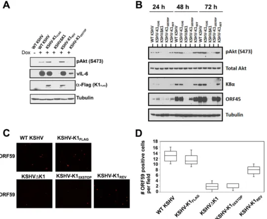
K1 is required for Akt phosphorylation.
We next investigated
the biology of the K1 mutant viruses in the doxycycline-inducible
KSHV-infected iSLK line, which was described by Myoung and
Ganem (
29
). Previously, we reported that the PI3K/Akt/mTOR
pathway is activated in cells expressing K1 (
18
,
22
). To examine
this pathway in cells infected with the recombinant viruses
de-scribed above, we investigated phosphorylation of Akt by Western
blotting. We found that Akt was phosphorylated at serine 473
(S473) at 72 h postreactivation (with 1.5
g/ml of doxycycline) in
cells latently infected with WT KSHV and KSHV-K1
FLAGbut not
in cells infected with the recombinant viruses KSHV
⌬
K1 and
KSHV-K1
5⫻STOP(
Fig. 2A
). In KSHV-K1
REV-infected cells, we
found that the level of phosphorylated Akt at S473 was restored
after reactivation from latency (
Fig. 2A
). As a control for efficient
reactivation, we also performed Western blotting for KSHV
vIL-6 and found that cells infected with WT KSHV and
KSHV-K1
FLAGexpressed much higher levels of vIL-6 than did cells
infected with the recombinant viruses KSHV
⌬
K1 and
KSHV-K1
5⫻STOP(
Fig. 2A
).
To elucidate our observations further, we compared Akt
phos-phorylation and viral lytic gene expression 24, 48, and 72 h after
reactivation with 1.5
g/ml of doxycycline. We found that the
level of phosphorylated Akt at S473 increased at several time
points following reactivation for WT KSHV-, KSHV-K1
FLAG-,
and KSHV-K1
REV-infected cells but not for KSHV
⌬
K1- and
KSHV-K1
5⫻STOP-infected cells. Following reactivation, the lytic
K8
␣
and ORF45 proteins were detectable in WT ,
KSHV-K1
FLAG-, and KSHV-K1
REV-infected cells but were barely
detect-able in KSHV
⌬
K1- and KSHV-K1
5⫻STOP-infected cells (
Fig. 2B
).
To further confirm that the K1 gene augments viral lytic
repli-cation, we performed an immunofluorescence assay with an
anti-body against the viral lytic protein ORF59. The number of red
fluorescence-labeled ORF59 cells was distinctly higher for WT
KSHV-, KSHV-K1
FLAG-, and KSHV-K1
REV-infected cells than for
KSHV
⌬
K1- and KSHV-K1
5⫻STOP-infected cells at 72 h
postreac-tivation (
Fig. 2C
and
D
).
To determine whether K1 could be detected at the protein level
in uninduced iSLK cells, we performed immunoprecipitation
from infected cells using anti-FLAG to pull down K1
FLAG.
Immu-noprecipitates using FLAG antibody from iSLK cells latently
in-fected with KSHV-K1
FLAG, KSHV-
⌬
K1, and KSHV-K1
REVwere
run on SDS-PAGE gels and subjected to Western blot analysis
with an anti-FLAG antibody. As shown in
Fig. 3
, we were able to
detect K1 expression in cells following immunoprecipitation. Our
results are concordant with those reported by Chandriani and
Ganem, who reported that K1 can be detected in latently infected
cells (
14
).
K1 deletion from KSHV results in reduced lytic replication of
KSHV.
To test the role of K1 during viral replication, we
reacti-vated iSLK cells with doxycycline. We observed greater total GFP
fluorescence in cells harboring WT KSHV, KSHV-K1
FLAG, and
KSHV-K1
REVthan in KSHV
⌬
K1- and KSHV-K1
5⫻STOP-infected
iSLK cells following reactivation (
Fig. 4A
and
B
). The GFP
fluo-rescence in WT KSHV-, KSHV-K1
FLAG-, and KSHV-K1
REV-in-fected cells ranged from 4- to 8-fold higher than that in
KSHV
⌬
K1- and KSHV-K1
5⫻STOP-infected cells (
Fig. 4C
).
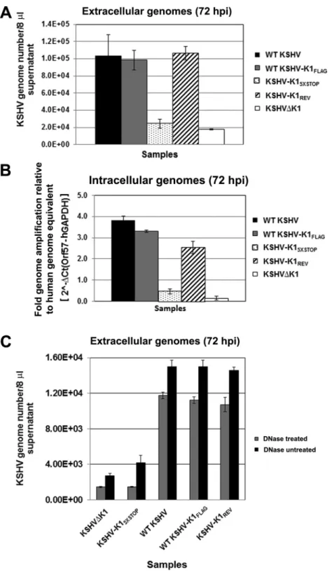
To quantify viral genomes, we extracted DNA from cell-free
supernatants and lysates of recombinant virus-infected cells
fol-lowing reactivation. We performed quantitative real-time PCR to
determine the
C
Tvalues and genome copy numbers. In the
super-natants, the genome copy numbers of released viruses from
in-duced cells harboring WT KSHV, KSHV-K1
FLAG, and
K1
REVwere markedly higher than those from cells harboring
KSHV-K1
5⫻STOPand KSHV
⌬
K1 (
Fig. 5A
). Following
reactiva-tion, WT KSHV, KSHV-K1
FLAG, and KSHV-K1
REVdisplayed a
5-fold increase in virion production, as measured by viral
ge-nomes, compared to KSHV
⌬
K1 and KSHV-K1
5⫻STOP(1
⫻
10
5genome copies versus 2
⫻
10
4genome copies) (
Fig. 5A
). Similar
results were observed for intracellular genome copy numbers in
reactivated cell lysates from cells that harbored WT KSHV,
KSHV-K1
FLAG, and KSHV-K1
REVcompared to cells harboring
KSHV-K1
5⫻STOPand KSHV
⌬
K1 (
Fig. 5B
).
In order to measure capsid-protected viral DNA, WT KSHV-,
KSHV-K1
FLAG-, KSHV-K1
REV-, KSHV
⌬
K1-, and KSHV-K1
5⫻STOP-infected iSLK cells were reactivated as described above. The
super-natants were harvested and subjected to DNase treatment for 1 h,
after which viral genomic DNA was harvested and quantitated.
Once again, we observed that WT KSHV, KSHV-K1
FLAG, and
KSHV-K1
REVdisplayed a 5- to 6-fold increase in virion
produc-tion (as measured by viral genomes) compared to KSHV
⌬
K1 and
KSHV-K1
5⫻STOP(
Fig. 5C
).
K1 deletion from KSHV results in decreased production of
infectious virions.
To test the infectivity of viruses produced from
reactivated cells, cell-free supernatants from reactivated cells were
used to infect naive Vero cells in triplicate, as described in
Mate-rials and Methods. GFP-positive cells were observed at 72 h
postinfection (
Fig. 6A
). We observed more GFP-positive, i.e.,
in-fected, cells from supernatants of WT KSHV-, KSHV-K1
FLAG-,
and KSHV-K1
REV-infected cells than from KSHV
⌬
K1- and
KSHV-K1
5⫻STOP-infected cells. The number of GFP-positive cells
FIG 2K1 is required for Akt phosphorylation and lytic gene expression in reactivated cells. (A and B) A total of 5⫻105WT KSHV-, KSHV-K1 FLAG-, KSHV-K15⫻STOP-, KSHV-K1REV-, and KSHV⌬K1-infected iSLK cells were seeded overnight and then treated with 1.5g/ml of doxycycline for 72 h (A) or for 24, 48, and 72 h (B). Cells were harvested, lysed, and subjected to Western blot analysis with an anti-FLAG antibody to detect K1, an anti-pAkt (S473) antibody to detect phosphorylated Akt, an anti-vIL-6 antibody to detect vIL-6, an anti-K8␣antibody to detect K8␣, and an anti-ORF45 antibody to detect ORF45. (C) A total of 2⫻105cells were seeded overnight and then treated with 3g/ml of doxycycline for 72 h. Cells were fixed with 3.7% formaldehyde and subjected to an immunofluorescence assay with primary anti-ORF59 antibody (1:500; Advanced Biotechnologies) and secondary anti-mouse IgG antibody conjugated to tetramethyl rhodamine isocyanate (1:500). (D) Graph depicting the number of quantitated ORF59-positive cells per field following reactivation.
in six random fields for each recombinant virus-infected sample
was quantified (
Fig. 6B
). The numbers of infectious virions from
reactivated cells harboring WT KSHV, KSHV-K1
FLAG, and
KSHV-K1
REVwere found to be dramatically higher than those
from cells harboring KSHV-K1
5⫻STOPand KSHV
⌬
K1.
To further assess the production of infectious virus and
quan-tify the number of KSHV-infected Vero cells, infected cells were
also subjected to flow cytometry analysis.
Figure 6C
shows that the
percentages of GFP-positive Vero cells were 12.5%, 10.8%, 3.0%,
3.2%, and 14.1% for WT KSHV, KSHV-K1
FLAG, KSHV
⌬
K1,
KSHV-K1
5⫻STOP, and KSHV-K1
REV, respectively, indicating that,
similar to our microscopic quantitation described above, the
per-centages of KSHV-infected Vero cells were higher with WT
KSHV, KSHV-K1
FLAG, and KSHV-K1
REVviruses than with
KSHV
⌬
K1 and KSHV-K1
5⫻STOPviruses.
To confirm the optical readout, we harvested infected Vero
cells as described in the legend of
Fig. 6A
, extracted DNA, and
performed real-time qPCR for viral genomes. We found that the
numbers of viral genomes were higher in reactivated cells
harbor-ing WT KSHV, KSHV-K1
FLAG, and KSHV-K1
REVthan in cells
harboring KSHV-K1
5⫻STOPand KSHV
⌬
K1 viruses (
Fig. 6D
).
To further assess the infectivity of these viruses on a more
rel-evant cell line, we harvested supernatants from reactivated
KSHV-infected iSLK cells and KSHV-infected primary HUVECs. Similar to the
case with Vero cells, the numbers of GFP-positive primary
HUVECs were significantly higher following infection with WT
KSHV, KSHV-K1
FLAG, and KSHV-K1
REVthan with KSHV
⌬
K1
and KSHV-K1
5⫻STOP(
Fig. 7A
and
B
).
DISCUSSION
The KSHV K1 gene has a dramatic impact on cell signaling in
single-gene assays and, in the context of herpesvirus saimiri
(HVS), can replace the HVS-transforming protein, STP (
30
). In
order to investigate the role of the KSHV K1 ORF in the life cycle
of KSHV, we constructed a set of K1 deletion or K1 mutant
re-combinant viruses, including KSHV-K1
FLAG, KSHV-K1
5⫻STOP,
KSHV-K1
REV, and KSHV
⌬
K1. JSC-1-derived recombinant
KSHV BAC16 (
28
) was used as the parental BAC for the
construc-tion of K1 deleconstruc-tion or mutant recombinant viruses. The integrity
and sequences of all the WT KSHV, KSHV-K1
FLAG,
KSHV-K1
5⫻STOP, KSHV-K1
REV, and KSHV
⌬
K1 genomes in this study
were identified and verified by multiple independent methods,
including whole-genome sequencing. We also epitope tagged the
K1 gene with the FLAG epitope in the context of the virus to use it
as a tool for investigating K1 function during the KSHV life cycle.
Previously, our laboratory and other groups studied K1 signal
transduction events (
4
,
14
,
17-20
,
22
). We reported that K1
acti-vates the PI3K/Akt/mTOR pathway and provides a survival
ad-vantage to K1-expressing cells (
18
,
22
). Here, we extended these
studies and found that cells that were infected with K1 deletion or
mutant viruses (KSHV-K1
5⫻STOPand KSHV-
⌬
K1) displayed
lower phospho-Akt levels than cells infected with viruses
containg WT K1. We detected low levels of K1 expression in cells
in-fected with KSHV-K1
FLAGusing coimmunoprecipitation. This
finding correlates with data from previous reports suggesting that
K1 can be expressed under latency conditions (
14
,
22
). However,
we cannot rule out the possibility that the K1 that we are detecting
is due to a low level of lytic replication in infected iSLK cells.
All iSLK cells harboring WT KSHV, KSHV-K1
FLAG,
KSHV-K1
5⫻STOP, KSHV-K1
REV, or KSHV
⌬
K1 were able to produce
infectious virus in response to doxycycline induction; however,
viruses lacking the K1 gene, including KSHV-K1
5⫻STOPand
KSHV-
⌬
K1, were significantly attenuated for viral reactivation in
response to RTA induction. Accumulating evidence suggests that
lytic replication may play a prominent role in KS (
5
,
31–34
), and
our data suggest that K1 is involved in enhancing lytic reactivation
and/or replication. Several groups have investigated the link
be-tween K1 signal transduction and KSHV reactivation by the
over-expression of K1 or deletion mutants (
26
,
27
). Lagunoff et al.
reported that the expression of lytic cycle genes was diminished up
to 80% in the presence of a K1 dominant negative mutant, which
inhibited wild-type K1 signal transduction in BCBL-1 cells that
were induced into reactivation by the ectopic expression of the
KSHV ORF50 transactivator (
26
). Our results demonstrate that
deletion of K1 using the deletion virus KSHV
⌬
K1 as well as the
recombinant virus KSHV-K1
5⫻STOPdisplayed reduced KSHV
re-activation as measured by vIL-6 lytic gene expression, KSHV
ge-nome copy numbers, and the production of infectious viruses. In
contrast, a revertant virus, where mutant K1 was restored back to
WT K1, grew similarly to KSHV-K1
FLAG. Collectively, our data
indicate that the lack of K1 protein expression leads to decreased
production of KSHV infectious virions. However, the exact
mech-anism by which this occurs is yet to be determined. In conclusion,
we have shown that KSHV K1 plays an important role in the
KSHV viral life cycle.
ACKNOWLEDGMENTS
We thank Jae Jung for the BAC16 construct and Shogo Misumi for pcDNA3.1D/V5-hGAPDH.
B.D. is a Leukemia and Lymphoma Society Scholar and a Burroughs Wellcome Fund Investigator in Infectious Disease.
FUNDING INFORMATION
This work, including the efforts of Blossom Damania, was funded by HHS | National Institutes of Health (NIH) (CA096500, AI107810, and DE018281). This work, including the efforts of Dirk P. Dittmer, was funded by HHS | National Institutes of Health (NIH) (CA019014, CA163217, and CA01608).
The UNC Vironomics Core is supported by NCI core grant P30 CA01608 to the UNC Lineberger Comprehensive Cancer Center.
REFERENCES
1.Chang Y, Cesarman E, Pessin MS, Lee F, Culpepper J, Knowles DM, Moore PS. 1994. Identification of herpesvirus-like DNA sequences in AIDS-associated Kaposi’s sarcoma. Science266:1865–1869.http://dx.doi .org/10.1126/science.7997879.
2.Cesarman E, Chang Y, Moore PS, Said JW, Knowles DM.1995. Kaposi’s sarcoma-associated herpesvirus-like DNA sequences in AIDS-related body-cavity-based lymphomas. N Engl J Med332:1186 –1191.http://dx .doi.org/10.1056/NEJM199505043321802.
3.Soulier J, Grollet L, Oksenhendler E, Cacoub P, Cazals-Hatem D, Babinet P, d’Agay MF, Clauvel JP, Raphael M, Degos L, Sigaux F.1995. Kaposi’s sarcoma-associated herpesvirus-like DNA sequences in multi-centric Castleman’s disease. Blood86:1276 –1280.
4.Wen KW, Damania B.2010. Hsp90 and Hsp40/Erdj3 are required for the expression and anti-apoptotic function of KSHV K1. Oncogene29:3532– 3544.http://dx.doi.org/10.1038/onc.2010.124.
5.Grundhoff A, Ganem D.2004. Inefficient establishment of KSHV latency suggests an additional role for continued lytic replication in Kaposi sar-coma pathogenesis. J Clin Invest113:124 –136.http://dx.doi.org/10.1172 /JCI200417803.
6.Dillon PJ, Gregory SM, Tamburro K, Sanders MK, Johnson GL, Raab-FIG 7KSHV K1 is required for efficient infectious virion production and
Traub N, Dittmer DP, Damania B.2013. Tousled-like kinases modulate reactivation of gammaherpesviruses from latency. Cell Host Microbe13: 204 –214.http://dx.doi.org/10.1016/j.chom.2012.12.005.
7.Gregory SM, West JA, Dillon PJ, Hilscher C, Dittmer DP, Damania B. 2009. Toll-like receptor signaling controls reactivation of KSHV from la-tency. Proc Natl Acad Sci U S A106:11725–11730.http://dx.doi.org/10 .1073/pnas.0905316106.
8.Gradoville L, Gerlach J, Grogan E, Shedd D, Nikiforow S, Metroka C, Miller G.2000. Kaposi’s sarcoma-associated herpesvirus open reading frame 50/Rta protein activates the entire viral lytic cycle in the HH-B2 primary effusion lymphoma cell line. J Virol74:6207– 6212.http://dx.doi .org/10.1128/JVI.74.13.6207-6212.2000.
9.Lukac DM, Kirshner JR, Ganem D.1999. Transcriptional activation by the product of open reading frame 50 of Kaposi’s sarcoma-associated herpesvirus is required for lytic viral reactivation in B cells. J Virol73: 9348 –9361.
10. Renne R, Zhong W, Herndier B, McGrath M, Abbey N, Kedes D, Ganem D.1996. Lytic growth of Kaposi’s sarcoma-associated herpesvirus (human herpesvirus 8) in culture. Nat Med2:342–346.http://dx.doi.org /10.1038/nm0396-342.
11. Cheng F, Weidner-Glunde M, Varjosalo M, Rainio EM, Lehtonen A, Schulz TF, Koskinen PJ, Taipale J, Ojala PM.2009. KSHV reactivation from latency requires Pim-1 and Pim-3 kinases to inactivate the latency-associated nuclear antigen LANA. PLoS Pathog5:e1000324.http://dx.doi .org/10.1371/journal.ppat.1000324.
12. Yu F, Harada JN, Brown HJ, Deng H, Song MJ, Wu TT, Kato-Stankiewicz J, Nelson CG, Vieira J, Tamanoi F, Chanda SK, Sun R. 2007. Systematic identification of cellular signals reactivating Kaposi sar-coma-associated herpesvirus. PLoS Pathog 3:e44. http://dx.doi.org/10 .1371/journal.ppat.0030044.
13. Lagunoff M, Ganem D.1997. The structure and coding organization of the genomic termini of Kaposi’s sarcoma-associated herpesvirus. Virology 236:147–154.http://dx.doi.org/10.1006/viro.1997.8713.
14. Chandriani S, Ganem D. 2010. Array-based transcript profiling and limiting-dilution reverse transcription-PCR analysis identify additional latent genes in Kaposi’s sarcoma-associated herpesvirus. J Virol84:5565– 5573.http://dx.doi.org/10.1128/JVI.02723-09.
15. Bowser BS, DeWire SM, Damania B.2002. Transcriptional regulation of the K1 gene product of Kaposi’s sarcoma-associated herpesvirus. J Virol 76:12574 –12583.http://dx.doi.org/10.1128/JVI.76.24.12574-12583.2002. 16. Lee BS, Connole M, Tang Z, Harris NL, Jung JU. 2003. Structural analysis of the Kaposi’s sarcoma-associated herpesvirus K1 protein. J Virol 77:8072– 8086.http://dx.doi.org/10.1128/JVI.77.14.8072-8086.2003. 17. Samaniego F, Pati S, Karp JE, Prakash O, Bose D. 2001. Human
herpesvirus 8 K1-associated nuclear factor-kappa B-dependent promoter activity: role in Kaposi’s sarcoma inflammation? J Natl Cancer Inst Monogr2001:15–23.
18. Tomlinson CC, Damania B.2004. The K1 protein of Kaposi’s sarcoma-associated herpesvirus activates the Akt signaling pathway. J Virol78: 1918 –1927.http://dx.doi.org/10.1128/JVI.78.4.1918-1927.2004. 19. Lagunoff M, Majeti R, Weiss A, Ganem D. 1999. Deregulated signal
transduction by the K1 gene product of Kaposi’s sarcoma-associated her-pesvirus. Proc Natl Acad Sci U S A96:5704 –5709.http://dx.doi.org/10 .1073/pnas.96.10.5704.
20. Wang S, Wang S, Maeng H, Young DP, Prakash O, Fayad LE, Younes A, Samaniego F.2007. K1 protein of human herpesvirus 8 suppresses lymphoma cell Fas-mediated apoptosis. Blood109:2174 –2182.http://dx .doi.org/10.1182/blood-2006-02-003178.
21. Lee H, Guo J, Li M, Choi JK, DeMaria M, Rosenzweig M, Jung JU.1998.
Identification of an immunoreceptor tyrosine-based activation motif of K1 transforming protein of Kaposi’s sarcoma-associated herpesvirus. Mol Cell Biol18:5219 –5228.http://dx.doi.org/10.1128/MCB.18.9.5219. 22. Wang L, Dittmer DP, Tomlinson CC, Fakhari FD, Damania B.2006.
Immortalization of primary endothelial cells by the K1 protein of Kaposi’s sarcoma-associated herpesvirus. Cancer Res66:3658 –3666.http://dx.doi .org/10.1158/0008-5472.CAN-05-3680.
23. Bhatt AP, Damania B.2012. AKTivation of PI3K/AKT/mTOR signaling pathway by KSHV. Front Immunol 3:401. http://dx.doi.org/10.3389 /fimmu.2012.00401.
24. Wang L, Wakisaka N, Tomlinson CC, DeWire SM, Krall S, Pagano JS, Damania B.2004. The Kaposi’s sarcoma-associated herpesvirus (KSHV/ HHV-8) K1 protein induces expression of angiogenic and invasion fac-tors. Cancer Res 64:2774 –2781. http://dx.doi.org/10.1158/0008-5472 .CAN-03-3653.
25. Tomlinson CC, Damania B.2008. Critical role for endocytosis in the regulation of signaling by the Kaposi’s sarcoma-associated herpesvirus K1 protein. J Virol82:6514 – 6523.http://dx.doi.org/10.1128/JVI.02637-07. 26. Lagunoff M, Lukac DM, Ganem D.2001. Immunoreceptor
tyrosine-based activation motif-dependent signaling by Kaposi’s sarcoma-associated herpesvirus K1 protein: effects on lytic viral replication. J Virol 75:5891–5898.http://dx.doi.org/10.1128/JVI.75.13.5891-5898.2001. 27. Lee BS, Paulose-Murphy M, Chung YH, Connlole M, Zeichner S, Jung
JU.2002. Suppression of tetradecanoyl phorbol acetate-induced lytic re-activation of Kaposi’s sarcoma-associated herpesvirus by K1 signal trans-duction. J Virol 76:12185–12199. http://dx.doi.org/10.1128/JVI.76.23 .12185-12199.2002.
28. Brulois KF, Chang H, Lee AS, Ensser A, Wong LY, Toth Z, Lee SH, Lee HR, Myoung J, Ganem D, Oh TK, Kim JF, Gao SJ, Jung JU.2012. Construction and manipulation of a new Kaposi’s sarcoma-associated herpesvirus bacterial artificial chromosome clone. J Virol86:9708 –9720. http://dx.doi.org/10.1128/JVI.01019-12.
29. Myoung J, Ganem D.2011. Generation of a doxycycline-inducible KSHV producer cell line of endothelial origin: maintenance of tight latency with efficient reactivation upon induction. J Virol Methods174:12–21.http: //dx.doi.org/10.1016/j.jviromet.2011.03.012.
30. Lee H, Veazey R, Williams K, Li M, Guo J, Neipel F, Fleckenstein B, Lackner A, Desrosiers RC, Jung JU.1998. Deregulation of cell growth by the K1 gene of Kaposi’s sarcoma-associated herpesvirus. Nat Med4:435– 440.http://dx.doi.org/10.1038/nm0498-435.
31. Kedes DH, Ganem D.1997. Sensitivity of Kaposi’s sarcoma-associated herpesvirus replication to antiviral drugs. Implications for potential ther-apy. J Clin Invest99:2082–2086.
32. Martin DF, Kuppermann BD, Wolitz RA, Palestine AG, Li H, Robinson CA.1999. Oral ganciclovir for patients with cytomegalovirus retinitis treated with a ganciclovir implant. Roche Ganciclovir Study Group. N Engl J Med340:1063–1070.
33. Campbell TB, Borok M, Gwanzura L, MaWhinney S, White IE, Nde-mera B, Gudza I, Fitzpatrick L, Schooley RT. 2000. Relationship of human herpesvirus 8 peripheral blood virus load and Kaposi’s sarcoma clinical stage. AIDS14:2109 –2116.http://dx.doi.org/10.1097/00002030 -200009290-00006.