Conditional deletion of
Pip5k1c
in sensory
ganglia and effects on nociception and
inflammatory sensitization
Lipin Loo
1and Mark J Zylka
1Abstract
Phosphatidylinositol 4-phosphate 5-kinase type 1 gamma (Pip5k1c) generates phosphatidylinositol 4,5-bisphosphate, also known as PI(4,5)P2or PIP2. Many pronociceptive signaling pathways and receptor tyrosine kinases signal via PIP2hydrolysis. Previously, we found that pain signaling and pain sensitization were reduced in Pip5k1cþ/ global heterozygous knockout mice. Here, we sought to evaluate the extent to which dorsal root ganglia selective deletion ofPip5k1caffected nociception in mice. Initially, we crossed sensory neuron-selectiveAdvillin-Cre mice with a conditional Pip5k1c knockout (cKO) allele (Pip5k1cfl/fl). However, these mice displayed an early onset proprioceptive deficit. To bypass this early onset phenotype, we used two different tamoxifen-inducible Cre lines (Brn3a-Cre-ERT2andAdvillin-Cre-ERT2) to conditionally deletePip5k1cin adults. Tamoxifen induced high efficiency deletion of PIP5K1C in dorsal root ganglia and slightly reduced PIP5K1C in spinal cord and brain inBrn3a-Cre-ERT2Pip5k1cfl/fl(Brn3a cKO) mice while PIP5K1C was selectively deleted in dorsal root ganglia with no changes in spinal cord or brain in Advillin-Cre-ERT2 Pip5k1cfl/fl (Advil cKO) mice. Acute thermosensation and mechanosensation were not altered in either line relative to wild-type mice. However, thermal hypersensitivity and mech-anical allodynia recovered more rapidly in Brn3a cKO mice, but not Advil cKO mice, following hind paw inflammation. These data collectively suggest that PIP5K1C regulates nociceptive sensitization in more regions of the nervous system than dorsal root ganglia alone.
Keywords
Lipid kinase, PIP5K1C,Brn3a-Cre-ERT2, Advillin-Cre-ERT2, nociception, inflammatory pain
Date received: 5 September 2017; accepted: 13 September 2017
Introduction
Nociceptive or physiological pain alerts an individual of potential threats such as injury or disease. However, acute pain can transform into pathophysiological stimu-lus-uncoupled chronic pain. Current pain management involves the use of opioids and nonsteroidal anti-inflam-matory drugs, but chronic administration of these drugs have serious side effects, highlighting a major unmet medical need for new pain treatments.
Nociceptor sensitization contributes to chronic pain.1 Pronociceptive factors in the inflammatory soup activate receptors such as G-protein-coupled receptors and recep-tor tyrosine kinases (RTKs). Downstream signaling cascades potentiate the activity and expression of a variety of ion channels and receptors, driving the increase in neuronal excitability. Inhibiting individual pronociceptive receptors and kinases have worked in
animal models but have shown modest to no effects in humans.2–5An alternative to these strategies is to target signaling molecules immediately downstream of multiple pronociceptive receptors.
Phosphatidylinositol 4,5-bisphosphate, also known as PI(4,5)P2 or PIP2, has important roles in cell signaling
and is immediately downstream of many pronociceptive signaling pathways, despite only accounting for 0.5% to 1% of the phospholipid molecules in cells.6,7 Type 1 phosphotidylinositol 4-phosphate 5-kinases (PIP5KIs)
1
Department of Cell Biology and Physiology, UNC Neuroscience Center, The University of North Carolina, Chapel Hill, NC, USA
Corresponding author:
Mark J Zylka, Department of Cell Biology and Physiology, UNC Neuroscience Center, The University of North Carolina, Chapel Hill, NC 27599, USA.
Email: [email protected]
Creative Commons Non Commercial CC BY-NC: This article is distributed under the terms of the Creative Commons Attribution-NonCommercial 4.0 License (http://www.creativecommons.org/licenses/by-nc/4.0/) which permits non-commercial use, reproduction and distribution of the work without further permission provided the original work is attributed as specified on the SAGE and Open Access pages (https:// us.sagepub.com/en-us/nam/open-access-at-sage).
Molecular Pain Volume 13: 1–8
synthesize PIP2 by phosphorylating the large pools of
phosphatidylinositol 4-phosphate in cells. There are three Pip5k1 genes, two of which are ubiquitously expressed (Pip5k1aandPip5k1b), while the third (phos-phatidylinositol 4-phosphate 5-kinase type 1 gamma,
Pip5k1c) is expressed predominantly in neuronal tis-sues.6,8,9We previously found that global heterozygous deletion of Pip5k1creduced PIP2in dorsal root ganglia
(DRG) and reduced pain signaling and inflammatory sensitization.10 While these studies suggested that it might be possible to reduce pronociceptive signaling by selectively inhibiting a lipid kinase that generates PIP2
in DRG neurons, whether the decrease observed in pain behavior was due to a reduction in the enzyme expression specifically in sensory neurons or globally throughout the nervous system was unresolved. Here, we sought to evaluate the extent to which sensory neuron-selective deletion of Pip5k1c reduced acute and chronic pain sensitivity.
Materials and methods
Animals
All procedures involving vertebrate animals were approved by the Institutional Animal Care and Use Committee at the University of North Carolina at Chapel Hill. Mice were raised on a 12:12 h light:dark cycle, had ad libitum access to food and water, and were tested during the light phase. Estrous cycle was not monitored in females.Pip5k1cfl/flmice11were crossed with Advillin-Cre mice,12 Brn3a-Cre-ERT2,13 and
Advillin-Cre-ERT2mice14to generate conditional knock-outs. All mice were backcrossed to C57BL/6J mice for at least eight generations.
Injections and behavioral assays
Tamoxifen was prepared fresh daily by dissolving in 10% ethanol and 90% corn oil and sonicated for half an hour at room temperature. Intraperitoneal injections of 120 mg/kg tamoxifen were administered for seven con-secutive days in mice aged six to eight weeks, and base-line behavioral testings were performed 10 days after the last injection. Mice were acclimated to the testing room, equipment, and experimenter one to three days before behavioral testing. Behavioral assays were performed as described previously.15
Western blot analysis
Tissue lysates were prepared as described10 from wild-type (WT) and conditional knockout (cKO) mice after completion of all behavioral testing. Briefly, DRG, spinal cord, and cerebral cortex were dissected and
sonicated in radioimmunoprecipitation assay buffer. Protein concentration was determined via bicinchoninic acid assay, and 50mg protein are loaded onto gels, which were subsequently transferred onto polyvinylidene difluoride membranes. Primary antibodies (1:4000 for PIP5K1C, generously provided by Hara et al.16 and 1:3000 for b-actin; Abcam, AB6276; served as loading control) were prepared in Licor blocking buffer. Secondary antibodies (1:10,000; Li-Cor IRDye 680 and 800) were prepared in Licor blocking buffer. Blots were imaged on a LiCor gel imaging machine and analyzed using ImageJ. PIP5K1C band intensities were normal-ized to their respectiveb-actin controls.
Results
We first crossed Advillin-Cre (Advil-Cre) mice with
Pip5k1c floxed (Pip5k1cfl/fl) mice to selectively delete
Pip5k1c in the DRG.11,15,17 Advil-Cre is expressed in trigeminal ganglia by embryonic day 16.5 and in DRGs by postnatal day 1.12,17 Beginning around two months of age, we observed hind limb clasping and abnormal gait phenotypes in Advil-CrePip5k1cfl/fl
mice (data not shown), suggestive of an early-onset pro-prioceptive deficit.18–20Hind limb clasping prevented us from probing the hind paw using standard nociceptive sensory assays.
To circumvent this early-onset proprioceptive pheno-type, we crossed Brn3a-Cre-ERT2 mice withPip5k1cfl/fl
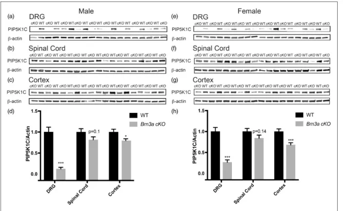
mice (henceforth called Brn3a cKO mice) to condition-ally deletePip5k1cin sensory neurons of adult mice.13,21 Tamoxifen administration greatly reduced PIP5K1C protein levels in the DRG of male and female Brn3a cKO mice (Figure 1). Non-Cre recombinase-expressing
Pip5k1cfl/fl mice, also tamoxifen-treated, served as WT controls. Although Brn3a-Cre-ERT2 mice were previ-ously used to delete genes in DRG,13,21 we observed a nonsignificant reduction of PIP5K1C in the spinal cord (male, p¼0.1; female, p¼0.14) and a small but signifi-cant reduction of PIP5K1C in the cerebral cortex (male, p<0.05; female, p<0.005).
Tamoxifen-treated WT andBrn3a cKOmice were also tested with the hotplate (55C), tail immersion (10C,
46.5C, 49C), radiant hind paw heating (Hargreaves),
cold plantar,22and electronic von Frey assays to probe for differences in noxious thermal and mechanical sensi-tivity. No significant differences relative to tamoxifen-treated WT mice were observed in any of these assays (Table 1). Tamoxifen-treated mice were then injected with complete Freund’s adjuvant (CFA) to model inflammatory pain. The contralateral paw served as the control. No significant differences in thermal or mechanical sensitivity were observed between WT and
Brn3a cKOmice at early time points post-CFA injection; however, both male and female Brn3a cKO mice
exhibited faster recovery to baseline withdrawal latencies and thresholds (Figure 2).
Since tamoxifen-treated Brn3a cKO mice showed a small but significant reduction of PIP5K1C in cerebral cortex, we sought to more selectively delete Pip5k1c in sensory ganglia of adults. To accomplish this, we crossed
Pip5k1cfl/fl mice with Advil-Cre-ERT2 mice (Advil cKO
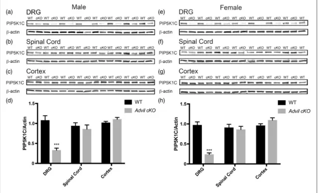
mice).14 Tamoxifen administration significantly reduced PIP5K1C protein levels in the DRG of male and female
Advil cKO mice but did not alter levels of PIP5K1C in spinal cord or cerebral cortex (Figure 3). TheAdvil cKO
line thus more selectively deleted PIP5K1C in adult Figure 1. Deletion of PIP5K1C in DRG and partial deletion in the cerebral cortex usingBrn3a-Cre-ERT2line. (a)–(c), PIP5K1C protein levels in the indicated tissues of male mice and (d) Western blot quantification relative tob-actin. Tissue harvested 18 to 20 days post tamoxifen injections.n¼10 WT and 14cKOmale mice. (e)–(g) PIP5K1C protein levels in the indicated tissues of female mice and (h) Western blot quantification relative tob-actin. Tissue harvested 18 to 20 days post tamoxifen injections.n¼14 WT and 15cKOfemale mice. All data are meanSEM. Asterisks indicate significant difference between WT andcKOmice byttest. *p<0.05, **p<0.001, and ***p<0.0005.
Table 1. Noxious heat, mechanical, and cold behavioral assays withBrn3a cKOmice.
Male Female
Assay/genotype WT Brn3a cKO p value WT Brn3a cKO p value
Hot plate (55C) 21.71.5 s 18.71.7 s 0.17 19.50.6 s 21.31.2 s 0.21
Tail immersion (46.5C) 4.20.6 s 4.10.4 s 0.89 4.00.4 s 4.20.3 s 0.75
Tail immersion (49C) 2.10.2 s 2.30.3 s 0.48 2.50.2 s 2.60.4 s 0.66
Tail immersion (10C) 15.52.3 s 14.62.5 s 0.79 18.72.6 s 15.71.5 s 0.15
Hargreaves (radiant heat) 10.00.6 s 10.70.5 s 0.26 9.80.3 s 100.4 s 0.63
Cold plantar 13.50.7 s 140.6 s 0.59 120.6 s 12.60.4 s 0.38
von Frey (mechanical) 5.30.3 g 5.40.3 g 0.71 4.10.2 g 4.00.2 g 0.55
DRG than theBrn3a cKOline. No significant differences were observed between tamoxifen-treated WT mice and tamoxifen-treated Advil cKO mice in assays of noxious thermal or mechanical sensitivity (Table 2), nor were sig-nificant differences observed following CFA-inflamma-tion (Figure 4). The discrepancy between Advil cKO
and Brn3a cKO mice in the CFA inflammatory pain
model could relate to less complete deletion of PIP5K1C in DRG of Advil cKO mice and/or partial deletion of PIP5K1C in cerebral cortex of Brn3a cKO
mice (compare Figures 1 and 3).
Discussion
Our study is the first to suggest that sensory-neuron selective loss of Pip5k1c causes an early-onset proprio-ceptive deficit in mice. Global loss of PIP5K1C in humans causes lethal congenital contractural syndrome type 3,23 a syndrome featuring severe arthrogryposis (joint contractures) and death shortly before or immedi-ately after birth. Joint contractures are seen in other human disorders that impair proprioception,24including
biallelic loss of PIEZO2.25–27The proprioceptive deficits that we observed in Advil-CrePip5k1cfl/fl mice might relate to developmental loss ofPip5k1cin DRG, deple-tion of PIP2, and/or impaired neurotrophin-mediated
neurite outgrowth. Neurotrophin-mediated RTK signal-ing regulates many aspects of neurodevelopment.28One of the kinases involved downstream of RTK signaling is phosphoinositide-3 kinase (PI3K). PI3K phosphorylates PIP2 to generate PIP3 to allow for the binding of Akt
(protein kinase B). This leads to disinhibition of mTOR (mammalian target of Rapamycin), and this signaling axis regulates formation of axonal filopodia and den-dritic arborization.29–31 Furthermore, PIP5K1C regu-lates adhesion junction formation and neuronal cell migration.32 Future studies will be needed to resolve whether early loss of PIP5K1C impairs proprioceptive neuron development and/or maintenance.
The primary focus of our research was to evaluate how sensory neuron selective loss of PIP5K1C affects nociception. Thus, to bypass this early-onset propriocep-tive phenotype, we conditionally deletedPip5k1cin adult sensory ganglia using two different tamoxifen-inducible Figure 2. Early recovery of inflammation-induced thermal and mechanical hypersensitivity inBrn3a cKOmice. One hind paw of male (a and b) and female (c and d) mice was injected with the inflammatory agent CFA. The uninjected paw served as a control. (a) and (c) Thermal sensitivity measured before and several days after injection using the Hargreaves assay. (b) and (d) Mechanical sensitivity measured using an electronic von Frey apparatus.n¼10 WT and 14cKOfor males.n¼14 WT and 15cKOfor females. All data are meanSEM. Asterisks indicate significant difference between WT inflamed and cKO inflamed by two-way ANOVA, Dunnett’s post hoc test. *p<0.05, **p<0.005, and ***p<0.0005.
Cre lines. We generated tamoxifen-inducibleBrn3a cKO
andAdvil cKOmice, neither of which displayed evidence of motor impairment or hind limb clasping over the course of our studies (data not shown).
We initially evaluated nociceptive phenotypes in tamoxifen-treated WT and Brn3a cKO mice. Brn3a cKOmice exhibited no difference in thermal or mechan-ical hypersensitivity following CFA inflammation but showed a faster recovery to baseline levels. We cannot
conclude that this more rapid recovery was exclusively caused by the deletion ofPip5k1cin sensory neurons, as these Brn3a cKO mice also showed a significant reduc-tion of PIP5K1C in the cerebral cortex. Initially, we tried a tamoxifen regiment (1 mg of intraperitoneal injections for five consecutive days) as reported in a previous
Brn3a-Cre-ERT2study,21but observed very low deletion efficiency (10%–50% deletion in DRG, data not shown). To further increase the deletion efficiency in DRG, we Figure 3. Deletion of PIP5K1C in DRG but not in the spinal cord or cerebral cortex usingAdvil-Cre-ERT2line. (a)–(c) PIP5K1C protein levels in the indicated tissues of male mice and (d) Western blot quantification relative tob-actin. Tissue harvested 18 to 20 days post tamoxifen injections.n¼13 WT and 13cKOmale mice. (e)–(g) PIP5K1C protein levels in the indicated tissues of female mice and (h) Western blot quantification relative tob-actin. Tissue harvested 18 to 20 days post tamoxifen injections.n¼12 WT and 9cKOfemale mice. All data are meanSEM. Asterisks indicates significant difference between WT andcKOmice byttest. ***p<0.0005.
Table 2. Noxious heat, mechanical, and cold behavioral assays withAdvil cKOmice.
Male Female
Assay/genotype WT Advil cKO p value WT Advil cKO p value
Hot plate (55C) 24.41.1 s 22.91.0 s 0.31 18.91.2 s 21.60.7 s 0.06
Tail immersion (46.5C) 4.90.3 s 5.00.5 s 0.91 5.10.3 s 5.40.3 s 0.54
Tail immersion (49C) 3.40.2 s 3.20.4 s 0.75 2.60.1 s 2.90.2 s 0.33
Tail immersion (10C) 13.11.4 s 14.11.9 s 0.68 16.01.6 s 16.21.4 s 0.96
Hargreaves (radiant heat) 10.30.7 s 9.70.5 s 0.44 9.50.4 s 9.640.4 s 0.86 Cold plantar 14.71.2 s 14.00.9 s 0.63 16.31.0 s 15.21.2 s 0.46 von Frey (mechanical) 4.50.2 g 4.30.2 g 0.70 4.30.2 g 3.70.2 g 0.08
tried a higher tamoxifen dose (120mg/g body weight, which is approximately 3 mg for a 25 g mouse, for seven consecutive days). However, this dosing schedule led to deletion of PIP5K1C in the spinal cord and cere-bral cortex.
Brn3a is expressed in the dorsal horn of the spinal cord.33Additionally, conditional reporter gene targeting revealed Brn3a-positive neuronal projections from the superior colliculus into the cerebral cortex but no expres-sion within the cerebral cortex.34,35The slight decrease in PIP5K1C observed in our cortical Western blots may reflect low-level recombination in the cerebral cortex or, less likely, unintentional sampling of the superior col-liculus, which is located just below the cerebral cortex. Interestingly, a LacZ reporter study showed laminar expression of Brn3ain the superior colliculus and mes-encephalic central gray, also known as the periaqueduc-tal gray (PAG).35 The PAG is known to regulate ascending and descending pain pathways.36–38It is there-fore possible that PIP5K1C was also deleted in the PAG of Brn3a cKO mice. The more rapid recovery ofBrn3a
cKO mice following CFA-induced inflammation might thus relate to a loss of Pip5k1c in more regions of the nervous system than DRG alone. In support of this pos-sibility,Advil cKOmice showed a more selective loss of
PIP5K1C in DRG but did not show a more rapid recov-ery following CFA-induced inflammation. Homozygous
Pip5k1c knockout mice (Pip5k1c/) showed major reduction of PIP2(50%) in the brain, and Pip5k1c
/
cortical neurons have impaired synaptic transmission, although these animals die at birth.39 It was thus not possible to study how global loss of Pip5k1c affected nociceptive behaviors in adults.
Collectively, our findings suggest that many of the nociceptive sensory phenotypes that we identified in
Pip5k1cþ/
mice are likely due to PIP5K1C haploinsuf-ficiency in more regions of the body and nervous system than DRG alone. Support for this conclusion comes from our observation that Advil cKO mice show a
>50% loss of PIP5K1C protein in DRG (which is greater than the 50% loss we reported in Pip5k1cþ/
mice) but showed no sensory phenotypes. Moreover,
Brn3a cKO mice showed faster recovery following CFA-induced inflammation, mirroring one of the pheno-types we identified in Pip5k1cþ/ mice, but PIP5K1C was reduced in other regions of the nervous system besides DRG. Our study highlights the importance of evaluating multiple mutant lines (global, conditional, and adult-specific conditional) before drawing strong conclusions as to whether a gene regulates nociception Figure 4. Inflammation-induced thermal and mechanical hypersensitivity inAdvil cKOmice is equivalent to WT mice. (a) and (b) One hind paw of male and (c) and (d) female mice was injected with the inflammatory agent CFA. The uninjected paw served as a control. (a) and (c) Thermal sensitivity measured before and several days after injection using the Hargreaves assay. (b) and (d) Mechanical sensitivity measured using an electronic von Frey apparatus.n¼13 WT and 13cKOfor males.n¼12 WT and 9cKOfor females. All data are meanSEM. No significant difference between WT inflamed andcKOinflamed by two-way ANOVA, Dunnett’s post hoc test.
exclusively via effects in sensory ganglia or via effects in other parts of the nervous system. Moreover, future work with additional Cre driver lines, such as spinal cord and/or brain specific, could be used to further val-idate the findings in this study.
Acknowledgments
The authors thank Brittany Schuck for her work in generating and characterizing the phenotypes ofAdvillin-CrePip5k1cfl/fl
mice, Hiroyuki Sakagami for providing PIP5K1C antibody, William Snider for providing the Brn3a-Cre-ERT2 mice, Ardem Patapoutian for providing the Advillin-Cre-ERT2 mice, and Eric McCoy for performing CFA injections.
Authors’ Contributions
LL performed the experiments. LL and MJZ designed the experiments, interpreted the data, and wrote the manuscript. Both authors read and approved the final manuscript.
Declaration of Conflicting Interests
The author(s) declared no potential conflicts of interest with respect to the research, authorship, and/or publication of this article.
Funding
The author(s) disclosed receipt of the following financial sup-port for the research, authorship, and/or publication of this article: This work was supported by a grant to MJZ from NINDS (R01NS081127).
References
1. Gold MS and Gebhart GF. Nociceptor sensitization in pain pathogenesis.Nat Med2010; 16: 1248–1257.
2. Anand P, Shenoy R, Palmer JE, et al. Clinical trial of the p38 MAP kinase inhibitor dilmapimod in neuropathic pain following nerve injury.Eur J Pain2011; 15: 1040–1048. 3. Cousins MJ, Pickthorn K, Huang S, et al. The safety and
efficacy of KAI-1678 – an inhibitor of epsilon protein kinase C (epsilonPKC)-versus lidocaine and placebo for the treat-ment of postherpetic neuralgia: a crossover study design.
Pain Med2013; 14: 533–540.
4. Ostenfeld T, Krishen A, Lai RY, et al. Analgesic efficacy and safety of the novel p38 MAP kinase inhibitor, losmapi-mod, in patients with neuropathic pain following peripheral nerve injury: a double-blind, placebo-controlled study.Eur J Pain2013; 17: 844–857.
5. Tong SE, Daniels SE, Black P, et al. Novel p38alpha mito-gen-activated protein kinase inhibitor shows analgesic effi-cacy in acute postsurgical dental pain. J Clin Pharmacol
2012; 52: 717–728.
6. Loo L, Wright BD and Zylka MJ. Lipid kinases as thera-peutic targets for chronic pain. Pain 2015; 156(Suppl 1): S2–S10.
7. Lemmon MA. Membrane recognition by phospholipid-binding domains.Nat Rev Mol Cell Biol2008; 9: 99–111. 8. Wenk MR and De Camilli P. Protein-lipid interactions and
phosphoinositide metabolism in membrane traffic: insights
from vesicle recycling in nerve terminals.Proc Natl Acad Sci U S A2004; 101: 8262–8269.
9. Wenk MR, Pellegrini L, Klenchin VA, et al. PIP kinase Igamma is the major PI(4,5)P(2) synthesizing enzyme at the synapse.Neuron2001; 32: 79–88.
10. Wright BD, Loo L, Street SE, et al. The lipid kinase PIP5K1C regulates pain signaling and sensitization.
Neuron2014; 82: 836–847.
11. White JK, Gerdin AK, Karp NA, et al. Genome-wide gen-eration and systematic phenotyping of knockout mice reveals new roles for many genes.Cell2013; 154: 452–464. 12. Zurborg S, Piszczek A, Martinez C, et al. Generation and characterization of anAdvillin-Credriver mouse line.Mol Pain2011; 7: 66.
13. O’Donovan KJ, Ma K, Guo H, et al. B-RAF kinase drives developmental axon growth and promotes axon regener-ation in the injured mature CNS.J Exp Med 2014; 211: 801–814.
14. Lau J, Minett MS, Zhao J, et al. Temporal control of gene deletion in sensory ganglia using a tamoxifen-inducible
Advillin-Cre-ERT2recombinase mouse.Mol Pain2011; 7:
100.
15. McCoy ES, Taylor-Blake B, Street SE, et al. Peptidergic CGRPaprimary sensory neurons encode heat and itch and tonically suppress sensitivity to cold. Neuron 2013; 78: 138–151.
16. Hara Y, Fukaya M, Tamaki H, et al. Type I phosphatidy-linositol 4-phosphate 5-kinase gamma is required for neur-onal migration in the mouse developing cerebral cortex.
Eur J Neurosci2013; 38: 2659–2671.
17. Hasegawa H, Abbott S, Han BX, et al. Analyzing somato-sensory axon projections with the somato-sensory neuron-specific
Advillingene.J Neurosci2007; 27: 14404–14414.
18. Lalonde R and Strazielle C. Brain regions and genes affecting limb-clasping responses.Brain Res Rev2011; 67: 252–259. 19. Guyenet SJ, Furrer SA, Damian VM, et al. A simple
com-posite phenotype scoring system for evaluating mouse models of cerebellar ataxia.J Vis Exp2010; 39: 1787. 20. Lin CH, Tallaksen-Greene S, Chien WM, et al.
Neurological abnormalities in a knock-in mouse model of Huntington’s disease.Hum Mol Genet2001; 10: 137–144. 21. Latremoliere A, Latini A, Andrews N, et al. Reduction of
neuropathic and inflammatory pain through inhibition of the tetrahydrobiopterin pathway.Neuron2015; 86: 1393–1406. 22. Brenner DS, Golden JP and Gereau RWt. A novel
behav-ioral assay for measuring cold sensation in mice.PLoS One
2012; 7: e39765.
23. Narkis G, Ofir R, Landau D, et al. Lethal contractural syndrome type 3 (LCCS3) is caused by a mutation in PIP5K1C, which encodes PIPKI gamma of the phophati-dylinsitol pathway.Am J Hum Genet2007; 81: 530–539. 24. Shibasaki H, Hitomi T, Mezaki T, et al. A new form of
congenital proprioceptive sensory neuropathy associated with arthrogryposis multiplex. J Neurol 2004; 251: 1340–1344.
26. Coste B, Houge G, Murray MF, et al. Gain-of-function mutations in the mechanically activated ion channel PIEZO2 cause a subtype of distal arthrogryposis. Proc Natl Acad Sci U S A2013; 110: 4667–4672.
27. Mahmud AA, Nahid NA, Nassif C, et al. Loss of the pro-prioception and touch sensation channel PIEZO2 in sib-lings with a progressive form of contractures.Clin Genet
2017; 91: 470–475.
28. Huang EJ and Reichardt LF. Neurotrophins: roles in neur-onal development and function.Annu Rev Neurosci2001; 24: 677–736.
29. Luikart BW, Zhang W, Wayman GA, et al. Neurotrophin-dependent dendritic filopodial motility: a convergence on PI3K signaling.J Neurosci2008; 28: 7006–7012.
30. Jaworski J, Spangler S, Seeburg DP, et al. Control of den-dritic arborization by the phosphoinositide-30 -kinase-Akt-mammalian target of rapamycin pathway.J Neurosci2005; 25: 11300–11312.
31. Abe N, Borson SH, Gambello MJ, et al. Mammalian target of rapamycin (mTOR) activation increases axonal growth capacity of injured peripheral nerves.J Biol Chem
2010; 285: 28034–28043.
32. Wang Y, Lian L, Golden JA, et al. PIP5KI gamma is required for cardiovascular and neuronal development.
Proc Natl Acad Sci U S A2007; 104: 11748–11753.
33. Ninkina NN, Stevens GE, Wood JN, et al. A novel Brn3-like POU transcription factor expressed in subsets of rat sensory and spinal cord neurons.Nucleic Acids Res1993; 21: 3175–3182.
34. Badea TC, Cahill H, Ecker J, et al. Distinct roles of tran-scription factors brn3a and brn3b in controlling the devel-opment, morphology, and function of retinal ganglion cells.Neuron2009; 61: 852–864.
35. Quina LA, Pak W, Lanier J, et al. Brn3a-expressing ret-inal ganglion cells project specifically to thalamocortical and collicular visual pathways. J Neurosci 2005; 25: 11595–11604.
36. Behbehani MM. Functional characteristics of the midbrain periaqueductal gray.Prog Neurobiol1995; 46: 575–605. 37. Reynolds DV. Surgery in the rat during electrical analgesia
induced by focal brain stimulation. Science 1969; 164: 444–445.
38. Boadas-Vaello P, Castany S, Homs J, et al. Neuroplasticity of ascending and descending pathways after somatosen-sory system injury: reviewing knowledge to identify neuro-pathic pain therapeutic targets. Spinal Cord 2016; 54: 330–340.
39. Di Paolo G, Moskowitz HS, Gipson K, et al. Impaired PtdIns(4,5)P2 synthesis in nerve terminals produces defects in synaptic vesicle trafficking.Nature2004; 431: 415–422.