15
Evaluating the effect of exercise and nutrition on bone density in rats , Ardeshir 4
, Kourosh Sayehmiri 3
, Siamak Derakhshan 2
Fatemeh Piri¹, Afra Khosravi
Moayeri¹*
1. Department of Anatomy, Faculty of Medicine, Ilam University of Medical Sciences, Ilam, Iran 2. Department of Immunology, Faculty of Medicine, Ilam University of Medical Sciences, Ilam,
Iran
3. Department of Nuclear Medicine, Faculty of Medicine, Kourdestan University of Medical Sciences, Kourdestan, Iran
4. Department of Epidemiology, Faculty of Medicine, Ilam University of Medical Sciences, Ilam, Iran
Abstract
Introduction: The aim of this research was to study the effect of a 6-week supplemented diet on increasing bone density by measuring calcium, phosphorus and bone mineral density (BMD) in male rats.
Materials and methods: In this experimental study, 24 Wistar male rats, aged between 15 and 20 days, were selected. The rats were randomly divided into 3 groups: immune system supplement (ISS), ISS plus exercise and control. Daily swimming was performed by time-increasing (starting from ten minutes and ten minutes was added each day and was fixed at sixty minutes). Supplements strengthening the immune system containing calcium and phosphorus were given to rats (5g/kg/day). After six weeks, BMD was measured using bone densitometer. Animals were anesthetized with ketamine and blood samples were gathered in order to separate their serum. The serum samples were used to measure calcium, phosphorus by the ELISA method.
Results: The results showed that in both groups (ISS plus exercise and ISS), BMD was higher than the control group. The highest level of calcium, phosphorus and BMD was seen in the group whose members were ISS plus exercise group. In contrast, the least amount of the mentioned markers was reported in the control group.
Conclusion: The results indicate a small but positive effect of ISS on whole body BMD in male rat; also results indicate the combination of exercise and proper nutrition was more effective on increasing the bone density in comparison with the proper nutrition sedentary group.
Keywords: Calcium, BMD, Phosphorus, Exercise Introduction
Bone tissue is continuously remodeled under physiological conditions (1). Environmental factors such as nutrition and exercise play important roles in increasing bone mineral density (BMD) (2). It is generally accepted that mechanical loading plays an important role
in the regulation of bone mass, and that rapidly growing bone appears to be more sensitive to increased mechanical loading than mature bone(3). Bone density increases gradually during childhood to reach its maximum value Based on a mechanical theory, bones respond to the
*Corresponding author: Tel: +98 8432235713 Fax: +98 84332227136
Address: Department of Anatomy, Faculty of Medicine, Ilam University of Medical Sciences, Ilam, Iran E-mail: [email protected]
Received; 2015/06/29 revised; 2015/07/28 accepted; 2015/09/13
16
loads on them by adding minerals (4). However, the majority of studies concerning peak bone mass have focused on adolescents, and little attention has been paid to the effects of exercise on bone mass in childhood, the period of rapid growth. Therefore, we were mainly interested in the influence of physical activity in childhood on bone mass (5) .Numerous observational and cross-sectional studies have indicated the beneficial effects of physical training on bones. The results of the observational studies on exercising individuals show that they have higher BMD and lower fracture risk than non-exercising people (6). The largest effects of physical activity on BMD have been similarly reported in children and teenagers (7). The nutritional factors are as well considered to be of specific importance to bone health because they are potentially modifiable. One aspect of diet that has received relatively little attention to date is the potential it has to influence the body’s acid-base balance (8). It is stated that plant-based diet produces less acid in the body; however, foods with animal origins increase acid production in the body (9). Increasing acid from the diet can have an effect on the skeletal health through increased calcium excretion and increased bone resorption (10, 11). A decrease in pH even in the normal range prevents osteoblast activity and enhances the activity of osteoblasts and, thus causes decreased bone formation and increased resorption (12.13). Bone and immune cells share the same progenitors residing in the bone marrow and are being affected by the same cytokines which has an impact on hematopoiesis, local immune responses and the bone cells as well. In addition, different immune cells such as macrophages, B lymphocytes, mast cells, natural killer cells (NK), etc. have been shown to influence the bone cells. Together with their prominent role in immune response; T cells also can affect bone remodeling. Moreover, resting T cells have been shown to blunt osteoclast
(OC) formation in vitro (14) and they may contribute to diminish bone resorption in vivo (15). In fact, the depletion of CD4+ and CD8+ T lymphocytes in mice in vivo enhances OC formation by a mechanism involving the complete suppression of osteoprotegerin production by B cells (16). The aim of this research is to study the effect of a 6-week supplemented diet in increasing bone density by measuring calcium, Phosphorus and BMD in male rats.
Materials and method
Subjects and classifications: The subjects of this study consisted of 24 male rats of Wistar origin between the ages of 15 and 20 days. The rats were divided into 3 groups. The first received group immune system supplement (ISS), whilst the second group was supplemented with ISS plus exercise. The third group were to be the control group. Each group consisted of 8 rats, kept in separate cages, which were tested for six weeks. All subjects had free access to water in special bottles. The regular food of the subjects were weighed on a weekly basis. (The air temperature was regulated to be 22 ± 2c and the light to dark cycle of 12: 12h with a humidity of 50 ± 5 percent).
Strengthening the immune system diet protocol: Foods rich in minerals, calcium, phosphorus and vitamins (such as almonds, broccoli, cauliflower, cabbage, celery and lettuce) were prepared and the ingredients were completely mixed and grinded as the normal food of rats and daily intake (10 g per kg body weight) was provided to rats (17).
Swimming protocol: subjects of this group were swimming on a daily for six weeks, and 5 days a week in swimming pools with a height of 70 cm and diameters of 45-50 cm and water temperature of 32 ± 2 °C. Length of the training session started from and ten minutes were added every day to sixty minutes at the sixth session and then they were swimming for an hour daily to the end of the experiment (18).
17
Bone densitometry: After the subjects were tested for six weeks, the BMD was measured in grams per square centimeter, using Norland densitometer made in America, at the Kurdistan center of Nuclear Medicine with a speed of 60 mm per second, and a Resolution of 1.0 x 1.0 mm. At the end, rats were anesthetized with chloroform and blood sampling and serum separation were performed, followed by the ELISA test.
Statistical analysis
To compare the subjects’ weight before and after the test in each group, paired t-test was used. For estrogen and calcium, parametric tests were employed. Variations between the groups were analyzed using one-way ANOVA analysis. Dunnett's test was performed in order to compare each group with the control
group. Level of statistical significance was defined as at P< 0.05
Results
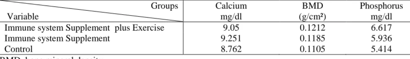
There was no significant statistical difference in the weight of subjects at the beginning of the study. At the end of the study, there a significant difference was observed between subjects and within the groups (P < 0.001). The groups of ISS plus exercise and ISS showed increase in BMD compared to the control group compared (Table 1). The group’s ISS plus exercise and ISS showed increase in serum calcium compared to the control group compared. The group’s ISS plus exercise and ISS showed increase in serum Phosphorus compared to the control group compared (Table 1). In the group ISS plus exercise showed increase in serum calcium and phosphorus and increase in BMD compared to the ISS group.
Table 1. The statue of phosphorus, calcium and bone mineral density (BMD) in the studied rats. Groups
Variable
Calcium mg/dl
BMD (g/cm²)
Phosphorus mg/dl
Immune system Supplement plus Exercise 9.05 0.1212 6.617
Immune system Supplement 9.251 0.1185 5.936
Control 8.762 0.1105 5.414
BMD, bone mineral density. Discussion
Several studies have shown that obtaining high bone density depends on physiological factors, such as genetic factors, hormonal changes, environmental factors, physical activity, nutrition (especially calcium in the diet) endocrine factors (sex steroids, active vitamin D3 and insulin-like growth factor) (19-21). T cells and B cells produce large amounts of cytokines which regulate bone resorption and bone formation. These factors play a critical role in the regulation of bone turnover in health and disease. In addition, immune cells of the bone marrow regulate bone homeostasis by cross-talking with bone marrow stromal cells and osteoblastic cells via cell surface molecules. These regulatory mechanisms are particularly
relevant to postmenopausal osteoporosis and hyperparathyroidism, two common forms of bone loss caused primarily by an expansion of the osteoclastic pool only particularly compensated by a stimulation of bone formation (22). It is stated that vegetables are natural sources of alkalinity used to neutralize the acid caused by metabolism of other nutrients in the body. In the acute phase of metabolic acidosis, if the alkaline food is not provided by the consumed foods, sodium and potassium in the intracellular and the extracellular fluid of the bones are used to neutralize the acid; however, in a chronic case; calcium, carbonate and citrate of bone crystals will be used to neutralize the acid, causing bone mineral loss (23). Research
18
conducted on animals and humans have confirmed these observations. Jones et al. for the first time reported a positive association between vegetable consumption and BMD performed on 10-year-old girls. In the same vein, another study confirmed that vegetables have significant predictability for the increase in total body bone mass (BMC) in boys between the ages of 8 and 20. Therefore, the positive effect of nutrition on bone health has been demonstrated (24). Reports in adults examined for this relationship revealed significant associations between vegetable intake and BMD (25), furthermore a recent study among 8- and 13-year-old girls (N=56) showed a significant relationship between bone size and high intakes of vegetables. In this study, the BMD was higher in the group with six weeks of exercise as compared with the sedentary control group; however, the BMD rate of the whole body did not differ between two groups. Studies have also demonstrated that exercise during the growth period has an effect on bone modeling process locally in the areas under pressure. Sport activities are as stimulators of bone formation that lead to a reduced risk of bone fractures through accumulation of minerals, strengthening of muscles, and improvement of the individual’s balance (26). During a study carried out on young children, it was concluded that high-intensity exercises lead to excessive stretching of the muscles connected to the bone and the bone undergoes high tension and pressure. This also stimulates bone formation and consequently increases bone density (27). Results of another research also indicated that running on a moving bar rails for eight weeks significantly prevents bone loss and leads to bone strength (28). In the same study; it was found that women who
exercised before the start of puberty had plenty of calories and calcium intake which leads to an increase in the bone mineral content and its longitudinal growth (29). Yeh et al. showed that exercise in young rats stimulated bone growth, resulting in an increased demand for minerals that was satisfied by an increase in serum 1, 25-dihydroxyvitamin D3 level as well as increased intestinal absorption of calcium with depressed parathyroid hormone production. Their findings suggested that part of the stimulation of bone growth may be associated with systemic actions of calcitropic hormones (30). On the other hand, Yeh and colleagues demonstrated that exercise in aged female rats decreased the ratio of erosion to mineralizing surface on the trabecular bone of the proximal tibia (31). Many observational and cross-sectional studies have indicated the beneficial effects of physical training on bones. The results of the observational studies on exercising individuals have shown that they have higher BMD and lower fracture risk than non-exercising individuals (32). Conclusion
The present study showed that regular exercise and physical activity including swimming therapy, and proper nutrition (rich in minerals) are among the factors that correlate with the current whole body BMD among male infant rats. The results also indicated a small but positive effect of the ISS on the whole body BMD in male rats, likewise the results suggested that a combination of exercise and proper nutrition is more effective on the increase in bone density in the experimental group in comparison with the proper nutrition in the sedentary control group.
19
References
1. Wagner EF, Karsenty G. Genetic control of skeletal development. Curr Opin Genet Dev. 2001; 11(5):527-32. 2. Anderson JJ. The important role of
physical activity in skeletal development: how exercise may counter low calcium intake. Am J Clin Nutr. 2000; 71(6):1384-6.
3. Carter DR. Mechanical loading histories and cortical bone remodeling. Calcif Tissue Int. 1984; 36(1):19-24 . 4. Davies JH, Evans BAJ, Gregory JW.
Bone mass acquisition in healthy children. Arch Dis Child. 2005; 90(4): 373-8.
5. Cassell C, Benedict M, Specker B. Bone mineral density in elite 7- to 9-yr-old female gymnasts and swimmers. Med Sci Sports Exerc. 1996; 28(10):1243-6.
6. Moreira LD, Longo de Oliveira M. Effects of different types of exercises on bone and physical function of postmenopausal women. Arq Bras Endocrinol. 2014; 58(5):514-22.
7. Chowdhur B. Effects of exercise training on bone mineral density of different aged postmenopausal women in India. Int J Healthcare Biomed Res. 2014; 2(3):180-85.
8. Bushinsky DA. Acid-base imbalance and the skeleton. Eur J Nutr. 2001; 40(1):238-44.
9. Remer T, Manz F. Potential renal acid load of foods and its influence on urine pH. Am J Dietetic Assoc. 1995; 95(7):791-7.
10.Buclin T, Cosma M, Appenzeller M, Jacquet A.F. Diet acids and alkalis influence calcium retention in bone. Osteoporos Int. 2001;12(1):493-9.
11.Maurer M, Riesen W, Muser J. Neutralization of western diet inhibits bone resorption independently of K intake and reduces cortisol secretion in humans. Am J Physiol. 2003; 284(1):32-40.
12.Mühlbauer RC. Effect of vegetableg on bone metabolism. Nature. 1999; 40(1): 343-4.
13.Mühlbauer RC, Lozano A, Reinli A. Various selected vegetables, fruits, mushrooms and red wine residue inhibit bone resorption in rats. J Nutt. 2003; 133(11):3592-7.
14.John V, Hock JM, Short LL, Glasebrook AL, Galvin RJ. A role for CD8+ T lymphocytes in osteoclast differentiation in vitro. Endocrinology. 1996;137(6):2457-63.
15.Li Y, Toraldo G, Li A, Yang X, Zhang H, Qian WP, Weitzmann MN. B cells and T cells are critical for the preservation of bone homeostasis and attainment of peak bone mass in vivo. Blood. 2007; 521(9):3839-48.
16.Grcević D, Lee SK, Marusić A, Lorenzo JA. Depletion of CD4 and CD8 T lymphocytes in mice in vivo enhances 1, 25-dihydroxyvitamin D3-stimulated osteoclast-like cell formation in vitro by a mechanism that is dependent on prostaglandin
synthesis. J Immunol.
2000;165(8):4231-8.
17.Vento PJ, Swartz ME, Martin LB, Food intake in laboratory rats provided standard and fenbendazole-supplemented diets. J Am Assoc Lab Anim Sci. 2008;47(6):46-50.
18.Shen J, Fox LE, Cheng J. Swim therapy reduces mechanical allodynia and thermal hyperalgesia induced by chronic constriction nerve injury in rats. Pain Med. 2013; 14(4): 516-25. 19.Anderson JJ, Tylavsky FA, Halioua L.
Determinants of peak bone mass in young adult women. J Bone Miner Metab. 2005; 23(6):470-5.
20.Heyse SP, Sartori L, Crepaldi G. Epidemiology of osteoporosis: a study of fracture mortality in Italy. Calcif Tissue Int. 1990; 46(5): 289-93.
21.Obrant KJ, Bengner U, Johnell O. Increasing age-adjusted risk of fragility
20
fractures: a sign of increasing osteoporosis in successive generations? Calcif Tissue Int. 1989;44(3):157-67. 22.Pacifici R. The immune system and
bone. Arch Biochem Biophys. 2010;503(1):41-53.
23.Feskanich D, Singh V, Willette WC, Colditz GA. Vitamin A intake and hip fractures among postmenopausal women. J Am Med Assoc. 2002; 287(1):47-54.
24.Melhus H, Michaelsson K. Excessive dietary intake of vitamin A is associated with reduced bone mineral density and increased risk for hip fracture. An Int Med. 1998; 129(10):770-8.
25.Tucker KL, Hannan MT, Kiel DP. The acid-base hypothesis: diet and bone in the Framingham Osteoporosis Study. Eur J Nutr. 2001; 40(5):231–7.
26.Borrer KT. Physical activity in the prevention and amelioration of osteoporosis in women. Sport med. 2005; 35 (9):779-830.
27.Choktanasiri. Bone mineral density in primary and secondary amenorrhea. J Med. 2000; 83(12): 243-2.
28.Gibson JH. Nutritional and exercise-related determination of loose: Density elite female runners. Osteoporosis Int. 2004; 15(12):611-8.
29.Kemmler W, Lauber D, Weineck J. Benefits of 2 years of intense exercise on bone density physical fitness and blood lipids in early post postmenopausal osteopenc women. Arch Intern Med. 2004;164(10):1084-91.
30.Yeh JK, Aloia JF. Effect of physical activity on calcitropic hormones and calcium balance in rats. Am J Physiol. 1990;258(2 Pt 1):E263-8.
31.Chen MM, Yeh JK, Aloia JF, Tierney JM, Sprintz S. Effect of treadmill exercise on tibial cortical bone in aged female rats: a histomorphometry and dual energy x-ray absorptiometry study. Bone. 1994;15(3):313-9.
32.Smith EL. Role of physical activity in regulation and maintenance of bone. Am Soc Bone Mineral Res. 2009; 55(1):323-26.