Evaluating the Efficacy of Grafting the Facial Gap at Immediately
Placed Implants in the Anterior Maxilla: A Systematic Review
and Randomized Controlled Trial
Bryan P. Jacobs DMD
A thesis submitted to the faculty of the University of North Carolina at Chapel Hill in partial fulfillment of the requirements for the degree of Master of Science in the School of Dentistry
(Prosthodontics).
Chapel Hill 2013
Approved By:
iii ABSTRACT
BRYAN P. JACOBS DMD: Evaluating the Efficacy of Grafting the Facial Gap at Immediately Placed Implants in the Anterior Maxilla: A Systematic
Review and Randomized Controlled Trial (Under the direction of Lyndon Cooper DDS PhD)
iv
TABLE OF CONTENTS
List of Tables ………….………..….. v
List of Figures ………….………..……… vi
Chapter 1: A Systematic Review of Grafting at the Time of Immediate Implant Placement in the Anterior Maxilla—Esthetic Outcomes ………. 1
Abstract ………. 1
Introduction ……….. 2
Materials and Methods ………. 3
Results ……… 5
Conclusion ……… 8
References ………. 10
Chapter 2: A Randomized Controlled Trial Evaluating the Efficacy of Grafting the Facial Gap at Immediately Placed Implants in the Anterior Maxilla ………. 13
Abstract ……….… 13
Introduction ……… 14
Materials and Methods ………..….... 15
Results ……… 25
Discussion ……… 27
Conclusion ……….. 31
v LIST OF TABLES
Table 2.1. Description of Study Subjects ………. 25
Table 2.2. Number of Implants, by Length ………... 26
Table 2.3. Changes in Peri-Implant Soft Tissue Levels ……… 26
Table 2.4. Pink Esthetic Score at Final Follow-Up ………. 26
vi
LIST OF FIGURES
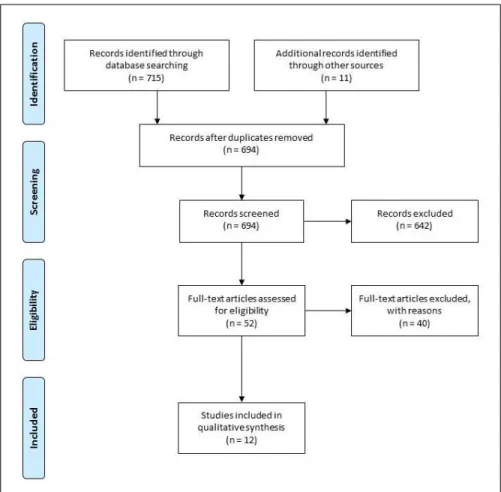
Figure 1.1. Search strategy flow diagram ………..……… 5
Figure 2.1. Sloped implant and positioning ………..…… 17
Figure 2.2. Pre-operative facial bone measurements ……… 18
Figure 2.3. Digital planning of bonded pontic provisional ………..……… 19
Figure 2.4. Flow chart of patient visits ……….…… 19
Figure 2.5. Facial bone evaluation ………..…… 20
Figure 2.6. Temporary abutment fabrication ………..… 21
Figure 2.7. Gold-shaded titanium CAD/CAM abutment ………..…… 22
Figure 2.8. Photographic soft tissue measurements ………..…… 23
Figure 2.9. Peri-implant facial bone measurements ………..……… 24
CHAPTER 1.
A Systematic Review of Grafting at the Time of Immediate Implant Placement in the Anterior Maxilla—Esthetic Outcomes
Abstract
Objectives: Immediate implant placement is an accepted and widely utilized treatment modality for tooth replacement following extraction. Survival and success are comparable to implant placement in healed ridges. However, the best methods for achieving predictable soft tissue esthetics are still up for debate. Contemporary surgical strategy often leaves a facial gap anterior to the implant, between it and the buccal plate. The ideal management of this gap—with regards to grafting or not—has not been clearly defined. The aim of this review is to identify immediate implant literature that examines esthetic outcomes based on facial gap grafting status.
Materials and Methods: An electronic search of Medline (via PubMed) and EMBASE was carried out in October 2012. To be included, studies required immediate placement in >10 patients in the maxillary esthetic zone, 6 month follow-up, and had to report on esthetic outcomes. Titles and abstracts of all resulting publications were reviewed, and those that met inclusion were selected for full text review, which was carried out in duplicate. Esthetic outcomes data were then extracted from the final list of selected publications.
Results: A total of 694 unique abstracts were reviewed. Full text evaluation was carried out on 52 publications—12 of which met the criteria for inclusion in this review. No randomized controlled trial (RCT) directly comparing grafting the facial gap versus not grafting was identified. The 12 included studies were organized into the following: related RCTs (n=3), cohort studies (n=2), case series (n=2), grafting only (n=3), no-grafting only (n=2).
2 Introduction
Immediate implant placement (type I, per Hammerle et al. 2004) is an accepted and widely utilized treatment modality for tooth replacement following extraction. Benefits may include decreased treatment duration, a decreased number of procedures, avoidance of an interim removable prosthesis, and the psychosocial benefit of having a lost tooth immediately replaced.
Numerous studies have shown that immediate implant placement is a biologically sound method for tooth replacement. High initial stability can routinely be achieved (Becker et al. 2005). Survival and success are comparable to implant placement in healed ridges (De Bruyn et al. 2013). In studies with ≥5 years follow-up, median survival of 95% was observed (Chen & Buser 2009). A recent review of 46 prospective studies found a 2 year survival rate of 98.4% (95% CI: 97.3-99%) (Lang et al. 2012). However, in an age where patients desire an exact replica of the tooth being removed—and want it to remain unchanged over time—the best methods for achieving predictable soft tissue esthetics are still up for debate.
In that regard, immediate implant placement has begun to provide encouraging esthetic results when compared to traditional placement in healed ridges (van Kesteren et al. 2010). It has been observed that, in immediate placement situations, immediate provisionalization may further improve preservation of hard and soft tissues (De Rouck et al. 2009). A flapless technique may be beneficial as well (Chu et al. 2012). The use of the platform switching concept for implant design also appears to be promising, in terms of contributing to the overall picture of an esthetically-appealing, stable result (Canullo et al. 2009a). In the anterior maxilla, placement of immediate implants with a bias to the palatal aspect of the extraction socket—a so called lingual or palatal shoulder position—also appears to be advantageous (Evans & Chen 2008). This surgical strategy, however, typically leaves a “facial gap” anterior to the implant, between it and the buccal plate. The question that then arises is: what is the ideal management strategy for addressing this gap?
If one momentarily sets aside the myriad of treatment combinations in terms of staging, abutment selection, flap or flapless approach, and provisionalization, the question of gap management can be distilled into three key elements:
Graft status – choose to graft or not to graft If choose to graft, what material
Gap size – i.e. is there a threshold one sets for which a graft applies only when the gap is at least that large (typically measured from the anterior surface of the implant to the palatal aspect of the buccal plate; also called jumping distance)
When grafting is elected for all sites or based on a gap size threshold, various types of bone, substitute materials, and barriers have been utilized, including:
Hard tissue replacement polymer alloplast (Ashman 1990, Ashman et al. 1995) Hydroxyapatite (Block & Kent 1991)
3
Antigen-extracted allogeneic bone (Block & Kent 1991)
Antigen-extracted allogeneic + autogenous bone (Block & Kent 1991) e-PTFE non-resorbable membrane (Becker et al. 1994)
Autogenous bone with cadaveric dura mater membrane (Peleg et al. 1999) Hydroxyapatite cement (Lew et al. 2000)
Resorbable hydroxyapatite-collagen chondroitin sulfate (Rebaudi et al. 2003) Enamel matrix derivative (Cangini & Cornelini 2005)
Collagen membrane only (Cangini & Cornelini 2005) Bovine bone + porcine collagen (Canullo et al. 2009a)
Calcium phosphate bone grafting cement (Taschieri et al. 2010) Freeze-dried bone allograft (Van Kesteren 2010)
Autogenous bone only (Bogaerde et al. 2010, Noelken et al. 2013) Bovine bone ± collagen membrane (Benic et al. 2012, Bilhan et al. 2011). Synthetic biphasic calcium phosphate (Levin 2011)
However, the objective value of augmenting the implant facial gap versus not doing so has not been clearly defined. The aim of this paper is to review the literature pertaining to implants placed immediately into extraction sockets that compares esthetic outcomes based on facial gap grafting status. Specifically, in PICO format:
Population: patients being treated with immediate implant placement (type I), for single tooth replacement in the anterior maxilla
Intervention: grafting the facial gap anterior to the implant Comparison: no grafting
Outcome: esthetic outcomes, including: a. mid-facial recession
b. change in mesial and/or distal papilla height c. pink esthetic score (PES) (Furhauser et al. 2005)
d. patient based outcomes (OHIP questionnaire, visual analog scale [VAS])
This review was carried out in accordance with the PRISMA 2009 guidelines for systematic reviews and meta-analyses (Moher et al. 2009).
Materials and Methods
Search Strategy and Study Selection
An electronic search of Medline (via PubMed) and EMBASE was carried out in October 2012. The search was limited to articles in the English language, in peer-reviewed publications. The
reference lists of recent reviews were also searched for relevant citations. Specific inclusion criteria were as follows:
4
Single-tooth immediate (type I) implant placement with/without immediate provisionalization
Follow-up of at least 6 months post-extraction
Patients allocated to either receive graft or no graft at facial gap
Defined method for allocating between study groups (e.g. randomization, based on gap size threshold)
Maxillary anterior teeth (any subset of #4-13)
No connective tissue graft, palatal pedicle flap, or simultaneous sinus elevation Report on esthetic outcomes, including any of the following:
o Mid-facial recession o Papilla height change o PES
o Patient perception (e.g. OHIP questionnaire, VAS)
After search optimization, with assistance from a librarian specializing in searches of the biomedical literature, the following Medline (via PubMed) search was carried out:
(Dental Implants[mesh] OR Dental Implantation[mesh] OR Dental Prosthesis,
Implant-Supported[mesh] OR dental implant*[tw]) AND immediate* AND (Bone Substitutes[mesh] OR bone substitute*[tw] OR bone replacement*[tw] OR Bone Transplantation[mesh] OR bone
transplant*[tw] OR bone graft*[tw] OR augmentation[tw] OR bovine bone[tw] OR bio-oss[tw] OR autologous bone[tw] OR puros[tw] OR bone ceramic*[tw] OR boneceramic*[tw] OR xenograft[tw]).
A corresponding search of EMBASE was also carried out:
(“Tooth implant”:de,ti,ab OR “tooth implantation”:de,ti,ab OR “dental implant”:ti,ab OR “dental implanation”:ti,ab) AND Immediate*:ti,ab AND (“Bone graft”:de,ti,ab OR “bone allograft”:de,ti,ab OR “bone transplantation”:de,ti,ab OR xenograft:de,ti,ab OR augmentation:de,ti,ab OR “bovine bone”:de,ti,ab OR “autologous bone”:ti,ab OR “bone ceramic”:de,ti,ab OR bio-oss OR puros OR boneceramic).
Titles and abstracts (TIAB) of all resulting publications, along with those of the citations found in recent review papers, were reviewed against the aforementioned inclusion criteria. Those that appeared to meet the criteria or those where it was unclear after TIAB review were selected for full text review.
5 Results
Electronic search revealed 715 abstracts, with 32 of these being exact duplicates. Hand searching revealed an additional 11 papers—resulting in a total of 694 unique abstracts reviewed. Full text evaluation was carried out on 52 publications. Of these, 12 met the criteria for inclusion in this review (Figure 1.1). These are presented below by study type: related RCTs (n=3), cohort studies (n=2), case series (n=2), grafting only (n=3), no-grafting only (n=2).
Figure 1.1. Search strategy flow diagram (adapted from PRISMA, Moher et al. 2009).
Reasons for exclusion of the 40 other full texts included: no objective esthetic outcomes (n=20), review article (n=7), had simultaneous connective tissue grafting or pedicle flap (n=7), multi-unit (n=2), case report or <10 patients (n=2), animal study (n=1), and posterior teeth only (n=1). Many of these studies met multiple exclusion criteria.
6 Related Randomized Controlled Trials (RCTs)
Canullo (Canullo et al. 2009b), in a double-blind RCT with mean follow-up of 25 months, looked at 22 single tooth immediate implants—6 maxillary incisors and 16 premolars. The study compared platform-switched (PS) abutments versus controls. Surgeries were carried out in a flapless manner, with immediate provisionalization. If the facial gap was >1 mm (n=7 for each group), bovine bone with porcine collagen was placed, as well as a fibrin sponge with tranexamic acid. Baseline measurements were made immediately after implant placement, with all measurements rounded to the closest half mm. Mean mid-facial tissue changes were 0.18 ± 0.46 mm gain (PS) and 0.45 ± 0.27 mm loss (control). Mean papilla height change was 0.05 ± 0.28 mm gain (PS) and 0.88 ± 0.63 mm loss (control). No difference was observed based on the presence or absence of the graft. De Angelis (De Angelis et al. 2011) carried out a RCT examining treating the facial gap with a resorbable collagen membrane ± bovine bone graft in 80 patients. The exact number of implants in the #4-13 sites is not stated, but 50/80 implants were maxillary and 50/80 implants were incisors, canines, or premolars. Inclusion required a facial gap of >1 mm—the mean gap for the study was approximately 3 mm. A flap was raised at the time of surgery and healing was submerged. At 1 year, mean PES scores were 8.94 (membrane only) and 11.29 (membrane + bovine bone)—a significant difference (P<.001).
Van Kesteren (Van Kesteren et al. 2010) carried out a RCT comparing immediate versus delayed implant placement in the esthetic zone, with 6 month follow-up. Immediate implants made up 13 of the samples—12 of these at the #4-13 sites and a single mandibular premolar. Flaps were raised during the surgery, and healing was transmucosal (no provisionalization). Patients who had a facial extraction gap ≥2 mm were grafted with freeze-dried bone allograft. Mean mid-facial
recession was .05 mm (10/13 had <0.5 mm change), but mean papilla loss was >1 mm mesially and distally.
Cohort Studies
Benic (Benic et al. 2012) presented 7 year follow-up data on a prospective cohort of immediate implants. These patients had flap elevation at the time of surgery with transmucosal healing. The 14 (of original 24) patients who reported for the 7 year exam had implants at 3 incisors and 11 premolars (2 mandibular teeth). No graft was used if the gap was <.5 mm, otherwise bovine bone and a resorbable collagen membrane were placed (11/14). The esthetic observation noted was that the width of keratinized mucosa at the site decreased 1.2 ± 1.0 mm (P ≤ .01).
7
membrane. Soft tissue results at 1 year showed that complete papilla fill was present at 78% of sites, with 6% of sites having hyperplastic papilla.
Case Series
Cangini (Cangini & Cornelini 2005) presented a consecutive case series following 32
immediate implant patients over 12 months. Of these, 18 received enamel matrix derivative (EMD) in the buccal gap, while 14 received resorbable collagen membrane—patients were not randomized and those with bony defects were excluded. The sites were maxillary and mandibular incisors (n=15) and premolars (n=17), were flapped at the time of surgery, and covered with coronally positioned flaps. The esthetic outcome measured was the distance between the soft tissue margin and implant shoulder. There was no difference between groups at interproximal or lingual sites, but there was a significant difference buccally (EMD: 0.22 ± 1.47 mm, membrane: 0.90 ± 1.29 mm, p≤.05).
Spinato (Spinato et al. 2012) carried out a “retrospective comparative study” of 45 immediate implants in 41 patients with thick biotype, with a mean follow-up of 32 months. All implants were in #4-13 sites, placed in a flapless procedure, and immediately provisionalized. One of 5 graft types (autologous, bovine ± autologous, demineralized freeze-dried bone allograft ± autologous) was placed in 22 sites, and no graft was used in 23 sites. The mean facial gap widths were 2.25 mm (graft) and 2.03 mm (no graft). No difference was seen between grafting and not grafting when examining papilla fill or mid-facial tissue level. Papilla fill of >50% was seen in 95% of sites, with complete fill in 42%. The various graft materials were not compared to each other due to small sample sizes.
Studies with Grafting Only
Cosyn and colleagues (De Rouck et al. 2008a, Cosyn et al. 2011) looked at 1 and 3 years results of a consecutive case series of 30 patients each receiving an immediate implant in a #4-13 site—all having bovine bone placed in the facial extraction gap. Surgeries were carried out with minimal flap elevation, immediate provisionalization, and required a thick biotype and intact buccal plate. At 1 year, mean tissue loss in relation to the pre-operative status at three locations was: 0.41 mm (mesial papilla), 0.31 mm (distal papilla), and 0.53 mm (mid-facial). At 3 years (n=25), these same measurements were: 0.05 mm (mesial papilla), 0.08 mm (distal papilla), and 0.34 mm (mid-facial)—significant tissue rebound noted at both papilla. The same group also reported on PES at 3 years, with a mean PES of 10.48 ± 2.47. Four of 25 cases did not meet the arbitrarily set clinical acceptance level of PES > 7, and 9 of 25 had PES >11.
Noelken (Noelken et al. 2011) conducted a case series of 18 immediate implants in the esthetic zone—all with complete loss of the facial bony lamellae and all receiving autogenous grafting. Surgeries were done in a flapless manner, with immediate provisionalization, and a mean follow-up of 13 months. Pre-operative PES was 12.2 for the group, with follow-up PES of 12.5 (no significant difference). Of note, implants with a scalloped design were used in this study
8
Noelken (Noelken et al. 2013) also examined a subgroup of a prospective multi-center study, that was treated with immediate implant therapy in the anterior maxilla (#4-13), with autogenous grafting of the facial gap (20 patients, 37 implants). Of note, only 12/37 were single tooth restorations. Surgeries were done with a flapless approach, and the implants were immediately provisionalized and splinted to adjacent teeth. Sites both with and without facial defect were included: no hard or soft tissue defect (n=8), hard tissue defect only (n=11), combination recession and bony deficiency (n=18). PES values were measured pre-operatively (10.65 ± 1.96), at 1 year (11.94 ± 1.59), and at 2 years (11.3 ± 1.8), with increases or no change noted in 78% of sites.
Studies without Grafting Only
Raes (Raes et al. 2012) looked at 16 consecutive patients each treated with immediate implant therapy in the maxillary anterior (#4-13 sites). Of these, 11/16 were done with flapless surgery, all had immediate temporization, and none were grafted. Notably, the study excluded patients with thin-scalloped biotype or a facial implant gap >2.5 mm. Baseline was considered provisional crown cementation. At 12 months, mean mid-facial recession was 0.12 ± 0.78 mm, with 2 cases displaying a gain of tissue >1 mm. Mean papilla changes were 0.07 mm gain (mesial) and 0.38 mm loss (distal). Patient satisfaction, based on the OHIP-14 questionnaire, showed significant improvement in satisfaction and well-being from pre-operative to 1 year (p<.001).
Sanz (Sanz et al. 2013), in a RCT comparing 2 implant shapes, reported on 3 year outcomes of 95 patients receiving 101 immediate implants with no grafting of the facial gap. Implants were in the maxillary esthetic zone (#4-13) and underwent submerged healing. At the time of permanent restoration, only 3% of sites displayed complete papilla. At 3 years, 28% of mesial papilla and 22% of distal papilla were completely filled. The soft tissue margin (vs. time of definitive restoration) was unchanged in 43%, and showed ≥1 mm gain in 30% of sites.
Conclusion
At the outset, the goal of this review was to examine randomized, controlled trials that directly compared grafting and no grafting and that reported on esthetic outcomes. Due to the paucity of studies discovered, the inclusion criteria for this review evolved iteratively to become more generalized. The purpose of this evolution was, of course, to hopefully provide some insight as to what data does exist with regard to facial gap grafting and esthetics.
The 12 studies presented in the current review’s results show that as time has progressed, the emphasis on tracking esthetic outcomes has increased. However, trials with sufficiently large sample sizes and rigorously controlled study designs are lacking. While overall treatment success is demonstrated in patient trials, cohorts, and series—the individual elements that lead to that success are too heterogeneous to draw any definite conclusions, with respect to grafting protocols.
long-9
term RCTs are needed—trials that track both objective esthetic parameters, as well as patient-based outcomes. It does appear that when a protocol utilizing immediate placement, minimal flap
10 References
Ashman A. An immediate tooth root replacement: an implant cylinder and synthetic bone combination. J Oral Implantol 16(1): 28-38, 1990.
Ashman A, LoPinto J, Rosenlicht J. Ridge augmentation for immediate postextraction implants: eight year retrospective study. Pract Periodontics Aesthet Dent 7(2): 85-94, 1995.
Becker W, Dahlin C, Becker BE, Lekholm U, van Steenberghe D, Higuchi K, Kultje C. The use of e-PTFE barrier membranes for bone promotion around titanium implants placed into extraction sockets: a prospective multicenter study. Int J Oral Maxillofac Implants 9(1): 31-40, 1994. Becker W, Sennerby L, Bedrossian E, Becker BE, Lucchini JP. Implant stability measurements for implants placed at the time of extraction: a cohort, prospective clinical trial. J Periodontol 76(3): 391-7, 2005.
Benic GI, Mokti M, Chen CJ, Weber HP, Hämmerle CH, Gallucci GO. Dimensions of buccal bone and mucosa at immediately placed implants after 7 years: a clinical and cone beam computed
tomography study. Clin Oral Implants Res 23(5): 560-6, 2012.
Bilhan H, Mumcu E, Geçkili O, Atalay B. The evaluation of the success of immediately placed single implants: a retrospective clinical study. Implant Dent 20(3): 215-25, 2011.
Block MS, Kent JN. Placement of endosseous implants into tooth extraction sites. J Oral Maxillofac Surg 49(12): 1269-76, 1991.
Bogaerde, L. V., Pedretti, G., Sennerby, L. and Meredith, N. Immediate/Early function of Neoss implants placed in maxillas and posterior mandibles: an 18-month prospective case series study. Clin Implant Dent Relat Res 12 Suppl 1:e83-94, 2010.
Cangini F, Cornelini R. A comparison between enamel matrix derivative and a bioabsorbable membrane to enhance healing around transmucosal immediate post-extraction implants. J Periodontol 76(10): 1785-92, 2005.
Canullo L, Goglia G, Iurlaro G, Iannello G. Short-term bone level observations associated with platform switching in immediately placed and restored single maxillary implants: a preliminary report. Int J Prosthodont 22(3): 277-82, 2009a.
Canullo L, Iurlaro G, Iannello G. Double-blind randomized controlled trial study on post-extraction immediately restored implants using the switching platform concept: soft tissue response.
Preliminary report. Clin Oral Implants Res 20(4): 414-20, 2009b.
11
Chu SJ, Salama MA, Salama H, Garber DA, Saito H, Sarnachiaro GO, Tarnow DP. The dual-zone therapeutic concept of managing immediate implant placement and provisional restoration in anterior extraction sockets. Compend Contin Educ Dent 33(7): 524-32, 534, 2012.
Cosyn J, Eghbali A, De Bruyn H, Collys K, Cleymaet R, De Rouck T. Immediate single-tooth implants in the anterior maxilla: 3-year results of a case series on hard and soft tissue response and aesthetics. J Clin Periodontol 38(8): 746-53, 2011.
De Angelis N, Felice P, Pellegrino G, Camurati A, Gambino P, Esposito M. Guided bone regeneration with and without a bone substitute at single post-extractive implants: 1-year post-loading results from a pragmatic multicentre randomised controlled trial. Eur J Oral Implantol 4(4): 313-25, 2011. De Bruyn H, Raes F, Cooper LF, Reside G, Garriga JS, Tarrida LG, Wiltfang J, Kern M. Three-years clinical outcome of immediate provisionalization of single Osseospeed(™) implants in extraction sockets and healed ridges. Clin Oral Implants Res 24(2): 217-23, 2013.
De Rouck T, Collys K, Wyn I, Cosyn J. Instant provisionalization of immediate single-tooth implants is essential to optimize esthetic treatment outcome. Clin Oral Implants Res 20(6): 566-70, 2009. Evans CD, Chen ST. Esthetic outcomes of immediate implant placements. Clin Oral Implants Res 19(1): 73-80, 2008.
Fürhauser R, Florescu D, Benesch T, Haas R, Mailath G, Watzek G. Evaluation of soft tissue around single-tooth implant crowns: the pink esthetic score. Clin Oral Impl Res 16(6): 639-44, 2005.
Hämmerle CH, Chen ST, Wilson TG Jr. Consensus statements and recommended clinical procedures regarding the placement of implants in extraction sockets. Int J Oral Maxillofac Implants 19 Suppl: 26-8, 2004.
Lang NP, Pun L, Lau KY, Li KY, Wong MC. A systematic review on survival and success rates of implants placed immediately into fresh extraction sockets after at least 1 year. Clin Oral Implants Res 23 Suppl 5: 39-66, 2012.
Levin BP. Immediate temporization of immediate implants in the esthetic zone: evaluating survival and bone maintenance. Compend Contin Educ Dent 32(4): 52-6, 58-60, 62, 2011.
Lew D, Rubey T, Krizan K, Keller JC. Use of hydroxyapatite cement to support implants in extraction sockets. Implant Dent 9(1): 45-50, 2000.
Moher D, Liberati A, Tetzlaff J, Altman DG; PRISMA Group. Preferred reporting items for systematic reviews and meta-analyses: the PRISMA statement. PLoS Med 6(7): e1000097, 2009.
12
Noelken R, Kunkel M, Wagner W. Immediate implant placement and provisionalization after long-axis root fracture and complete loss of the facial bony lamella. Int J Periodontics Restorative Dent 31(2): 175-83, 2011.
Noelken R, Neffe BA, Kunkel M, Wagner W. Maintenance of marginal bone support and soft tissue esthetics at immediately provisionalized OsseoSpeed™ implants placed into extraction sites: 2-year results. Clin Oral Implants Res [Epub ahead of print]. Jan 14, 2013.
Peleg M, Chaushu G, Blinder D, Taicher S. Use of lyodura for bone augmentation of osseous defects around dental implants. J Periodontol 70(8): 853-60, 1999.
Raes F, Cosyn J, De Bruyn H. Clinical, Aesthetic, and Patient-Related Outcome of Immediately Loaded Single Implants in the Anterior Maxilla: A Prospective Study in Extraction Sockets, Healed Ridges, and Grafted Sites. Clin Implant Dent Relat Res. E-published ahead of print, Jan 17, 2012. Rebaudi A, Silvestrini P, Trisi P. Use of a resorbable hydroxyapatite-collagen chondroitin sulfate material on immediate postextraction sites: a clinical and histologic study. Int J Periodontics Restorative Dent 23(4): 371-9, 2003.
Sanz M, Cecchinato D, Ferrus J, Pjetursson EB, Lang NP, & Lindhe J. A prospective, randomized-controlled clinical trial to evaluate bone preservation using implants with different geometry placed into extraction sockets in the maxilla. Clin Oral Impl Res 21: 13-21, 2010.
Spinato S, Agnini A, Chiesi M, Agnini AM, Wang HL. Comparison between graft and no-graft in an immediate placed and immediate nonfunctional loaded implant. Implant Dent 21(2): 97-103, 2012. Taschieri S, Rosano G, Weinstein T, Del Fabbro M. Replacement of vertically root-fractured
endodontically treated teeth with immediate implants in conjunction with a synthetic bone cement. Implant Dent 19(6): 477-86, 2010.
Valentini P, Abensur D, Albertini JF, Rocchesani M. Immediate provisionalization of single
extraction-site implants in the esthetic zone: a clinical evaluation. Int J Periodontics Restorative Dent 30(1): 41-51, 2010.
CHAPTER 2.
A Randomized Controlled Trial Evaluating the Efficacy of Grafting the Facial Gap at Immediately Placed Implants in the Anterior Maxilla
Abstract
Objectives: A ‘gold standard’ protocol does not exist for immediate implant placement in the esthetic zone. Tooth removal leads to defined resorption of bundle bone and preferential loss of the facial plate. Neither implants nor autologous grafts counteract post-extraction bone
remodeling, however xenograft placement within the gap between the implant and buccal bone has shown promise in animal models. The purpose of this study is to evaluate whether grafting the facial gap between the implant and buccal bone at the time of immediate implant placement will improve tissue preservation when compared to a no-grafting protocol.
Materials and Methods: Twenty-two patients were enrolled and had initial records made. Each underwent extraction and immediate implant placement without loading. Randomization allocation to either receive xenograft or no graft was announced following sloped implant placement. Fixed provisionalization occurred at 3 months, followed by permanent restoration. Final records were repeated at 9-10 months follow-up. Soft tissue changes, gingival esthetics, and buccal bone thickness were evaluated.
Results: Eighteen patients with mean age 56.8 years (range 23-91) had definitive restorations placed and final records made—nine from each group. Mean values for facial bone thickness pre-extraction ranged from 0.60 to 0.93 mm, with no difference between groups. At follow-up, no statistically significant differences in soft tissue changes were detected between groups (P=.846). Soft tissue changes were significantly associated with patient age, regardless of group (P=.038). There was no difference in pink esthetic scores or buccal bone dimensions between groups. Mean implant buccal bone thickness at all locations was greater than 1 mm.
14 Introduction
Impending tooth loss in the esthetic zone is a relatively frequent situation encountered by dentists and dental specialists. Timely replacement of an extracted tooth is, understandably, a primary concern of patients—for both functional and esthetic purposes. However, a ‘gold standard’ protocol for implant-borne tooth replacement in these situations has not yet been universally defined. Accepted treatment sequences range from separate extraction, grafting, and implant surgeries to extraction through loading in the same visit. To add a layer of complexity, within each of these potential timelines, wide varieties of techniques and protocols have been put forth.
Immediate implant placement (type I, per Hammerle et al. 2004) is an accepted and widely utilized treatment modality for tooth replacement following extraction. Survival and success are comparable to placement into healed ridges (De Bruyn et al. 2013). In studies with five or more years follow-up, median survival of 95% was observed (Chen & Buser 2009). A recent review of 46 prospective studies found a two-year survival rate of 98.4% (95% CI: 97.3-99%) (Lang et al. 2012). However, unpredictable soft tissue outcomes in the esthetic zone following immediate placement are a concern for many practitioners. Biologically sound techniques, with data demonstrating consistently esthetic results, are needed.
An understanding of extraction biology is a prerequisite for addressing this problem. It is known that, following extraction, an edentulous site will undergo both quantitative and qualitative changes that are more pronounced on the buccal surface (Pietrokovsi & Massler 1967, Schropp et al. 2003, Sanz et al. 2010). Socket walls will be reduced both in height and width (Araujo et al. 2005, Araujo et al. 2006), with approximately 50% of the bucco-lingual dimension lost during the first year post-extraction (Schropp et al. 2003). A recent review including 20 studies calculated the mean hard tissue loss at extraction sites six months post-operatively to be 3.79 mm in the horizontal dimension and 1.24 mm in the vertical dimension (Tan et al. 2012). In this situation, placement of autologous bone will not change bone formation or prevent ridge resorption (Araujo & Lindhe 2011).
Why the buccal bone may be more affected is a question of great interest. One reason may have to do with bundle bone, a tooth-related tissue that disappears following tooth loss. The buccal bone wall often has a greater proportion of this bone type compared to the lingual cortical plate. It has been suggested that the reduction of the buccal bone wall is related to both its pre-surgical thickness and this loss of bundle bone (Araujo et al. 2005, Araujo & Lindhe 2005). Buccal bone may also be more susceptible to surgical trauma, and therefore resorption, than the lingual bone (Wilderman et al. 1960, Wood et al. 1972, Araujo et al. 2005).
15
implants without grafting or membranes measured horizontal resorption after 4 months of healing and found 56% reduction buccally and 30% reduction lingually/palatally (Botticelli et al. 2004). Mean vertical resorption around the implants ranged from 0.3 to 0.6 mm depending on the surface measured. Another prospective study of 93 immediately placed implants showed horizontal reduction in the buccal crest of 30-43% and in the palatal crest of 11-18% (Sanz et al. 2010). Of note, all three of the aforementioned studies elevated flaps at the surgical sites. It has been suggested that resorption can be reduced if the surgical and restorative procedures are all carried out without flap elevation (Chu et al. 2012).
The use of xenografts to slow or minimize extraction changes has been another avenue of research. One such material that has been evaluated is Bio-Oss (Osteohealth, Shirley, NY). Bio-Oss has been used to attempt preservation of the alveolar ridge following extraction with some reports of success and others equivocal (Nevins et al. 2006, Araujo & Lindhe 2009). For example, Bio-Oss promoted de novo hard tissue formation, in particular at the margins of extraction sites (Araujo et al. 2008). Dimensions of the hard tissue walls were maintained and ridge profile preserved.
The potential effectiveness of Bio-Oss may be due to altered healing. Bio-Oss grafted sites at three and six months seemed to show delayed healing but also less ridge resorption (Araujo et al. 2008, Araujo et al. 2009). This modified wound healing and bone remodeling may be influenced by the presence of multinucleated cells occurring in the tissue harboring the graft (Araujo et al. 2009). That is, Bio-Oss may require “surface cleaning” by osteoclasts prior to bone deposition (Jensen et al. 2006, Araujo et al. 2010).
In an attempt to have greater and more predictable tissue preservation at the time of immediate implant placement, one group has tested grafting of the buccal extraction gap with Bio-Oss in an animal model (Araujo et al. 2011). Bio-Bio-Oss was placed in the buccal extraction gap of immediately placed implants in dogs and showed: 1) modified healing, 2) additional amounts of hard tissue, and 3) an improved level of marginal bone-implant contact.
While this technique is regularly utilized in clinical practice, little high quality evidence exists for or against its use. The aim of this study is to carry out a randomized, controlled trial in humans evaluating extraction and immediate implant placement with the use of xenograft in the facial extraction gap versus the same treatment without grafting. It is hypothesized that there will be no clinically meaningful difference, based on evaluation of soft tissue changes, soft tissue esthetics, and implant buccal bone dimensions.
Materials and Methods
Patient Selection
16
the UNC-CH School of Dentistry, as well as the community at large who responded to an online posting on the School’s website. To be included in the study, patients had to meet the following criteria:
Have one or more of teeth #5-12 requiring extraction
Have natural teeth adjacent to proposed site
At least 18 years of age
Available for one year follow-up
Consent to trial.
Patients who met any of the following criteria were excluded from the study:
Untreated caries or periodontal disease
Smoked tobacco within the past 12 months
ASA Class 3 or higher or immune-compromised
Pregnant or plan to be pregnant within study period
History of bruxism
History of bisphosphonate use
Additionally, all subjects were informed that if, following extraction, the surgical site was found to be non-ideal, they would be excluded from the trial as well. Non-ideal was defined as any dehiscence, fenestration, or facial crestal bone level greater than 4 mm from the soft tissue margin. Clinical Protocol
Following consent, all subjects had the following pre-operative records made/taken at their first appointment:
Maxillary and mandibular irreversible hydrocolloid impressions (Jeltrate Plus, Dentsply, York, PA)
Polyvinyl siloxane (PVS) inter-occlusal record in maximum intercuspation (Imprint 3 Quick Step, 3M ESPE, Maplewood, MN; Regisil 2x, Dentsply, York, PA)
Standardized digital photographs
17
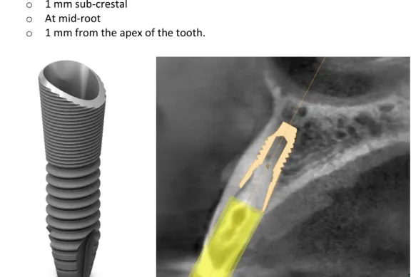
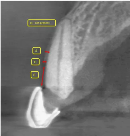
Implant surgeries were digitally planned for placement of 4.5 mm diameter sloped implants (AstraTech OsseoSpeed TX Profile, Dentsply, York, PA) using planning software (Simplant Pro, Materialise, Plymouth, MI). The general strategy for implant positioning was to engage the bone palatal to the planned extraction site, with the long axis through the incisal edge of the planned cement-retained crown (Figure 2.1). The facial aspect of the implant platform was aligned horizontally with the height of the facial plate. Facial bone measurements were also made at this stage. A representative two-dimensional slice (perpendicular to the panoramic curve) through the surgical site was selected and the following measurements were made (Figure 2.2):
Facial cement-enamel junction (CEJ) to the most coronal point of the facial bone crest
Bone thickness facial to the tooth, measured along a line perpendicular to the long axis of the root at the following points:
o 1 mm sub-crestal o At mid-root
o 1 mm from the apex of the tooth.
18
Figure 2.2. Representative two-dimensional slice through the surgical site showing reference measurements and measurements of a) facial CEJ to the most coronal point of the facial bone crest, b) facial plate 1 mm subcrestal, c) facial plate at mid-root, and d) facial plate 1 mm from the apex of the tooth.
19
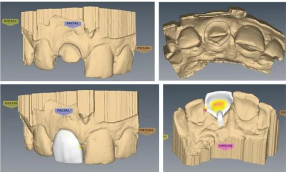
Figure 2.3. Screenshots displaying scanned cast with arbitrary preparation (top) and planned bonded pontic provisional with small palatal “wings” (bottom).
The second appointment consisted of surgery and provisionalization (see Figure 2.4 for flow chart of patient visits). A pre-operative dose of 2g amoxicillin (or 600 mg clindamycin for those with penicillin allergy), as well as 800mg ibuprofen, was given 30-60 minutes prior to surgery. Patients were also instructed to rinse for 90 seconds with an antimicrobial rinse (chlorhexidine gluconate 0.12%). Articaine with epinephrine (Septocaine, Septodont, Lancaster, PA) infiltration was used to achieve anesthesia.
20
Teeth were extracted in a minimally traumatic fashion using periotomes and root tip
forceps. The sockets were then inspected for defects that could lead to study exclusion (Figure 2.5). Two patients were excluded for facial fenestration and two for facial dehiscence (these four patients not included in the 22 study patients reported on in this study). For the remaining patients, the implant osteotomy preparation and placement were carried out per manufacturer’s
recommendations for soft bone, in a flapless manner. All implants were 4.5 mm diameter Astra Tech OsseoSpeed TX Profile implants—either 11, 13, or 15 mm in length.
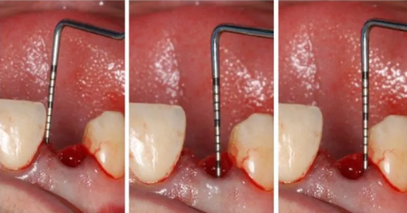
Figure 2.5. Using a periodontal probe to evaluate facial bone in relation to soft tissue margin.
Following placement, the individual patient’s randomization allocation was revealed (Random Allocation Softwaresee reference: Saghaei 2004). For patients assigned to receive no graft, a 4 mm healing abutment was placed, papilla (if separated; 5/10 patients) were sutured with vertical mattress sutures, and the provisional restoration was placed. For patients assigned to receive a graft, a cover screw was placed. Large particle Bio-Oss was then placed in the gap facial to the implant—to the level of the implant platform, followed by an absorbable collagen dressing (CollaPlug, Zimmer Dental, Carlsbad, CA). A figure-of-eight suture was used to secure the dressing and graft, followed by provisionalization. All sutures were 4-0 chromic gut (Ethicon, Johnson & Johnson, New Brunswick, NJ). A periapical radiograph was exposed to observe implant position. A one week post-operative course of amoxicillin (or clindamycin) was prescribed, as well as two weeks with twice daily chlorhexidine gluconate 0.12% rinses. Acetaminophen/hydrocodone or ibuprofen was prescribed for analgesia and homecare instructions were given.
A post-surgical follow-up visit was conducted at one week to ensure compliance with homecare instructions and to observe soft tissue healing. Additional visits were scheduled as necessary to adjust or rebond interim restorations.
21
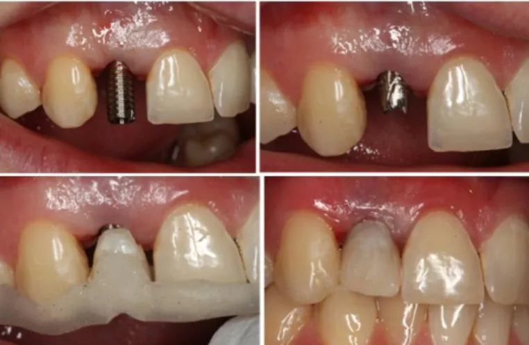
resin, maintaining access to the abutment screw. The assembly was then removed in one piece and modified in the laboratory until appropriate contours and finish were achieved. The provisional was then placed at 15 N-cm and the screw access covered with a cotton pellet and composite resin (Figure 2.6).
Figure 2.6. Temporary abutment before and after modification (top). Using jig to position pontic and checking final contours before polishing (bottom).
Final impressions were made approximately 6 weeks after provisionalization (mean for graft = 43 ± 10 days; mean for no graft = 42 ± 22 days). Full arch impressions were taken with PVS
material using a closed tray technique, without customization or modification of the impression transfer. Opposing arch impressions and inter-occlusal records in maximum intercuspation were made with PVS material as well. Intraoral photographs were taken with shade tabs to help guide the laboratory in definitive restoration fabrication.
Definitive restorations were delivered approximately seven months post-operatively (mean for graft = 232 ± 46 days; mean for no graft = 209 ± 47 days). The temporary restoration was
22
Figure 2.7. Facial view of gold-shaded titanium CAD/CAM abutment.
Post-Operative Records and Measurements
Final follow-up for the investigation took place 2-3 months after delivery (mean for graft = 79 ± 41 days; mean for no graft = 59 ± 38 days). This was approximately 9 to 10 months post-placement (mean for graft = 311 ± 66 days; mean for no graft = 269 ± 69 days). At this time, the following records were made/taken:
PVS impression of the maxillary anterior sextant (Imprint 3 Quick Step, 3M ESPE, Maplewood, MN)
Standardized digital photographs
Small volume cone-beam CT scan (Orthophos XG 3D, Sirona, Charlotte, NC).
23
Figure 2.8. Follow-up standardized photograph demonstrating soft tissue measurements.
Follow-up cone-beam CT scan analysis was similar to the pre-operative measurements that were made at the teeth to be extracted. A representative two-dimensional slice (perpendicular to the panoramic curve) through the implant was selected and the following measurements were made (Figure 2.9):
Facial crown-abutment interface to the most coronal point of the facial bone crest
Bone thickness facial to the implant, measured along a line perpendicular to the long axis of the implant at the following points:
o 1 mm sub-crestal o At mid-implant
24
Figure 2.9. Follow-up CBCT scan slice demonstrating locations of facial bone measurements.
Pink esthetic scores (PES) were assigned by a group of seven calibrated individuals (one prosthodontist, six prosthodontic residents) over the course of two sessions. Cases were presented by digital projection in a random order, using retracted frontal photos of the maxillary anterior teeth, in the manner described by Furhauser (Furhauser et al. 2005). Additionally—for lateral incisor, canine, and premolar sites—a photo perpendicular to the facial/buccal aspect of the crown in question was presented as well, so as to better visualize papilla. Each evaluator scored every case, without discussion amongst the group and blinded to each patient’s assignment. After all evaluations were complete, scores were compiled, the high and low score dropped for each case, and PES assigned as the mean of the five intermediate scores.
Statistical Analysis
25
Pre-operative descriptive statistics were compared between groups. The chi-square test was applied to look at the association between group and sex. The Wilcoxon rank-sum test was used to examine age differences between the two groups. Linear mixed models were used to detect differences in baseline CBCT values based on group assignment or age. Spearman correlation analysis was applied to the pre-operative CBCT values and age as well.
Follow-up data were evaluated in a number of ways. Linear mixed models were used to compare soft tissue changes based on group, age, and position along the tooth (mesial papilla, mid-facial, distal papilla). These were also used to examine differences in follow-up CBCT
measurements, based on group and age. PES was compared using the Wilcoxon rank-sum test. Spearman correlation analysis was applied to the follow-up soft tissue measurements and age, and separately to the follow-up CBCT measurements and age. For all tests, the level of significance was set at α = 0.05.
Results
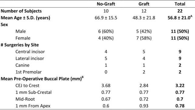
A total of 22 patients—11 men and 11 women—with a mean age of 56.8 ± 21.0 years were enrolled in the study (Table 2.1). Twelve patients, with mean age 48.3 years (range: 23-86 years), were assigned to the graft group. Ten patients, with mean age 66.9 years (range: 38-91 years), were assigned to the no-graft group. There was no association between group and sex (P=.392). There was a statistically significant difference in mean age between groups (P=.044).
Table 2.1. Description of Study Subjects
No-Graft Graft Total
Number of Subjects 10 12 22
Mean Age ± S.D. (years) 66.9 ± 15.5 48.3 ± 21.8 56.8 ± 21.0A Sex
Male 6 (60%) 5 (42%) 11 (50%)
Female 4 (40%) 7 (58%) 11 (50%)
# Surgeries by Site
Central incisor 4 5 9
Lateral incisor 5 4 9
Canine 1 1 2
1st Premolar 0 2 2
Mean Pre-Operative Buccal Plate (mm)B
CEJ to Crest 3.68 2.84 3.22
1 mm Sub-Crestal 0.77 0.77 0.77
Mid-Root 0.67 0.72 0.7
1 mm From Apex 0.6 0.93 0.78
A
Statistically significant difference between groups (P=.044)
B
26
Surgical sites included maxillary central incisors, lateral incisors, canines, and first premolars. Implants were placed in 9 central incisor sites, 9 lateral incisor sites, 2 canine sites, and 2 premolar sites. Twelve 13 mm implants and five each of 11 mm and 15 mm implants were placed (Table 2.2).
Table 2.2. Number of Implants, by Length
11 mm 13 mm 15 mm Total
No-Graft 1 6 3 10
Graft 4 6 2 12
Total: 5 12 5 22
Pre-operative CBCT measurements of the buccal plate showed a mean distance of 3.22 mm from the CEJ to the alveolar crest for the 22 patients. Mean values for facial bone thickness were all less than 1 mm (range: .60-.93 mm). There was no statistically significant difference in the pre-operative buccal plate measurements based on either group assignment or patient age. There was a significant correlation between facial bone thickness measured 1 mm sub-crestal and mid-root (P<.001).
Of the 22 patients, 18 were definitively restored and had follow-up records taken (n=17 for follow-up CBCT). The remaining 4 patients currently have screw-retained provisional restorations in place—their treatment being delayed due to a variety of external factors (long periods of travel, finances, etc.). One patient had implant mobility noted at the provisionalization stage. The implant was removed, the defect curetted, and a new implant placed at the same visit. This second implant subsequently healed uneventfully and was included in the study. Thus implant survival is 22/23, or 95.7% at 10 months. The follow-up analysis was carried out on the 18 completed patients.
No statistically significant differences in soft tissue changes (Table 2.3) were detected between groups (P=.846) or based on location along the tooth (P=.089). Soft tissue changes were significantly associated with patient age, regardless of group (P=.038). Specifically, age was correlated with mid-facial change (P=.040) and distal papilla change (P=.034)—as age increased, tissue loss increased at these sites. Mean PES for each group was 8.2, with an overall range of 4.4 to 13.4 (Table 2. 4). There was no difference in PES between the groups (P=.794).
Table 2.3. Changes in Peri-Implant Soft Tissue Levels (mean ± S.D., mm)A
Mid-Facial Mesial Papilla Distal Papilla
No-Graft 0.92 ± 0.67 0.57 ± 0.59 0.79 ± 0.75
Graft 0.94 ± 1.13 0.33 ± 0.46 0.49 ± 0.62
A
Positive values indicate apical movement of tissues
Table 2.4. Pink Esthetic Score at Final Follow-Up (mean ± S.D.)
No-Graft 8.2 ± 1.8
27
Follow-up CBCT measurements of implant buccal bone (Table 2.5) showed no differences between the two study groups (P=.998) or based on age (P=.845). Buccal bone thickness measured 1 mm sub-crestally was significantly correlated with the thickness measured at the mid-implant level (P<.001).
Table 2.5. Buccal Bone on Follow-Up CBCT (mean ± S.D., mm)
No Graft Graft
Crown Margin to Crest 2.08 ± 0.63 2.00 ± 1.35 1 mm Sub-crestal 1.43 ± 0.61 1.43 ± 0.70
Mid-Implant 1.39 ± 1.12 1.47 ± 1.08
1 mm from Apex 2.61 ± 2.11 2.28 ± 1.24
Of note are two study deviations and a number of new adjacent restorations done
concurrently with the study restorations. One patient had a successful, restored implant rather than a natural tooth adjacent to the surgical site. A second patient had a gingivectomy (approximately 1 mm) performed at the central incisor surgical site prior to final impressions so as to match the tissue level of the adjacent central incisor. In the graft group, adjacent restorations included a crown and a veneer (1 patient), a veneer (1 patient), and a composite resin addition (1 patient). In the no-graft group, adjacent restorations included two crowns (2 patients) and one crown (2 patients).
Discussion
The present study demonstrates that grafting of the facial gap at immediately placed implants is no better or worse than not doing so in terms of the evaluated outcomes. Facial soft tissue preservation was similar between the groups, though on average there was a net loss of tissue when compared to the pre-extraction levels. The mean mid-facial tissue loss of
approximately 0.9 mm is consistent with a recent review which found mean mid-facial recession to range from 0.5-0.9 mm across three studies (Chen & Buser 2009). The current study’s observed mean papilla loss of 0.33-0.79 mm is in line with the 0.5-0.6 mm mean loss reported in the same review.
28
somewhat greater than that of De Rouck et al. It is hypothesized that this may be due to inclusion of all biotypes or implant platform configuration (discussed below), though a host of factors may contribute to this discrepancy.
The same group also reported on PES in the aforementioned cohort at three years, with a mean PES of 10.48 ± 2.47 (Cosyn et al. 2011). Clinical acceptance was arbitrarily set at PES > 7, and scoring was carried out by a single clinician. The current study mean PES of 8.2 ± 2.1 across both groups is somewhat lower, but does meet the arbitrary threshold for esthetic success. Indeed, PES as an evaluation tool has been shown to be reproducible when used by the same group of
examiners over time, but significantly different based on different groups of examiners (Furhauser et al. 2005). Thus, head-to-head comparisons of PES across studies are not appropriate, unless the same evaluators are used. In the current study, PES was based on evaluations by the same group of seven calibrated clinicians for all cases. Regarding PES and patient-based outcomes, one study with a small sample size (n=28) has shown that PES is associated with patient subjective satisfaction on a visual analog scale (Luo et al. 2011).
In another study of consecutively treated patients with single tooth implants, 16/48 patients were treated with immediate implant therapy, no grafting, and immediate provisionalization, then followed up to 1 year (Raes et al. 2012). Patients with thin-scalloped biotype or a facial implant gap >2.5 mm were excluded. Baseline was considered provisional crown cementation, and 11/16 surgeries were done with a flapless approach. Mean mid-facial recession was 0.12 ± 0.78 mm, with 2 cases displaying gain of tissue >1 mm. Mean papilla changes were 0.07 mm gain (mesial) and 0.38 mm loss (distal). The stability of the mid-facial tissue and mesial papilla are in contrast to the current study, as well as the review by Chen and Buser (Chen & Buser 2009). Possible explanations for these differences may be the choice of baseline, the biotype inclusion criterion, or the exclusion of larger facial gaps, among others.
With regard to the buccal bone dimensions, it is commonly held, though not proven, that maintaining at least 1 mm of bone facial to implants is important for stability of the result (Belser et al. 2008). In the current study, follow-up buccal bone measurements of greater than 1 mm at all three points along the implants were observed in both groups. These results are similar to a clinical trial subgroup of 20 patients receiving 37 immediate implants in the anterior maxilla with a flapless approach, treated regardless of buccal plate defect (Noelken et al. 2013). All of these patients received autogenous bone graft anterior to the implant, were immediately provisionalized, and were splinted to adjacent teeth and/or implants. Pre-operative and 1 year post-surgical CBCT scans were taken to measure the thickness of facial bone at 1, 3, and 6 mm apical to the reference (projected healthy oral lamella). Pre-operative measurements were 0.05 mm, 0.5 mm, and 0.52 mm, respectively. Post-operative measurements were 1.22 mm, 1.3 mm, and 1.29 mm,
29
It is noteworthy that the healed implants and pre-extraction teeth shared a similar CBCT finding. For both sets of scans, the buccal bone measurements made 1 mm sub-crestally and mid-root/implant were significantly correlated. That is, facial bone thickness stays relatively constant from crest to mid-root/implant—both facial to teeth and to implants. Mean pre-operative buccal bone thickness was less than 1 mm at all points, consistent with previous findings (Vera et al. 2012).
Patient age—which was not one of the principle variables at the outset—was found to be an important factor. While a patient’s group assignment did not significantly affect their soft tissue outcomes, the outcomes were significantly correlated with age. This is interesting, as young patients are often considered the highest risk esthetic patients due to greater gingival/papillary display (Desai et al. 2009, Hochman et al. 2012). In a sense, the results here show that the age-associated risk is tempered by the age-age-associated soft tissue response.
Sloped implants have been indicated for use in healed ridges, where the buccal and lingual bone have differential heights, and the goal is to preserve as much bone as possible (Noelken et al. 2012). The justification for their use in anterior immediate sites stems from the fact that buccal bone resorption outpaces that of the palatal bone and thus they may better fit the anatomy of the future healed site. The difficulty in their use in immediate sites however stems from the slope itself.
30
Figure 2.10. CBCT sections displaying the effect of implant angulation (0°, 15°, 30°) on the vertical difference between the facial and palatal aspect of the sloped implant platform.
Another factor to be considered in the clinical scenario examined here is abutment design. For this trial, custom CAD/CAM-designed gold-shaded titanium abutments with concave sub-gingival emergence profiles were used (Option 5, Atlantis, Dentsply, York, PA). The gold-shading was
selected to both help combat gingival graying, as well as give a natural appearance underneath the definitive ceramic crown. The concave emergence was selected based on the fact that abutments with concave transmucosal profiles have shown promise in minimizing facial recession (Rompen et al. 2007). As another esthetic alternative to unshaded titanium, zirconia abutments could have been used in place of the gold-shaded titanium. It has been shown that titanium and zirconia abutments have similar bacterial colonization profiles (van Brakel et al. 2011, de Oliveira et al 2012). However, little long term data exists on clinical outcomes with zirconia abutments. Further research is needed to better understand the geometries and materials best suited for soft tissue
maintenance.
31
groups should have had one or the other. Indeed, immediate provisionalization for both groups might have been the most ideal (De Rouck et al. 2009).
Issues around the dimensions of the buccal extraction gap also somewhat limit this study. All patients were assigned to a group, regardless of gap size. In both groups there were patients with smaller and larger gaps. While not objectively measured in three dimensions, it was noted that it was difficult to find space to place graft particles in five of the twelve graft patients. Choosing to include patients with small gap sizes may, in essence, dilute any potential differences between the two study groups.
Soft tissues changes at sites where apical movement of tissue would be desirable were challenging to account for as well. For example, one patient had gingivectomy at a central incisor implant site so as to match the contralateral tooth—apical movement that was measured as soft tissue loss. Another patient’s surgical site had supporting tissues approximately 1.5 mm coronal to the ideal position. The implant was purposely placed deep and provisional contours used to position the tissue ideally. Here again, the apical movement will add up to soft tissue loss—even though improving esthetics at the particular site.
Future directions should further refine the study design, to account for contemporary protocols and planning. A three group model may be considered—gap width less than 2 mm without graft, gap greater than 2 mm with grafting, and gap greater than 2 mm without grafting. Additionally, immediately loading all stable implants would remove a potentially confounding
variable. An ideal abutment design will have to be selected and applied consistently to each patient. Finally, the use of a sloped implant in the immediate placement scenario may need to be
reconsidered. The standard, flat-top design, when placed at an angle, already gives a differential platform height from palatal to facial. Sloped implants may be best relegated to healed ridges where sloped anatomy may correspond nicely. This group has used them successfully in the posterior mandible, in particular.
Conclusion
32 References
Araujo MG, Liljenberg B, & Lindhe J. Dynamics of Bio-Oss Collagen incorporation in fresh extraction wounds: an experimental study in the dog. Clin Oral Impl Res 22: 55-64, 2010.
Araujo M, Linder E, & Lindhe J. Effect of a xenograft on early bone formation in extraction sockets: an experimental study in dog. Clin Oral Impl Res 20: 1-6, 2009.
Araujo MG, Linder E, & Lindhe J. Bio-Oss Collagen in the buccal gap at immediate implants: a 6-month study in the dog. Clin Oral Impl Res 22: 1-8, 2011.
Araújo M, Linder E, Wennström J, & Lindhe J. The influence of Bio-Oss Collagen on healing of an extraction socket: an experimental study in the dog. Int J Periodontics Restorative Dent 28(2): 123-35, 2008.
Araujo MG & Lindhe J. Dimensional ridge alterations following tooth extraction. An experimental study in the dog. J Clin Periodontol 32: 212-218, 2005.
Araujo MG & Lindhe J. Ridge preservation with the use of Bio-Oss collagen. A 6 month study in the dog. Clin Oral Impl Res 20: 433-440, 2009.
Araujo MG & Lindhe J. Socket grafting with the use of autologous bone: an experimental study in the dog. Clin Oral Impl Res 22: 9-13, 2011.
Araujo MG, Sukekava F, Wennstrom JL, & Lindhe J. Ridge alterations following implant placement in fresh extraction sockets: an experimental study in the dog. Clin Oral Impl Res 32: 645-652, 2005. Araujo MG, Sukekava F, Wennstrom JL, & Lindhe J. Tissue modeling following implant placement in fresh extraction sockets. Clin Oral Impl Res 17: 615-624, 2006.
Belser U, Bernard JP, & Buser D. Implants in the esthetic zone. Clinical Periodontology and Implant Dentistry 5: 1146–1174. 2008.
Botticelli D, Berglundh T, & Lindhe J. Hard tissue alterations following immediate implant placement in extraction sites. J Clin Periodontol 31: 820-828, 2004.
Chen S, Buser D. Clinical and Esthetic Outcomes of Implants Placed in Postextraction Sites. Int J Oral Maxillofac Implants 24 Suppl: 186-217, 2009.
Chu SJ, Salama MA, Salama H, Garber DA, Saito H, Sarnachiaro GO, Tarnow DP. The dual-zone therapeutic concept of managing immediate implant placement and provisional restoration in anterior extraction sockets. Compend Contin Educ Dent 33(7): 524-32, 534, 2012.
33
Covani U, Cornelini R, Barone A. Bucco-lingual bone remodeling around implants placed into immediate extraction sockets: A case series. J Periodontol 74: 268-273, 2003.
De Bruyn H, Raes F, Cooper LF, Reside G, Garriga JS, Tarrida LG, Wiltfang J, Kern M. Three-years clinical outcome of immediate provisionalization of single Osseospeed(™) implants in extraction sockets and healed ridges. Clin Oral Implants Res 24(2): 217-23, 2013.
de Oliveira GR, Pozzer L, Cavalieri-Pereira L, de Moraes PH, Olate S, de Albergaría Barbosa JR. Bacterial adhesion and colonization differences between zirconia and titanium implant abutments: an in vivo human study. J Periodontal Implant Sci 42(6): 217-23, 2012.
De Rouck T, Collys K, Cosyn J. Immediate single-tooth implants in the anterior maxilla: a 1-year case cohort study on hard and soft tissue response. J Clin Periodontol 35(7):649-57, 2008.
De Rouck T, Collys K, Wyn I, Cosyn J. Instant provisionalization of immediate single-tooth implants is essential to optimize esthetic treatment outcome. Clin Oral Implants Res 20(6): 566-70, 2009. Desai S, Upadhyay M, Nanda R. Dynamic smile analysis: changes with age. Am J Orthod Dentofacial Orthop 136(3): 310.e1-10, 2009.
Fürhauser R, Florescu D, Benesch T, Haas R, Mailath G, Watzek G. Evaluation of soft tissue around single-tooth implant crowns: the pink esthetic score. Clin Oral Impl Res 16(6): 639-44, 2005. Jensen SS, Broggini N, Hjorting-Hansen E, Schenk R, & Buser D. Bone healing and graft resorption of autograft, anorganic bovine bone and beta-tricalcium phosphate. A histologic and
histomorphometric study in the mandibles of minipigs. Clin Oral Impl Res 17: 237-243, 2006. Hämmerle CH, Chen ST, Wilson TG Jr. Consensus statements and recommended clinical procedures regarding the placement of implants in extraction sockets. Int J Oral Maxillofac Implants 19 Suppl: 26-8, 2004.
Hochman MN, Chu SJ, Tarnow DP. Maxillary anterior papilla display during smiling: a clinical study of the interdental smile line. Int J Periodontics Restorative Dent 32(4): 375-83, 2012.
Lang NP, Pun L, Lau KY, Li KY, Wong MC. A systematic review on survival and success rates of implants placed immediately into fresh extraction sockets after at least 1 year. Clin Oral Implants Res 23 Suppl 5: 39-66, 2012.
Luo Z, Zeng R, Luo Z, Chen Z. Single implants in the esthetic zone: analysis of recent peri-implant soft tissue alterations and patient satisfaction. A photographic study. Int J Oral Maxillofac Implants 26(3): 578-86, 2011.
34
Noelken R, Donati M, Fiorellini J, Gellrich NC, Parker W, Wada K, Berglundh T. Soft and hard tissue alterations around implants placed in an alveolar ridge with a sloped configuration. Clin Oral Implants Res [Epub ahead of print]. Dec 5, 2012.
Noelken R, Neffe BA, Kunkel M, Wagner W. Maintenance of marginal bone support and soft tissue esthetics at immediately provisionalized OsseoSpeed™ implants placed into extraction sites: 2-year results. Clin Oral Implants Res [Epub ahead of print]. Jan 14, 2013.
Pietrokovsi J & Massler M. Alveolar ridge resorption following tooth extraction. J Prosthetic Dent 17: 21-27, 1967.
Raes F, Cosyn J, De Bruyn H. Clinical, Aesthetic, and Patient-Related Outcome of Immediately Loaded Single Implants in the Anterior Maxilla: A Prospective Study in Extraction Sockets, Healed Ridges, and Grafted Sites. Clin Implant Dent Relat Res [E-pub ahead of print]. Jan 17, 2012.
Rompen E, Raepsaet N, Domken O, Touati B, Van Dooren E. Soft tissue stability at the facial aspect of gingivally converging abutments in the esthetic zone: a pilot clinical study. J Prosthet Dent 97(6 Suppl): S119-25, 2007.
Saghaei M. BMC Med Res Methodol 4: 26. Published online Nov 9, 2004 Available: http://mahmoodsaghaei.tripod.com/Softwares/randalloc.html#Version
Sanz M, Cecchinato D, Ferrus J, Pjetursson EB, Lang NP, & Lindhe J. A prospective, randomized-controlled clinical trial to evaluate bone preservation using implants with different geometry placed into extraction sockets in the maxilla. Clin Oral Impl Res 21: 13-21, 2010.
Schropp L, Wenzel A, Kostopoulos L, & Karring T. Bone healing and soft tissue contour changes following single-tooth extraction: a clinical and radiographic 12-month prospective study. Int J Periodontics Restorative Dent 23: 313-323, 2003.
Tan WL, Wong TL, Wong MC, Lang NP. A systematic review of post-extractional alveolar hard and soft tissue dimensional changes in humans. Clin Oral Implants Res 23 Suppl 5:1-21, 2012.
van Brakel R, Cune MS, van Winkelhoff AJ, de Putter C, Verhoeven JW, van der Reijden W. Early bacterial colonization and soft tissue health around zirconia and titanium abutments: an in vivo study in man. Clin Oral Implants Res 22(6): 571-7, 2011.
Vera C, De Kok IJ, Reinhold D, Limpiphipatanakorn P, Yap AK, Tyndall D, Cooper LF. Evaluation of buccal alveolar bone dimension of maxillary anterior and premolar teeth: a cone beam computed tomography investigation. Int J Oral Maxillofac Implants 27(6): 1514-9, 2012.
Wilderman MN, Wentz F, & Orban BJ. Histogenesis of repair after mucogingival surgery. J Periodontology 31: 282-299, 1960.