BIOCHEMICAL AND SINGLE-MOLECULE FLUORESCENCE CHARACTERIZATION OF MUTS AND MUTS HOMOLOG
PROTEIN-DNA INTERACTIONS
Vanessa Carolyn DeRocco
A dissertation submitted to the faculty of the University of North Carolina at Chapel Hill in partial fulfillment of the requirements for the degree of Doctor of Philosophy in the
Department of Chemistry.
Chapel Hill 2012
©2012
ABSTRACT
VANESSA CAROLYN DEROCCO: Biochemical and single-molecule fluorescence characterization of MutS and MutS homolog protein-DNA interactions
(Under the direction of Dr. Dorothy A. Erie)
MutS and MutS homologs are the proteins within the prokaryote and eukaryote DNA mismatch repair pathways that are the responsible for recognizing single base-base mismatches or insertion/deletion errors in newly replicated DNA. Specific interactions between MutS and these DNA defects trigger a cascade of protein-protein interactions that ultimately results in repair of the DNA error. Mutations in the homologs of the MutS and MutL repair proteins involved the recognition and initiation of post replicative DNA mismatch repair are associated with ~80% of Hereditary Nonpolyposis Colorectal Cancer (HNPCC) occurrences. The mechanism by which MutS recognizes mismatch DNA and initiates of downstream repair is not well understood. In this dissertation, I present biochemical and single molecule fluorescence studies of Thermus aquaticus (Taq) MutS as well as human and yeast MutS heterodimer homologs (hMSH2-MSH6 and yMsh2-Msh6) protein-DNA interactions in an effort to better understand DNA mismatch recognition and repair.
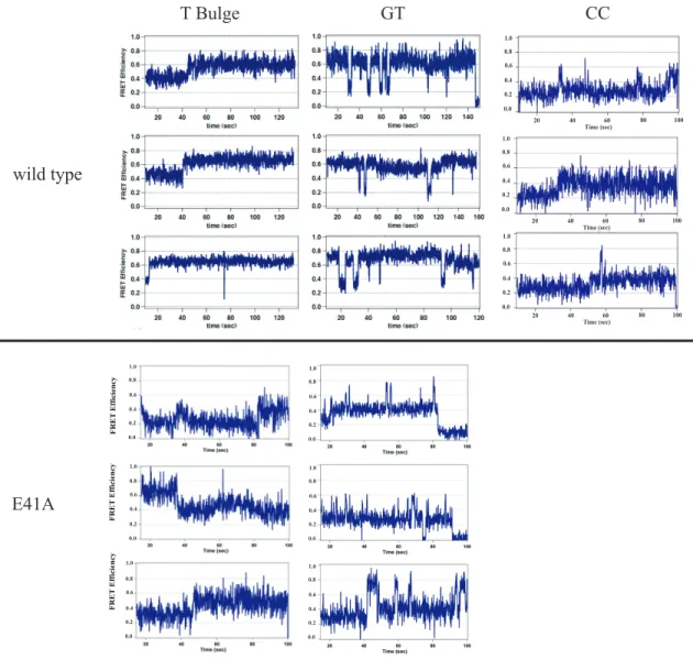
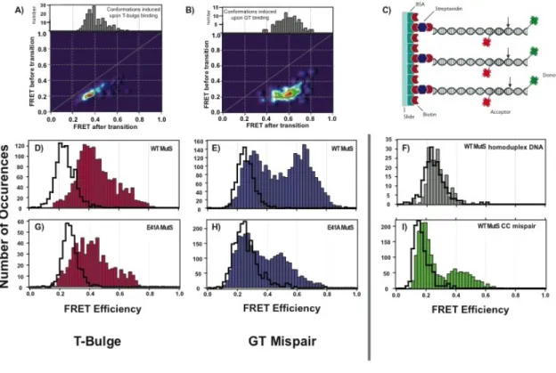
dominant states. In this work, the kinetics of interconversion among bending states was determined to vary widely for different mismatches. Further, the E41A mutant, which is known to have specific deficiencies in repair capability, demonstrates altered DNA bending kinetics on DNA mismatches that correlate with its repair deficiencies.
To my husband, Jason,
for all of his love, support, and understanding, To Rose,
for her strength, &
ACKNOWLEDGEMENTS
First and foremost, I’d like to thank my advisor, Dorothy Erie. No one could ask for a better student-mentor relationship. She has always supported every scientific and professional decision I have made as her student. She has given me the freedom to pursue the questions that I thought were most interesting even if that meant brief and often fruitless obsessions or insane quests to figure out why yeast cells hate me. Dorothy has been there for some of the biggest, saddest, and scariest events of my life and remained supportive through all of it. Most importantly, she’s taught me how to be a scientist. I cannot thank her enough for taking a chance on me. I’d like to think it has worked out for the both of us!
Enough cannot be said about the amazing people I have worked with in my graduate career. I’d like to thank my collaborators, Keith Weninger and Peggy Hsieh. Keith opened his lab to me and allowed me to monopolize his microscopes in my first few graduate years. I always felt that I had two advisors with Keith and Dorothy pushing me on. Peggy gave me the opportunity to collaborate with her on the human protein experiments. It has truly been a pleasure and a great learning experience working together.
establish the basis for everything I have learned. Thank you all for continuing to allow me bounce ideas and questions off of you regardless of where you have moved away to. I thank a special alumna of our lab, Hong, for taking the time to mentor me and do her best to advise me in my professional development. You are going to be an excellent PI in your own right, Hong! I thank Cherie for contributing her programming expertise at every turn. She made data analysis far less painful than it was when I first started this project and has always been ready to tweak programs based on analysis needs at the time. To Marc, Jackie, Dong, Dan, Matt, Kira, Jake, Zimeng, and Danielle- thank you for all of the Fridays. A special thanks to Jackie, Matt, Kira, and Jake for reading and editing
manuscripts, chapters, CVs, and cover letters. Your input and discussions have been very much appreciated. I cannot thank you enough. You all have helped keep me sane.
Thank you to the Weninger Lab - particularly to Brandon, John, Trevor and Ruoyi – for making me feel a part of their lab and for all of their helpful discussions. I thank the Hsieh Lab – especially Hui and Chunwei – for being no more than an e-mail away for questions and requests.
A very special thank you to my friends on the eighth floor. To Jen, Phil, Jake, and Nate for your ever-constant moral support, cynic humor, and often needed comic relief. You have helped make the labors of graduate school that much more bearable. I have learned entirely too much about RNA and RNA structure from you all, and somehow I enjoyed it. How strange!
Kady-Ann, Kevin, Brent, and Brad – there are no words. It is not often one finds friends as wonderful as you. You are my family here in North Carolina.
My wonderful husband – I would fill pages with the number of thank yous that you are owed. You have supported me throughout my endeavors to earn my doctorate and the bachelor’s degree before it. You have shared with me your family whose strength is infinite. You put up with the emotional roller coasters, the stress, the doubts, the fears, and the many, many tears. I cannot express how much I appreciate and love you. Thank you.
TABLE OF CONTENTS
LIST OF TABLES ... XIII
LIST OF FIGURES ... XIV
LIST OF ABBREVIATIONS ... XVI
CHAPTER 1 INTRODUCTION
Introduction ... 1
DNA Mismatch Repair ... 2
Prokaryotic Mismatch Repair ... 2
Eukaryotic Mismatch Repair ... 5
MutS in MMR Recognition and Initiation ... 6
MutS and its Homologs: A Structural Comparison ... 11
MutS(α) Mismatch DNA Binding – The Phe-X-Glu Motif ... 12
MutS and DNA Damage Response ... 20
MutS(α) ATPase activity and the sliding clamp ... 22
Bulk and Single-Molecule Fluorescence Techniques – The Biophysical Tools ... 25
Single-Molecule Fluorescence Resonance Energy Transfer (smFRET) ... 25
Their Best ... 32
Research Scope and Objectives ... 37
References ... 40
CHAPTER 2 DISTINCT DNA BENDING DYNAMICS ALLOW MUTS TO DISTINGUISH DIFFERENT DNA MISMATCHES Introduction ... 48
Results ... 53
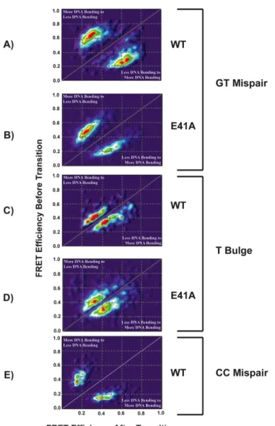
Wild type MutS-DNA conformations and conformational dynamics vary significantly from mismatch to mismatch ... 53
MutS binds the mismatch and directly induces unique conformational changes in the DNA ... 62
Taq E41A MutS-DNA complex adopts wild type-like conformations on T bulge and intermediately bent conformations on GT mismatch ... 64
Discussion ... 65
Glu41 stabilizes the MutS-mismatch DNA complex and also stabilizes DNA bending and unbending ... 67
DNA bending and unbending are essential in the ability for MutS to signal repair ... 69
Can DNA bending dynamics tell us something about mismatch repair in vivo? ... 70
Conclusions ... 72
Experimental Procedures ... 73
Protein and DNA substrates ... 74
Fluorescence microscopy ... 74
FRET data analysis ... 76
CHAPTER 3
FOUR-COLOR SINGLE MOLECULE FLUORESCENCE WITH NONCOVALENT DYE LABELING TO MONITOR DYNAMIC
MULTIMOLECULAR COMPLEXES
Introduction ... 81
Materials and Methods ... 85
Oligonucleotides ... 85
yMsh2-Msh6 protein ... 85
SNARE proteins ... 86
DNA Sample Preparation ... 87
SNARE Sample Preparation ... 87
tris-NTA-Oregon Green Labeling ... 88
Microscope ... 88
Observation protocols ... 90
Results and Discussion ... 91
References ... 103
CHAPTER 4 CHARACTERIZATION OF HUMAN MSH2-MSH6 NUCLEOTIDE OCCUPANCY Introduction ... 106
Materials and Methods ... 110
Preparation of human MSH2-MSH6 and its mutants ... 110
DNA substrates ... 111
UV-crosslinking of Nucleotide ... 111
WT, T1219D, and G674A preferentially bind ATP into
MSH6 over MSH2 under non-hydrolyzing conditions ... 117
DNA induces equal nucleotide binding into MSH2 and MSH6 of WT under hydrolyzing conditions ... 119
DNA induces a preferential binding of nucleotide into MSH2 for T1219D but into MSH6 for G674A under hydrolyzing conditions ... 120
WT, T1219D, and G674A bind ATP preferentially in MSH6 over MSH2 under non-hydrolyzing conditions ... 122
Discussion ... 125
T1219D is not deficient for nucleotide binding ... 126
G674A is a nucleotide binding deficient in MSH2 ... 128
Conclusions ... 129
References ... 130
CHAPTER 5 CHARACTERIZATION OF THE PROTEIN-DNA INTERACTIONS OF HUMAN MSH2-MSH6 Introduction ... 134
Materials and Methods ... 137
Preparation of human MSH2-MSH6 and its mutants ... 137
DNA Substrates ... 137
Fluorescence Anisotropy ... 139
TIRF Microscope ... 140
Results ... 141
Anisotropy binding constants support sliding clamp deficiency in T1219D and G674A ... 141
bent interaction ... 144
HNPCC mutant T1219D does not dissociate from a GT mismatch. ... 152
HNPCC mutant G674A adopts a conformation similar to that of sliding clamp in WT in the presence of a GT mismatch ... 159
Discussion ... 162
Non-dynamic DNA binding of wild type MSH2-MSH6 may be stabilized by the N-terminal region of MSH6 ... 162
G674A and T1219D are trapped in separate points in the nucleotide processing mechanism prior to sliding clamp formation. ... 164
Conclusions ... 168
APPENDIX A SINGLE-MOLECULE FRET DATA ANALYSIS PROTOCOL Part 1 – Conversion of .pma files to .traces files ... 171
Part 2 – Conversion of .trace files to an .itx file ... 176
Tips on saving molecules: ... 180
Part 3 – Analyzing the .itx files ... 183
LIST OF TABLES
Table 1.1– Mismatch repair proteins and their eukaryotic
homologs ... 3 Table 4.1 - DNA oligonucleotide sequences used for cross-linking
DNA substrate construction. ... 112 Table 5.1 - PCR primers used in 500mer DNA substrate
construction. ... 138 Table 5.2 - Dissociation constants derived from anisotropy binding
LIST OF FIGURES
Figure 1.1- Human DNA mismatch repair pathway. ... 8 Figure 1.2 - Crystal Structure of T. aquaticus MutS. ... 13 Figure 1.3 - Crystal Structure of human Msh2-Msh6. ... 14 Figure 1.4 - Distance dependence of donor to acceptor fluorescence
resonance energy transfer. ... 31 Figure 1.5 – Through Prism Total Internal Reflectance Fluorescence
(TIRF) Microscopy. ... 35 Figure 1.6 - Bulk Fluorescence Anisotropy L-format Instrumentation. ... 38 Figure 2.1 – Example WT MutS and E41A MutS FRET traces in the
presence of DNA substrates. ... 55 Figure 2.2 – WT MutS and E41A MutS smFRET histogram
distributions ... 57 Figure 2.3 - Transition density plots for WT MutS and E41A MutS
in the presence of GT, T bulge, and CC mismatch DNA. ... 61 Figure 3.1 – Four-color single molecule TIRF microscope schematic. ... 83 Figure 3.2 - yMutSα smFRET experiments. ... 93 Figure 3.3 - In situ labeling of SNARE proteins reconstituted in a
supported lipid bilayer using tris-NTA dyes. ... 97 Figure 3.4 - SNARE smFRET experiments. ... 99 Figure 4.1 – UV cross-linking experimental design. ... 113 Figure 4.2 – SDS-PAGE of UV cross-linked nucleotide into subunits
of WT, G674A, and T1219D in the absence of DNA under hydrolyzing
conditions. ... 116 Figure 4.3 - SDS-PAGE of UV cross-linked nucleotide into subunits
of WT, G674A, and T1219D in the absence of DNA under non-
appropriate DNA under hydrolyzing conditions. ... 121 Figure 4.5 - SDS-PAGE of UV cross-linked nucleotide into subunits
of WT, G674A, and T1219D in the presence of 10 nM of the
appropriate DNA under non-hydrolyzing conditions. ... 123 Figure 5.1 - Example of single-molecule fluorescent intensities and
FRET efficiencies. ... 145 Figure 5.2 - Example WT MutSα smFRET traces. ... 147 Figure 5.3 - Histogram distributions of smFRET states for WT,
T1219D, and G674A in the absence of nucleotide. ... 148 Figure 5.4 - Histogram distributions of smFRET values for WT,
T1219D, and G674A in the presence of 1 mM ADP. ... 150 Figure 5.5 - Histogram distributions of smFRET values for WT,
T1219D, and G674A in the presence of 1 mM ATP. ... 153 Figure 5.6 - Histogram distributions of smFRET values for WT,
T1219D, and G674A in the presence of 1 mM ATPγS. ... 155 Figure 5.7 - Example T1219D MutSα smFRET traces. ... 158 Figure 5.8 - Example G674A MutSα smFRET traces. ... 160 Figure 5.9 – MutS(α) MMR initiation model with trapped G674A
LIST OF ABBREVIATIONS
° Degree
γ Gamma correction factor
ηA Detection efficiency of an acceptor dye ηD Detection efficiency of a donor dye
θ1 Angle of incidence
θc Critical angle
λ Wavelength
µM micromolar
ΦA Quantum yield of an acceptor fluorophore ΦD Quantum yield of a donor fluorophore 8-oxo-G:A 8-oxo-guanine to adenine mismatch
a Anisotropy
Å Angstrom
ABC ATP binding cassette
ADP Adenine diphosphate
AFM Atomic force microscopy
Ala Alanine
ATP Adenine triphosphate
bp Base pair
BSA Bovine serum albumin
CC Cytosine-cytosine mismatch
d Evanescent field depth
dcrx dichroic
DNA Deoxyribonucleic acid
DTT Dithiothreitol
E FRET efficiency
E. coli Escherichia coli
EMCCD Electron-multiplying charge coupled device
Exo I Exonuclease I
Exo VII Exonuclease VII
Exo X Exonuclease X
FA Phenylalanine to alanine mutation
FRAP Fluorescence recovery after photobleaching FRET Fluorescence resonance energy transfer
G Correction factor in anisotropy G674A hMSH2G674A-hMSH6WT mutant
GG Guanine-guanine mismatch
Glu Glutamic acid
GT Guanine-thymine mismatch
GUI Graphic user interface
HCl Hydrogen chloride
HEPES 4-(2-hydroxyethyl)-1-piperazineethanesulfonic acid
his Histidine
HNPCC Hereditary non-polyposis colorectal cancer
Hz Hertz
I Intensity
𝐼∥ Intensity of emission parallel to excitation polarization 𝐼! Intensity of emission horizontal to excitation polarization IA Emission intensity of an acceptor fluorophore
IAafter bleach Acceptor emission intensity after a bleach event IAbefore bleach Acceptor emission intensity before a bleach event ID Emission intensity of a donor fluorophore
IDafter bleach Donor emission intensity after a bleach event IDbefore bleach Donor emission intensity before a bleach event IDL Insertion deletion loop
𝐼!! Horizontal polarization intensity after horizontally polarized excitation 𝐼!" Horizontal polarization intensity after vertically polarized excitation
𝐼!! Vertical polarization intensity after vertically polarized excitation IRC Initial recognition complex
IT Total intensity
kcal Kilocalorie
kcat Catalysis rate constant
KCl Potassium chloride
Kd Dissociation constant
m Meter
M Molar
MEF Mouse embryonic fibroblasts
mg Milligram
MgCl2 Magnesium chloride
min Minute
mL Milliliter
MLH1 MutL homolog 1
Mlh1 Yeast MutL homolog 1
MLH1-MLH3 Heterodimer of MutL homologs 1 and 3
Mlh1-Pms1 Heterodimer of MutL homolog 1 and postmeiotic segregation increased 1
MLH3 MutL homolog 3
mM Millimolar
mmDNA Mismatch DNA
MMR Mismatch repair
MNNG N-methyl-N’-nitro-N-nitrosoguanidine
mol Mole
MSH2 MutS homolog 2
MSH2-MSH3 Heterodimer of MutS homologs 2 and 3 MSH2-MSH6 Heterodimer of MutS homologs 2 and 6
MSH3 MutS homolog 3
Msh4 MutS homolog 4
MSI Microsatellite instability
MutLγ Heterodimer of MutL homologs 1 and 3
MutS(α) MutS and MutSα
MutSα Heterodimer of MutS homologs 2 and 6 MutSβ Heterodimer of MutS homologs 2 and 3
mW Milliwatt
n Refractive index
NA Numerical aperture
NaCl Sodium chloride
NaOAc Sodium acetate
ND Neutral density filter
NHS N-Hydroxysuccinimide ester
Ni2+ Nickel (II)
NIDDK National Institute of Diabetes and Digestive and Kidney Diseases NIH National Institutes of Health
nm Nanometer
nM Nanomolar
No. Number
NTP Nucleotide triphosphate
NTR N-terminal region
O6-MeG O6-methyl guanine
O6-MeGT O6-methyl guanine to thymine mismatch
OG Oregon green
P Polarization
PAGE Polyacrylamide gel electrophoresis PALM Photoactivated light microscopy PCNA Proliferating cell nuclear antigen PDB ID Protein database identification
Phe Phenylalanine
pM Picomolar
Pms1 Yeast postmeiotic segregation increased 1 PMS2 Postmeiotic segregation increased 2 PMSF Phenylmethylsulfonyl fluoride
Pol III Polymerase III
Pol α Polymerase α
Pol δ Polymerase δ
r Radius between donor and acceptor fluorophore
RFC Replication factor C
Ro Förster distance
RPA Replication factor A
s Second
S. cerevisiae Saccharomyces cerevisiae
SDS Sodium dodecyl sulfate
SH Detection efficiency of horizontally polarized light smFRET Single-molecule fluorescence resonance energy transfer SNAP-25 Soluble NSF Attachment Protein-25
SNARE Soluble NSF Attachment Protein Receptor SSB Single stranded DNA binding protein
ssDNA Single stranded DNA
SV Detection efficiency of vertically polarized light T bulge Single thymine insert
T1219D hMSH2WT-hMSH6T1219D mutant
TAMRA Tetramethylrhodamine
Taq Thermus aquaticus
TCEP tris(2-carboxyethyl)phosphine TDP Transition density plot
TIR Total internal reflection
Tris tris(hydroxymethyl)aminomethane buffer tris-NTA tris-nitrilotriacetic acid
TROLOX 6-hydroxy-2,5,7,8-tetramethylchroman-2-carboxylic acid URC Ultimate recognition complex
UV Ultra-violet
UvrD E. coli helicase II
WT Wild type
yMutSα Yeast MSH2-MSH6
Chapter 1
INTRODUCTION
DNA mismatch repair and single-molecule fluorescence techniques.
Introduction
The fidelity of any genome relies on the ability of the involved DNA polymerases to correctly replicate the genetic code. Despite the relatively high fidelity of the
replicative DNA polymerases (Pol α and Pol δ in eukaryotes and Pol III in prokaryotes), approximately 1 in every 107 base pairs synthesized result in an error being generated in the DNA per round of replication (Kunkel and Erie 2005; Iyer, Pluciennik et al. 2006; Kunz, Saito et al. 2009). These errors consist of base-base mismatches, insertion/deletion loops (IDLs) that result from polymerase slippages, and DNA lesions as a result of damaged templates and nucleotide triphosphate (NTP) pools (Schofield and Hsieh 2003; Iyer, Pluciennik et al. 2006; Hsieh and Yamane 2008; Kunz, Saito et al. 2009). Errors in the genetic code must be corrected to prevent carcinogenesis and disease. Fortunately, there are various repair pathways that post-replicatively repair DNA errors. DNA
replication decreases 100 fold to about 1 in every 109 bases synthesized (Schofield and Hsieh 2003; Kunkel and Erie 2005; Iyer, Pluciennik et al. 2006; Jiricny 2006; Kunz, Saito et al. 2009). The focus of this work is to characterize the initiation of one of the three mentioned DNA repair pathways, MMR.
DNA Mismatch Repair
DNA mismatch repair is a bi-directional, post-replicative process that repairs the afore mentioned base-base mismatches and IDLs (Haber and Walker 1991; Schofield and Hsieh 2003; Kunkel and Erie 2005; Iyer, Pluciennik et al. 2006; Hsieh and Yamane 2008). MMR is a highly conserved process across both prokaryotes and eukaryotes (Table 1.1) (Kunkel and Erie 2005).
Prokaryotic Mismatch Repair
Table 1.1– Mismatch repair proteins and their eukaryotic homologs
E. coli Protein Function Eukaryotic
Homolog
Function
MutS Recognizes base-base mismatches and IDLs
MSH2-MSH6
(MutSα)
Recognizes base-base
mismatches and 1-2 base IDLs and initiates MMR
MSH2-MSH3
(MutSβ)
Recognizes 2 or more base IDLs and initiates repair
MSH4-MSH5 Involved in meiosis
MutL Interacts in an ATP dependent manner with MutS once a mismatch has been identified.
Increases MutS specificity
Together, MutL and MutS initiate MMR.
MLH1-PMS2
(MutLα)
(yeast Mlh1-Pms1)
Interacts in an ATP dependent
manner with MutSα once a
mismatch has been identified.
Together, MutLα and MutSα
initiate MMR.
MLH1-PMS1
(MutLβ)
(yeast Mlh1-Mlh2)
Human function unknown
Minor role in frameshift suppression
MLH1-MLH3
(MutLγ)
Involved in repair of some IDLs. Functions in meiosis
MutH Nicks the daughter strand at a hemi-methylated GATC site
No known homolog
Helicase II (UvrD)
Unwinds the DNA for MMR
No known homolog
β-clamp Participates in DNA replication and repair synthesis
Increases processivity of Pol III during replication
Proliferating cell nuclear antigen (PCNA)
Participates in DNA
replication and repair synthesis
Has known interactions with
MutSα, MutSβ, MutLα, Exo I,
Pol δ, and Pol ε.
Recruits MMR proteins to DNA
Increases mismatch DNA binding specificity of Msh2-Msh6
Participates in DNA excision
γ – δ complex Loads β clamp onto the DNA
Replication Factor C
(RFC complex)
Loads PCNA onto the DNA
Exo I
Exo X
Required for 3′ to 5′
excision
EXO 1 5′ to 3′ exonuclease involved
in MMR excision
Deletion of EXO I results in only a mild mutator phenotype
Exo VII,
RecJ
Required for 5′ to 3′
excision Single stranded DNA Binding Protein (SSB) Binds ssDNA
Prevents self annealing of ssDNA
Participates in both DNA synthesis and repair
Replication Protein A (RPA)
Binds ssDNA in the gap
Prevents self annealing of ssDNA
Participates in both DNA synthesis and repair
Pol III DNA polymerase required for DNA resynthesis after excision
Polδ/ε DNA polymerase required for
DNA resynthesis after excision
Ligase Seals any nicks in the DNA after resynthesis
Ligase Seals any nicks in the DNA
after resynthesis
DNA beyond the error. Pol III resynthesizes a new DNA strand to fill the DNA gap, and DNA ligase seals any nicks. This process is bidirectional, meaning that MMR proceeds in both the 5′ to 3′ and the 3′ to 5′ directions (Hsieh 2001; Schofield and Hsieh 2003;
Kunkel and Erie 2005; Iyer, Pluciennik et al. 2006; Jiricny 2006; Hsieh and Yamane 2008).
Eukaryotic Mismatch Repair
Eukaryotic MMR is conserved from the prokaryotic system with almost every prokaryotic MMR protein having a eukaryotic homolog (Table 1.1). Unlike prokaryotic MMR, however, there is no known MutH or UvrD homologs in the eukaryotic repair pathway. Mismatch recognition responsibilities are shared by the MutS homolog
heterodimers, MSH2-MSH6 (MutSα) and MSH2-MSH3 (MutSβ). MutSα is responsible for recognizing base-base mismatches as well as small (1-2 nucleotide) IDLs. MutSβ is responsible for recognizing larger IDLs comprised of up to 16 nucleotides. MMR is initiated by MutSα or MutSβ recognizing a base-base mismatch or IDL error in the newly synthesized DNA strand. MutSα then interacts with the MutL homolog heterodimer MutLα (MLH1-PMS2 in humans or Mlh1-Pms1 in yeast) in an ATP dependent manner. MutLα has been shown to have its own endonuclease activity that is mismatch dependent (Kadyrov, Dzantiev et al. 2006; Kadyrov, Holmes et al. 2007; Kadyrov, Genschel et al. 2009). MutLα nicks the daughter strand and the MutSα-MutLα complex initiates MMR. How the protein complex differentiates the nascent DNA strand from the newly
A (RPA) binds and stabilizes the ssDNA-gapped region, and the DNA is resynthesized by Pol δ. Finally, DNA ligase seals any nicks in the new DNA strand.
MutS in MMR Recognition and Initiation
Unfortunately, there is no clearly defined mechanism by which MutS or MutSα (MutS(α) when referring to both the prokaryote and eukaryote) recognizes a mismatch and initiates repair of the DNA. Recognition of DNA mismatches is speculated to be related to the local flexibility of the DNA at the mismatch due to the local distortion effects of the incorrect pairing (Rajski, Jackson et al. 2000; Hsieh 2001; Schofield and Hsieh 2003; Kunkel and Erie 2005). The local flexibility of the DNA would differ at a mismatch versus homoduplex DNA. However, the thermodynamic stability of each mismatch does not correlate well with the relative repair efficiency of each mismatch. In fact, there is an apparent inverse correlation in the repair efficiency of a mismatch and the stability of that mismatch such that the more thermodynamically stable mismatches appear to be better repaired than the less stable mismatches (Peyret, Seneviratne et al. 1999; Wang, Yang et al. 2003; Kunkel and Erie 2005). The most stable mismatches, guanine-guanine (GG) or guanine-thymine (GT) mismatch, are the most efficiently repaired, with the least stable mismatch, cytosine-cytosine (CC) mismatch, being the least efficiently repaired (Su and Modrich 1986; Peyret, Seneviratne et al. 1999). This inverse correlation would suggest that structural factors must play role in mismatch
discrimination.
5’ 5’ 5’ 5’ 5’ 5’ 5’ 5’ 5’ 5’ 5’ 5’ PCNA RFC MSH2 MSH6 MLH1 PMS2 RPA Pol Ligase Mismatch Generated Recognition MMR Iniation & Nicking
Excision of MM DNA
Resynthesis
Ligation
ATP
Figure 1.1- Human DNA mismatch repair pathway.
binding properties of E. coli and Thermus aquaticus (Taq) MutS in the presence of a single thymine insertion (T bulge), or a GT mismatch, and homoduplex DNA (Wang, Yang et al. 2003). The AFM results indicated the presence of both bent (~42° bend for
Taq and ~74° bend for E. coli) and unbent (a ~0° bend for both Taq and E. coli)
populations on heteroduplex DNA with MutS localized at the DNA bend (Wang, Yang et al. 2003). The unbent population was only observed at the position of the mismatch or IDL (Wang, Yang et al. 2003). The distribution of states in the presence of homoduplex DNA indicated the presence of a single bent population comparable to the bent
population observed in the presence of heteroduplex DNA (a ~40-60° bend) (Wang, Yang et al. 2003). Given that there is no crystal structure of MutSα in complex with homoduplex DNA, this is one of the first structural insights into the interaction differences between MutSα and mismatch or homoduplex DNA.
repair of the identified mismatch or IDL (Wang, Yang et al. 2003; Kunkel and Erie 2005). Recent fluorescence resonance energy transfer (FRET) studies of MutS DNA bending dynamics and their associated kinetics lends further support to this DNA bending model as the discriminatory mechanism between homoduplex and heteroduplex DNA (DeRocco, Anderson et al. 2010; Sass, Lanyi et al. 2010).
The steps following DNA error recognition remain unclear. Three models have been proposed to explain the MMR initiation mechanism. The translocation model suggests that upon recognizing a mismatch, ATP hydrolysis by MutS(α) drives the protein to travel along the DNA bidirectionally away from the mismatch (Allen, Makhov et al. 1997; Blackwell, Bjornson et al. 1998; Gradia, Subramanian et al. 1999; Blackwell, Bjornson et al. 2001; Acharya, Foster et al. 2003; Gorman, Chowdhury et al. 2007). The function of MutS traveling away from the mismatch is proposed to be the recruitment of other mismatch repair proteins and the coordination of downstream repair events (Allen, Makhov et al. 1997; Blackwell, Bjornson et al. 1998; Gradia, Subramanian et al. 1999; Blackwell, Bjornson et al. 2001; Acharya, Foster et al. 2003; Gorman, Plys et al. 2010). The molecular switch model suggests that a combination of factors must occur prior to MutS(α) initiated repair. MutS(α) must recognize a mismatch and then bind ATP into both subunits. The rate limiting exchange of ADP to ATP in Msh2 (or subunit 2 in Taq
mismatch site (Habraken, Sung et al. 1998; Wang, Lawrence et al. 1999; Junop, Obmolova et al. 2001; Schofield, Nayak et al. 2001; Selmane, Schofield et al. 2003; Geng, Sakato et al. 2012). There is evidence to support each of the proposed MMR initiation models. The mechanism of MutS(α) action remains an area of great interest in the MMR field.
MutS and its Homologs: A Structural Comparison
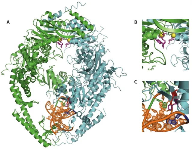
MutS exists as a homodimer in prokaryotes and a heterodimer of MSH2 and MSH6 (MutSα) or MSH2 and MSH3 (MutSβ) in eukaryotes. For the purpose of this work, we will exclude MutSβ from this discussion. MutS(α) contains a DNA binding domain at the N terminus and an ATPase domain at the C terminus of each subunit (Lamers, Perrakis et al. 2000; Obmolova, Ban et al. 2000; Natrajan, Lamers et al. 2003). The overall structure of the protein is conserved from E. coli to Taq (Figure 1.2) (Lamers, Perrakis et al. 2000; Obmolova, Ban et al. 2000; Warren, Pohlhaus et al. 2007). Human MSH2-MSH6 has also been crystallized (Figure 1.3) as a truncation mutant with a deletion of 341 residues at the N terminus of MSH6 (Warren, Pohlhaus et al. 2007). The truncated structure is remarkably similar to that of the prokaryotic protein structures. In all structures, the DNA is kinked in the DNA binding domains of the MutS(α) protein [60o angle in Taq (Obmolova, Ban et al. 2000) and E. coli (Lamers, Perrakis et al. 2000; Natrajan, Lamers et al. 2003), 45o angle in human MutSα (Warren, Pohlhaus et al. 2007)].
residue contacts that are specific to a mismatch in the DNA. The mismatch interaction motif will be discussed in detail within the next section of this chapter. Domain IV is often referred to as the clamp region since it is the region that encircles the DNA. Domain III is considered the core of the protein, as it maintains direct contacts with domains II, IV, and V. Domain II connects domain I to the core domain, domain III. Domain V acts as the dimerization interface for the two subunits. Once the two subunits dimerize, domain V forms the ATPase domain of each subunit. The ATPase sites require the two domains to be interfaced to be active (Obmolova, Ban et al. 2000; Junop,
Obmolova et al. 2001; Selmane, Schofield et al. 2003). Prior to DNA binding, domains I and IV exist as an unstructured globular region. Upon DNA binding, the domains I and IV form the structured DNA clamp region (Obmolova, Ban et al. 2000). Eukaryotic MutSα also has an additional 340 amino acid region located at the N-terminus of the MSH6 subunit. This additional region in MSH6 contains a PCNA interaction peptide motif (PIP box) (Gu, Hong et al. 1998; Clark, Valle et al. 2000; Bowers, Tran et al. 2001; Kleczkowska, Marra et al. 2001; Lau and Kolodner 2003; Lee and Alani 2006; Shell, Putnam et al. 2007; Iyer, Pohlhaus et al. 2008).
MutS(α) Mismatch DNA Binding – The Phe-X-Glu Motif
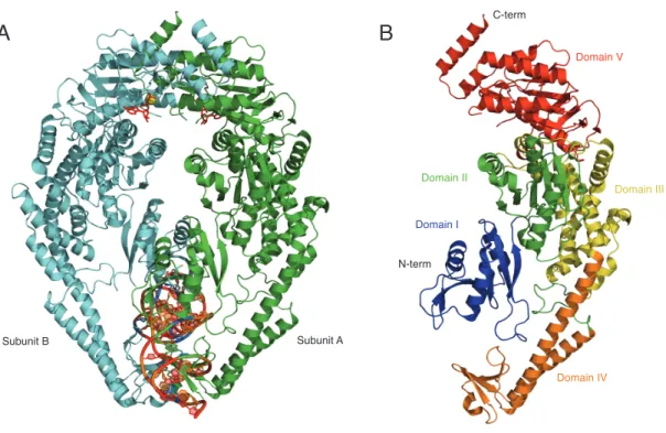
Figure 1.2 - Crystal Structure of T. aquaticus MutS.
(A) Front view of the MutS homodimer structure in complex with a single thymine insert (PDB ID: 1FW6). Subunit A contains the only specific contacts with the DNA mismatch. ADP is noted in the ATPase domains of each subunit in red stick representations.
Magnesium ligands involved in coordinating ADP binding in the ATPase domains are represented as yellow spheres. (B) Structural representation of each domain within each subunit of the MutS homodimer. Domain I, II, III, IV, and V are noted in blue, green, yellow, orange, and red respectively.
B
C-term
N-term
Domain I Domain II
Domain III
Domain IV
Domain V
Thermus Aquaticase MutS Homodimer
A
Figure 1.3 - Crystal Structure of human Msh2-Msh6.
subunit shown in green) and in domain I of MSH6 in the heterodimer (Figure 1.3). The Phe-X-Glu motif is evolutionarily conserved in the N terminus of the MutS protein family with the exception of Msh2, Msh3, Msh4, and Msh5 (Sachadyn 2010). The lack of the motif in Msh2 and presence in Msh6 is consistent with the asymmetric DNA binding observed in the Msh2-Msh6 protein. The lack of the motif in the DNA binding domain of Msh3 suggests the Msh2-Msh3 recognition mechanism of large IDLs differs from that of MSh2-Msh6. Msh4 and Msh5 have been shown to be actively involved in the meiotic DNA repair pathway, homologous recombination (HR) (Malkov, Biswas et al. 1997; Sachadyn 2010). Therefore, Msh4 and Msh5 may not have evolutionarily conserved the Phe-X-Glu contact due to the nature of their roles in DNA repair.
The exact roles of each residue in the Phe-X-Glu motif have been the subject of debate within the MMR field. However, there is no refuting the conserved stacking of the phenylalanine upon the mismatched thymine observed in the crystal structures of E. coli
MutS, Taq MutS, and human MSH2-MSH6 (Lamers, Perrakis et al. 2000; Obmolova, Ban et al. 2000; Warren, Pohlhaus et al. 2007). In each structure, the glutamate residue appears to form a hydrogen bond with the N3 of a mismatched thymine or the N7 of mismatched purines. Mutations in either Phe or Glu of the Phe-X-Glu motif result in significant changes in the MMR capabilities of each protein.
Mutation of the phenylalanine residue results in the elimination of both DNA binding by the protein and mismatch repair as a whole in E. coli (MutS F36A), Taq
Drotschmann, Yang et al. 2001; Tessmer, Yang et al. 2008). Each phenylalanine to alanine (FA) mutant MutS(α) exhibits dimerization stability and ATPase activity in the absence of DNA that is consistent with wild type MutS. In each case, the overall DNA binding affinity of FA MutS(α) is significantly weaker than that of wild type MutS(α) for both hetero- and homoduplex DNA substrates. Interestingly, the weakened FA MutS(α) binding affinity for heteroduplex DNA is on the same order of the FA MutS(α) binding affinity for homoduplex DNA indicating that the FA mutant is unable to discriminate between hetero- and homoduplex DNA. The combination of weakened DNA binding and loss of specificity would explain the elimination of MMR as a whole. (Malkov, Biswas et al. 1997; Bowers, Sokolsky et al. 1999; Das Gupta and Kolodner 2000; Dufner, Marra et al. 2000; Yamamoto, Schofield et al. 2000; Drotschmann, Yang et al. 2001; Tessmer, Yang et al. 2008)
Similar mutational analyses of the glutamate residue in the E. coli, Taq, and S. cerevisiae yielded inconclusive results. The glutamate residue has been proposed to facilitate DNA kinking via electrostatic repulsion of the DNA phosphate backbone. Various mutations to abolish both hydrogen bonding and charge repulsion have resulted in genome-wide mutation rates similar to that of a MutS null system in prokaryotes (Holmes, Scarpinato et al. 2007) and a weak mutator phenotype in S. cerevisiae (Das Gupta and Kolodner 2000; Drotschmann, Yang et al. 2001; Holmes, Scarpinato et al. 2007).
al. 2001; Lebbink, Georgijevic et al. 2006; Tessmer, Yang et al. 2008). Notably, DNA binding affinities of the EA mutants for homoduplex DNA show improvement to almost that of the heteroduplex substrates. Studies using DNA analogs that have no hydrogen bond capabilities indicate that hydrogen bonding at the glutamate residue is not required for DNA binding (Schofield, Brownewell et al. 2001; Drotschmann, Topping et al. 2004). MutS EA mutants in the presence of DNA containing base analogs exhibited similar binding affinities to that of typical heteroduplex DNA (Schofield, Brownewell et al. 2001; Drotschmann, Topping et al. 2004). This is consistent with a decreased
discrimination between homo- and heteroduplex DNA similar to that observed with the FA mutants.
ATPase studies of EA mutants in the presence of homoduplex, T bulge, and GT mismatch DNA exhibit a loss of the biphasic ATPase activity profile that is characteristic of WT MutS in the presence of DNA. Typically, WT MutS pre-steady state ATPase activity includes an initial fast burst phase followed by a slower linear phase. The slow ATPase phase is believed to be a result of rate-limiting ADP exchange. The EA mutants only exhibit the fast ATPase phase. There is no inhibition of ATPase due to ADP exchange. This, coupled with the observation that the EA mutant dissociates from DNA faster than WT MutS, suggests that while the EA mutants are able to bind DNA with a near wild type affinity, they are unable to adopt a secondary signaling conformation (largely believed to be the sliding clamp) as a result of ATP hydrolysis or ADP release (Lebbink, Georgijevic et al. 2006; Tessmer, Yang et al. 2008).
Scarpinato et al. 2007). The glutamate to alanine mutation in Msh2-Msh6 (Msh2-Msh6E339A) exhibits a weak mutator phenotype such that the mutant results in only an 8-fold increase in mutation rate over WT as compared to the 1000 8-fold increase in mutation rate seen in the Msh6 null strain (Drotschmann, Topping et al. 2004). This is
accompanied with both a slight loss in DNA binding affinity and a loss of mispair
specificity (Drotschmann, Topping et al. 2004). In depth characterization of the mutation spectrum of Msh2-Msh6E339A indicates that the glutamate residue is dispensable for repair of IDLs while remaining important in the repair of base-base mismatches (Holmes, Scarpinato et al. 2007). Interestingly, evaluation of Msh2-Msh6E339A in an oxidative damage (8-oxo-guanine DNA damage) prone cell line indicates a specific role for the glutamate residue in recognizing 8-oxo-G:A mispairs (Holmes, Scarpinato et al. 2007). Overall, the differences in EA mutant phenotypes from prokaryotes to eukaryotes suggest a much more mismatch specific repair system is at work in the higher organism.
DNA as compared to wild type while E41A exhibited comparable DNA binding affinities to wild type for homo- and heteroduplex DNA substrates (Malkov, Biswas et al. 1997; Yamamoto, Schofield et al. 2000; Tessmer, Yang et al. 2008). However, when the DNA conformations at a bound MutS protein were evaluated, differences between each mutant and wild type were observed.
As previously described in this chapter, wild type MutS bends homoduplex DNA into a single bend angle population (~60°) while MutS bends heteroduplex DNA into two populations (0° and ~40°). The unbent conformation is hypothesized to be the signaling conformation for downstream repair. In contrast, the bend angle distributions for F39A on homoduplex, GT mismatch, and T bulge DNA are each single populations centered on the same ~60° angle as is seen with WT MutS on homoduplex DNA. This result is
consistent with the F39A mutant being unable to discriminate between homo- and heteroduplex DNA. Neither the F39A GT mispair distribution nor the T bulge
distribution exhibits the 0° bend angle population seen with WT MutS on the same DNA substrates. The loss of the 0° bend angle suggests that the phenylalanine residue plays a role in either forming or stabilizing the unbent DNA conformation that is unique to MMR DNA substrates (Tessmer, Yang et al. 2008).
MutS and DNA Damage Response
Mutations in MMR proteins have been identified in HNPCC patients.
MutSα is believed to play a role in the initiation of DNA damage induced apoptosis as a result of the presence of alkylated or cisplatinated nucleic acids in newly replicated DNA. Patients with mutations in the MMR protein alleles often display a resistance to the affectivity of certain chemotherapeutic agents such as N-methyl-N’-nitro-N-nitrosoguanidine (MNNG) and cisplatin.
MMR deficient cells were found to resist the effects of methylating agents used for chemotherapy while MMR proficient cells did not have the same resistive phenotype (Stojic, Brun et al. 2004). These methylating agents include such active components as temozolomide (TMZ), dacarbazine and procarbazine, and MNNG. MNNG exposure results in the most common DNA damage, O6-methylguanine (O6-MeG) (Stojic, Brun et al. 2004). In general, the presence of O6-MeGT mismatches is believed to trigger
apoptosis (Fishel 1999; Karran 2001; Kunkel and Erie 2005).
The mechanism by which apoptosis is signaled is not conclusively known. However, two theories have been proposed (Fishel 1999; Karran 2001). The “futile repair” hypothesis proposes that MutSα binds to the O6-MeGT mismatch and signals MMR. If the parent strand contains the methylated base, MMR results in the removal of the thymine. DNA resynthesis results in a reinsertion of a thymine opposite to the O6 -MeG. This process continually repeats because the damaged portion of the DNA resides in the parent strand. Eventually, this excision and resynthesis leads to apoptosis triggered by double strand breaks formed in the DNA during replication. Alternately, the
mechanism by which MutS(α) signals apoptosis rather than repair may be better understood.
MutS(α) ATPase activity and the sliding clamp
The ATPase domains of MutS located in domain V of each subunit are essential for DNA repair (Haber and Walker 1991; Wu and Marinus 1994; Studamire, Quach et al. 1998; Drotschmann, Clark et al. 1999; Lamers, Perrakis et al. 2000; Biswas, Obmolova et al. 2001). Each ATPase site is comprised of a highly conserved Walker A and Walker B nucleotide binding motif as well as a set of six motifs that are a signature of ATP-binding cassette transporters (ABC transporters) at the C terminus of the subunit (Gorbalenya and Koonin 1990; Lamers, Perrakis et al. 2000; Obmolova, Ban et al. 2000; Junop, Obmolova et al. 2001; Alani, Lee et al. 2003). MutS and its homologs therefore belong to the ABC ATPase superfamily (Gorbalenya and Koonin 1990). The two ATPase regions of the protein exist at the interface between subunits as is characteristic of this superfamily. Each subunit of MutS(α) site contains five of the ABC motifs while the sixth ABC ATPase motif is provided by the interfacing subunit. The MutS(α) subunit dimerization results in two functional composite ATPase sites (Junop, Obmolova et al. 2001).
studies (Haber and Walker 1991; Gradia, Acharya et al. 1997; Blackwell, Bjornson et al. 1998; Blackwell, Martik et al. 1998; Gradia, Subramanian et al. 1999; Gradia, Acharya et al. 2000; Drotschmann, Hall et al. 2002; Joshi and Rao 2002; Antony and Hingorani 2003; Selmane, Schofield et al. 2003; Lamers, Georgijevic et al. 2004; Antony, Khubchandani et al. 2006; Mazur, Mendillo et al. 2006; Jacobs-Palmer and Hingorani 2007; Cyr 2008; Zhai and Hingorani 2010; Heinen, Cyr et al. 2011). Progress has been made in understanding the role of nucleotide occupancy in the formation of MutS(α)-MutL(α) ternary complexes and signaling downstream repair. Studies have shown that while the presence of ATP is a requirement for MutS(α)-MutL(α)-mismatch DNA (mmDNA) complex formation, ATP hydrolysis by either protein is not necessary
Remarkably, MMR deficiency is not a result of loss of ATP hydrolysis or of a loss in ternary complex formation with MutL. Rather, MMR deficiency has been shown to be a result of loss of downstream signaling effects.
Other mutations in the ATPase domains result in separation-of-function mutant MutSα proteins which are deficient in MMR but are able to participate in apoptotic responses due to DNA damage. For example, a mutation of a glycine residue in the Walker A motif of Msh2 (GXXXGK(S/T)) (MSH2G674A-MSH6WT in mouse and human systems) results in a deficiency in MMR while maintaining DNA damage response (Yang, Scherer et al. 2004; Geng, Sakato et al. 2012). Mouse embryonic fibrobalsts (MEFs) containing the Msh2G674A-Msh6 protein exhibit the same mutagenic spectrum of cancer development as that of MutSα null cells (Yang, Scherer et al. 2004). However, the Msh2G674A-Msh6 MEFs have a longer lifespan than that of the Msh2 null mice and exhibit the same susceptibility to chemotherapeutic treatment that WT Msh2-Msh6 mice had (Yang, Scherer et al. 2004) suggesting that the Msh2G674A-Msh6 cells are inducing apoptosis in a similar manner to that of WT Msh2-Msh6 cells.
hallmark of cancer susceptibility) while maintaining wild type-like cellular vulnerability to DNA damage (Yang, Scherer et al. 2004).
Interestingly, both separation-of-function mutants described above have been shown in vitro to be able to bind mmDNA like wild type protein, but are unable to form the ternary complex with MutLα that is required for MMR to proceed (Hess, Gupta et al. 2002; Hess, Mendillo et al. 2006; Geng, Sakato et al. 2012) suggesting a role for the MutSα-MutLα ternary complex in MMR signaling and some divergent function of MutSα in apoptotic response.
Bulk and Single-Molecule Fluorescence Techniques – The Biophysical Tools More and more, fluorescence-based techniques are being used to answer biological questions. The low-noise characteristic of fluorescence spectroscopy is attractive for use with complicated biological systems. The ability to use fluorescence at little to no risk to the integrity of the biological systems being probed is also highly appealing. The improvement of detection methods for fluorescence systems, in
conjunction with the discovery of fluorescent proteins and the engineering of new, longer lifetime fluorescent proteins and organic dyes has pushed fluorescent technologies to the forefront of biological application. Technologies such as fluorescence resonance energy transfer (FRET), photoactivated light microscopy (PALM), fluorescence recovery after photobleaching (FRAP), and multi-photon fluorescence microscopy have allowed for high resolution of biological processes both in vivo and in vitro.
about the average of all states present. However, bulk studies do not provide information about the individual characteristics of subpopulations of conformations which may be present but unresolvable within the ensemble average (Gell, Brockwell et al. 2006). Based on these same principles, if two states exist in approximately equal occurrence in bulk then both states are averaged to converge on a single mean value that may not actually represent either of the major states. These bulk experiments, therefore, disregard the heterogeneity of a sample by either assuming that the dominant states are the only states involved in a process or by not representing two equal states at all (Gell, Brockwell et al. 2006; Roy, Hohng et al. 2008). Single-molecule experiments allow an experimenter to distinguish between conformations that may be present at any given point in time given the time resolution of the technique being utilized.
Single-molecule fluorescence is a low noise technique that allows for detection limits on the order of 1.66 x 10⁻²⁴ mole (or molecule) of the analyte of interest (Gell, Brockwell et al. 2006; Lakowicz 2006). In addition to very low limits of detection, single-molecule fluorescence techniques allow for very high spatial resolution of the probe of interest and the environment surrounding that probe (Gell, Brockwell et al. 2006; Lakowicz 2006; Roy, Hohng et al. 2008). The combination of low limits of detection, high resolution, and current advances in nanotechnology and electronics has advanced techniques like single-molecule FRET (Gell, Brockwell et al. 2006).
the ratio of the energy transfer rate to the sum of all processes resulting in the relaxation of the donor molecule (Gell, Brockwell et al. 2006; Lakowicz 2006). Efficiency of energy transfer is dependent on several properties of the donor and acceptor dye pair selected: (1) the degree of spectral overlap between the donor emission spectrum and the acceptor absorption spectrum, (2) the quantum yield of the donor molecule, which is defined as the number of photons emitted relative to the number of photons absorbed by the molecule, (3) the relative orientation of the donor and acceptor transition dipoles, and (4) the physical distance between the donor and acceptor molecules (Lakowicz 2006). Energy transfer efficiency (FRET efficiency, E) is defined as Eq. 1.1:
𝐸 = 1
1+ 𝑟!
𝑅!!
Eq. 1.1 where r is the distance between the donor and acceptor probe and Ro is the Forster
and acceptor dye (Figure 1.4). Measureable changes in positions are typically on the order of 10-9 m.
In single-molecule experiments, FRET efficiency can be expressed (Eq. 1.2) as an intensity ratio such that
𝐸= 𝐼!
𝐼!+𝐼!
Eq. 1.2 where IA is the intensity of fluorescence being emitted by the acceptor molecule and ID is the intensity of the donor fluorescence (Gell, Brockwell et al. 2006). In this case, only the donor molecule is excited and the transfer to the acceptor molecule is monitored in terms of fluorescence intensity. More correctly, experimental FRET efficiency is expressed as Eq. 1.3:
𝐸= 𝐼!
𝛾𝐼!+𝐼!
Eq. 1.3 where γ is a correction factor that accounts for quantum yields and detection efficiencies of each dye for a particular experimental system. The γ factor (Eq. 1.4) allows
experimental normalization of results from microscope to microscope where
𝛾 = Φ!𝜂!
Φ!𝜂!
Total Internal Reflection Fluorescence Microscopy – TIRFing For
Single-molecule Detection
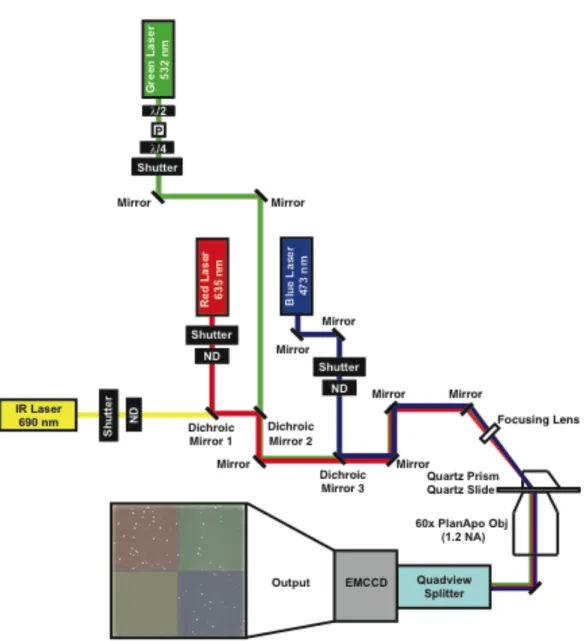
Single-molecule detection has been made possible because of low noise excitation methods such as total internal reflection fluorescence (TIRF) as well as the improvement in electronic systems for detection. The increase in the number of researchers performing single-molecule fluorescence methodologies is due to the rise of commercially available (and relatively inexpensive) microscope systems for TIRF based excitation. There are two modes of TIRF excitation – through objective and through prism (Figure 1.5). Both utilize the same principle of total internal reflection to generate an evanescent wave in order to excite a sample. For the purpose of this work, I will describe TIR as it applies to the through prism instrument design shown in Figure 1.5.
Total internal reflection occurs when the angle of incidence (θ1) of a beam at the interface between two materials with different refractive indices (n1 and n2) is equal to or greater than the critical angle (θc) (Figure 1.5) (Gell, Brockwell et al. 2006; Lakowicz 2006). The critical angle is defined as the angle at which all incident light is reflected and is calculated by Snell’s law as (Eq. 1.5)
𝜃! =sin!! 𝑛!
𝑛!
Eq. 1.5 The intensity of the reflected light is also somewhat dependent on the polarization of the incident light.
index (n2 in Figure 1.5). The propagation intensity decay has a z direction dependence described by Eq. 1.6:
𝐼 𝑧 = 𝐼 0 𝑒𝑥𝑝 −𝑧 𝑑
Eq. 1.6 where I(z) is the field intensity in the z direction into the lower refractive index material (n2), I(0) is the intensity of the evanescent field at the interface, and d is the penetration depth of the evanescent field. The penetration depth of the field is dependent on the wavelength of the incident beam (λ), the angle of incidence, and the refractive indices of the two materials at the interface. This relationship is described by Eq. 1.7.
𝑑 = 𝜆
4𝜋 𝑛!!sin!𝜃 !−𝑛!!
Figure 1.4 - Distance dependence of donor to acceptor fluorescence resonance energy transfer.
Regardless of the excitation wavelength used, the overall sample excitation is restricted to ~200 nm at the interface between the two materials. This limited excitation range allows for low noise, single-molecule detection at the interface as only the
fluorophores within this 200 nm range will be excited. The use of through-prism TIRF microscopy for smFRET excitation and detection of the DNA binding and bending properties of Taq, S. cerevisiae, and human MutS(α) for homoduplex, GT, T bulge, and O6MeGT DNA will be extensively discussed in this dissertation.
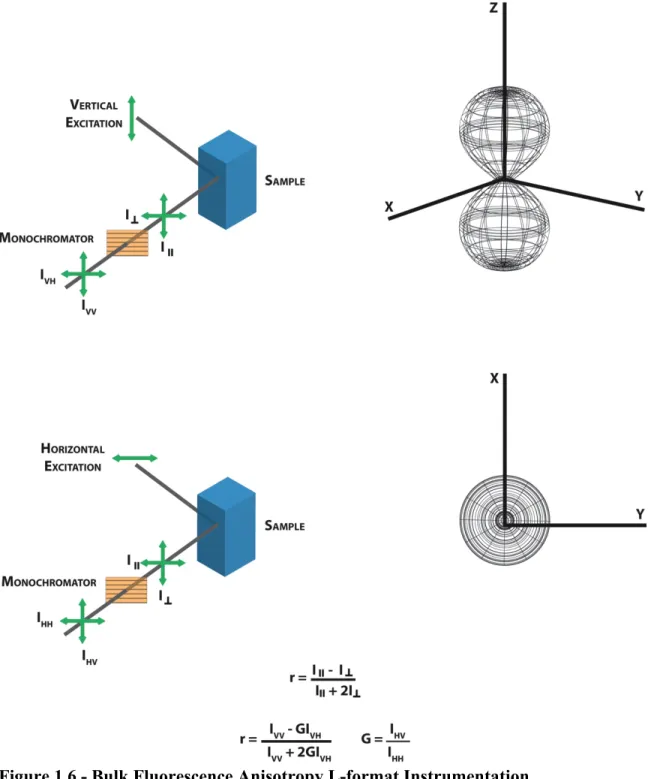
Fluorescence Anisotropy – Bulk Measurements at Their Best
Fluorescence anisotropy is a standard bulk fluorescence technique capable of measuring the binding of a fluorescent ligand or substrate by biological macromolecules. The technique takes advantage of principles of light polarization. When a fluorescent sample is excited with a polarized light, the sample emission is also polarized. The degree of polarization of the sample emission is described as anisotropy. Changes in the degree of polarization of a sample emission result in changes in the sample anisotropy, which reflect the overall heterogeneity of that sample.
Measurement of anisotropy values in an L-format fluorimeter is depicted in Figure 1.6. Excitation with both vertically and horizontally polarized light yields two emission components, 𝐼∥ (intensity of emission parallel to excitation polarization) and
𝐼!(intensity of emission perpendicular to excitation polarization). Anisotropy is a
ratiometric measurement of 𝐼∥ and 𝐼! with respect to the total intensity (𝐼!) of the sample emission.
Eq. 1.8 The 𝐼! term accounts for intensity of the sample in the x direction (𝐼!), the y direction (𝐼!), and the z direction (𝐼∥). Anisotropy is therefore expressed as shown in Eq. 1.9
𝑎 = 𝐼∥−𝐼! 𝐼∥+2𝐼!
Eq. 1.9 where a is the anisotropy of the sample. This is not the same as a polarization
measurement. Polarization of a sample and anisotropy of a sample are related, but not directly interchangeable terms. Polarization of a sample is not measured with respect to the total sample emission intensity as it does not account for both x and y 𝐼! terms (Eq.
1.10).
𝑃= 𝐼∥−𝐼!
𝐼∥+𝐼!
Eq. 1.10 In terms of experimental measurements, anisotropy is calculated from four
measureable intensities: 𝐼!! (excitation with vertically polarized light resulting in vertically polarized emission intensity), 𝐼!" (excitation with vertically polarized light
Figure 1.5 – Through Prism Total Internal Reflectance Fluorescence (TIRF) Microscopy.
polarized light resulting in vertically polarized emission intensity), and 𝐼!! (excitation with horizontally polarized light resulting in horizontally polarized emission intensity) ( Figure 1.6). Experimental anisotropy is calculated as:
𝑎 = 𝐼!!−𝐺𝐼!"
𝐼!!+2𝐺𝐼!"
Eq. 1.11 where 𝐺 is a factor that accounts for differences in the efficiency of instrumental
detection of vertical or horizontally polarized light.
𝐺 =𝑆𝑆! ! =
𝐼!" 𝐼!!
Eq. 1.12 where 𝑆! and 𝑆! are the detection efficiencies of vertical and horizontal light
respectively. The G factor can also be expressed in terms of 𝐼!" and 𝐼!! as shown in Eq. 1.12.
population of protein-bound ligand. As the sample becomes a heterogeneous mixture containing protein-bound and unbound fluorescent ligand, the ensemble average anisotropy of the sample shifts away from the innate anisotropy of fluorescent ligand alone until the sample becomes a homogenous mixture of protein bound ligand. These changes in anisotropy can be used to construct a binding isotherm, which can be fit with the appropriate binding model to determine a protein binding affinity for the fluorescent ligand. We take advantage of this system to measure the DNA binding affinity of human MutSα for homoduplex DNA, a GT mismatch, and O6MeGT damaged DNA substrate.
Research Scope and Objectives
Figure 1.6 - Bulk Fluorescence Anisotropy L-format Instrumentation.
system partially due to the need to explore both DNA bending and protein colocalization at the mismatch simultaneously in the eukaryotic system. Finally, through a collaboration established with Peggy Hsieh at the NIDDK (NIH, Bethesda), we were given the
opportunity to explore the DNA binding properties of higher eukaryotes in analyses of human MutSα for both base-base mismatched DNA and the damaged DNA substrate O6MeGT. The latter work also includes analyses of two separation-of-function mutant proteins (MSH2WT-MSH6T1219D and MSH2G674A-MSH6WT) that have been shown to be deficient in MMR but proficient in apoptotic response to DNA damage.
During my research tenure, I have used smFRET in conjunction with bulk
fluorescence anisotropy to characterize the DNA binding and bending interactions of Taq (described in Chapter 2) and human MutS(α) (described in Chapter 5) with a variety of DNA substrates including homoduplex, GT mismatch, single thymine IDL, and O6MeGT damaged DNA. I have optimized a multi-color TIRF microscope and dye labeling system for simultaneous excitation and detection of up to four laser lines (described in Chapter 3). I have disassembled and assembled a two-color TIRF and a four-color TIRF
References
Acharya, S., P. L. Foster, et al. (2003). "The coordinated functions of the E. coli MutS and MutL proteins in mismatch repair." Mol Cell 12(1): 233-246.
Alani, E., J. Y. Lee, et al. (2003). "Crystal structure and biochemical analysis of the MutS.ADP.beryllium fluoride complex suggests a conserved mechanism for ATP interactions in mismatch repair." J Biol Chem 278(18): 16088-16094.
Allen, D., A. Makhov, et al. (1997). "MutS mediates heteroduplex loop formation by a translocation mechanism." The EMBO journal 16(14): 4467-4476.
Antony, E. and M. M. Hingorani (2003). "Mismatch recognition-coupled stabilization of Msh2-Msh6 in an ATP-bound state at the initiation of DNA repair." Biochemistry 42(25): 7682-7693.
Antony, E., S. Khubchandani, et al. (2006). "Contribution of Msh2 and Msh6 subunits to the asymmetric ATPase and DNA mismatch binding activities of Saccharomyces cerevisiae Msh2-Msh6 mismatch repair protein." DNA repair 5(2): 153-162. Berends, M. J., Y. Wu, et al. (2002). "Molecular and clinical characteristics of MSH6
variants: an analysis of 25 index carriers of a germline variant." Am J Hum Genet 70(1): 26-37.
Biswas, I., G. Obmolova, et al. (2001). "Disruption of the helix-u-turn-helix motif of MutS protein: loss of subunit dimerization, mismatch binding and ATP hydrolysis." J Mol Biol 305(4): 805-816.
Bjornson, K. P. and P. L. Modrich (2003). "Differential and simultaneous adenosine di- and triphosphate binding by MutS." The Journal of biological chemistry 278(20): 18557-18562.
Blackwell, L. J., K. Bjornson, et al. (1998). "DNA-dependent activation of the hMutSalpha ATPase." The Journal of biological chemistry 273(48): 32049-32054.
Blackwell, L. J., K. P. Bjornson, et al. (2001). "Distinct MutS DNA-binding modes that are differentially modulated by ATP binding and hydrolysis." The Journal of biological chemistry 276(36): 34339-34347.
Blackwell, L. J., D. Martik, et al. (1998). "Nucleotide-promoted release of hMutSalpha from heteroduplex DNA is consistent with an ATP-dependent translocation mechanism." The Journal of biological chemistry 273(48): 32055-32062. Bowers, J., T. Sokolsky, et al. (1999). "A mutation in the MSH6 subunit of the
Bowers, J., P. T. Tran, et al. (2001). "MSH-MLH complexes formed at a DNA mismatch are disrupted by the PCNA sliding clamp." Journal of molecular biology 306(5): 957-968.
Clark, A. B., F. Valle, et al. (2000). "Functional interaction of proliferating cell nuclear antigen with MSH2-MSH6 and MSH2-MSH3 complexes." The Journal of biological chemistry 275(47): 36498-36501.
Cyr, J. (2008). "Hereditary cancer-associated missense mutations in hMSH6 uncouple ATP hydrolysis from DNA mismatch binding." Journal of Biological Chemistry. Das Gupta, R. and R. D. Kolodner (2000). "Novel dominant mutations in Saccharomyces
cerevisiae MSH6." Nat Genet 24(1): 53-56.
DeRocco, V., T. Anderson, et al. (2010). "Four-color single-molecule fluorescence with noncovalent dye labeling to monitor dynamic multimolecular complexes." Biotechniques 49(5): 807-816.
Drotschmann, K., A. B. Clark, et al. (1999). "Mutator phenotypes of yeast strains
heterozygous for mutations in the MSH2 gene." Proc Natl Acad Sci U S A 96(6): 2970-2975.
Drotschmann, K., M. C. Hall, et al. (2002). "DNA binding properties of the yeast Msh2-Msh6 and Mlh1-Pms1 heterodimers." Biol Chem 383(6): 969-975.
Drotschmann, K., R. P. Topping, et al. (2004). "Mutations in the nucleotide-binding domain of MutS homologs uncouple cell death from cell survival." DNA Repair (Amst) 3(7): 729-742.
Drotschmann, K., W. Yang, et al. (2001). "Asymmetric recognition of DNA local distortion. Structure-based functional studies of eukaryotic Msh2-Msh6." J Biol Chem 276(49): 46225-46229.
Dufner, P., G. Marra, et al. (2000). "Mismatch recognition and DNA-dependent stimulation of the ATPase activity of hMutSalpha is abolished by a single mutation in the hMSH6 subunit." The Journal of biological chemistry 275(47): 36550-36555.
Fishel, R. (1999). Signaling mismatch repair in cancer. Nature medicine. 5: 1239-1241. Forster, T. (1959). "Transfer Mechanisms of electronic excitation." Discussions of the
Faraday Society 27: 7-17.
Geng, H., M. Sakato, et al. (2012). "Biochemical Analysis of the Human Mismatch Repair Proteins hMutS MSH2G674A-MSH6 and MSH2-MSH6T1219D." The Journal of biological chemistry 287(13): 9777-9791.
Gorbalenya, A. E. and E. V. Koonin (1990). "Superfamily of UvrA-related NTP-binding proteins. Implications for rational classification of recombination/repair systems." Journal of molecular biology 213(4): 583-591.
Gorman, J., A. Chowdhury, et al. (2007). "Dynamic basis for one-dimensional DNA scanning by the mismatch repair complex Msh2-Msh6." Mol Cell 28(3): 359-370. Gorman, J., A. J. Plys, et al. (2010). "Visualizing one-dimensional diffusion of eukaryotic
DNA repair factors along a chromatin lattice." Nature structural & molecular biology 17(8): 932-938.
Gradia, S., S. Acharya, et al. (1997). "The human mismatch recognition complex hMSH2-hMSH6 functions as a novel molecular switch." Cell 91(7): 995-1005. Gradia, S., S. Acharya, et al. (2000). "The role of mismatched nucleotides in activating
the hMSH2-hMSH6 molecular switch." J Biol Chem 275(6): 3922-3930. Gradia, S., D. Subramanian, et al. (1999). "hMSH2-hMSH6 forms a
hydrolysis-independent sliding clamp on mismatched DNA." Mol Cell 3(2): 255-261. Gu, L., Y. Hong, et al. (1998). "ATP-dependent interaction of human mismatch repair
proteins and dual role of PCNA in mismatch repair." Nucleic Acids Research 26(5): 1173-1178.
Haber, L. T. and G. C. Walker (1991). "Altering the conserved nucleotide binding motif in the Salmonella typhimurium MutS mismatch repair protein affects both its ATPase and mismatch binding activities." The EMBO journal 10(9): 2707-2715. Habraken, Y., P. Sung, et al. (1998). "ATP-dependent assembly of a ternary complex
consisting of a DNA mismatch and the yeast MSH2-MSH6 and MLH1-PMS1 protein complexes." J Biol Chem 273(16): 9837-9841.
Hargreaves, V. V., S. S. Shell, et al. (2010). "Interaction between the Msh2 and Msh6 Nucleotide-binding Sites in the Saccharomyces cerevisiae Msh2-Msh6 Complex." Journal of Biological Chemistry 285(12): 9301-9310.
Heinen, C. D., J. L. Cyr, et al. (2011). "Human MSH2 (hMSH2) Protein Controls ATP Processing by hMSH2-hMSH6." The Journal of biological chemistry 286(46): 40287-40295.
Hess, M. T., M. L. Mendillo, et al. (2006). "Biochemical basis for dominant mutations in the Saccharomyces cerevisiae MSH6 gene." Proc Natl Acad Sci U S A 103(3): 558-563.
Holmes, S. F., K. D. Scarpinato, et al. (2007). "Specialized mismatch repair function of Glu339 in the Phe-X-Glu motif of yeast Msh6." DNA Repair (Amst) 6(3): 293-303.
Hsieh, P. (2001). "Molecular mechanisms of DNA mismatch repair." Mutat Res 486(2): 71-87.
Hsieh, P. and K. Yamane (2008). "DNA mismatch repair: molecular mechanism, cancer, and ageing." Mechanisms of ageing and development 129(7-8): 391-407.
Iyer, R. R., A. Pluciennik, et al. (2006). "DNA mismatch repair: functions and mechanisms." Chem Rev 106(2): 302-323.
Iyer, R. R., T. J. Pohlhaus, et al. (2008). "The MutSalpha-proliferating cell nuclear antigen interaction in human DNA mismatch repair." J Biol Chem 283(19): 13310-13319.
Jacobs-Palmer, E. and M. M. Hingorani (2007). "The effects of nucleotides on MutS-DNA binding kinetics clarify the role of MutS ATPase activity in mismatch repair." J Mol Biol 366(4): 1087-1098.
Jiang, J., L. Bai, et al. (2005). "Detection of high-affinity and sliding clamp modes for MSH2-MSH6 by single-molecule unzipping force analysis." Molecular cell 20(5): 771-781.
Jiricny, J. (2006). "The multifaceted mismatch-repair system." Nature reviews Molecular cell biology 7(5): 335-346.
Joshi, A. and B. J. Rao (2002). "ATP hydrolysis induces expansion of MutS contacts on heteroduplex: a case for MutS treadmilling?" Biochemistry 41(11): 3654-3666. Junop, M., G. Obmolova, et al. (2001). "Composite Active Site of an ABC ATPase::
MutS Uses ATP to Verify Mismatch Recognition and Authorize DNA Repair." Molecular cell.
Kadyrov, F. A., L. Dzantiev, et al. (2006). "Endonucleolytic function of MutLalpha in human mismatch repair." Cell 126(2): 297-308.